. 2007 Oct 23;17(20):1752-8.
doi: 10.1016/j.cub.2007.09.039. Epub 2007 Oct 11.

Role of Septin cytoskeleton in spine morphogenesis and dendrite development in neurons

Affiliations

Role of Septin cytoskeleton in spine morphogenesis and dendrite development in neurons

Tomoko Tada et al. Curr Biol. .

Abstract

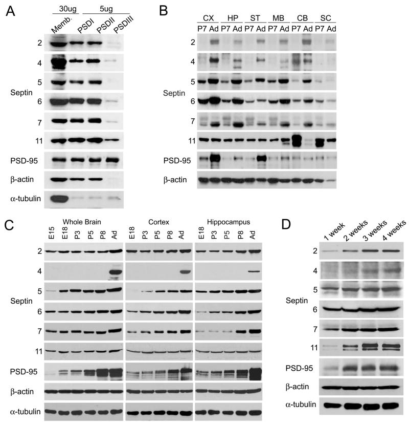
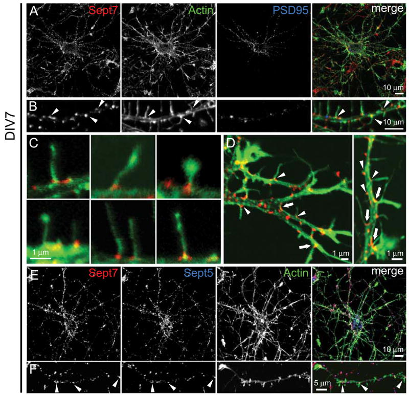
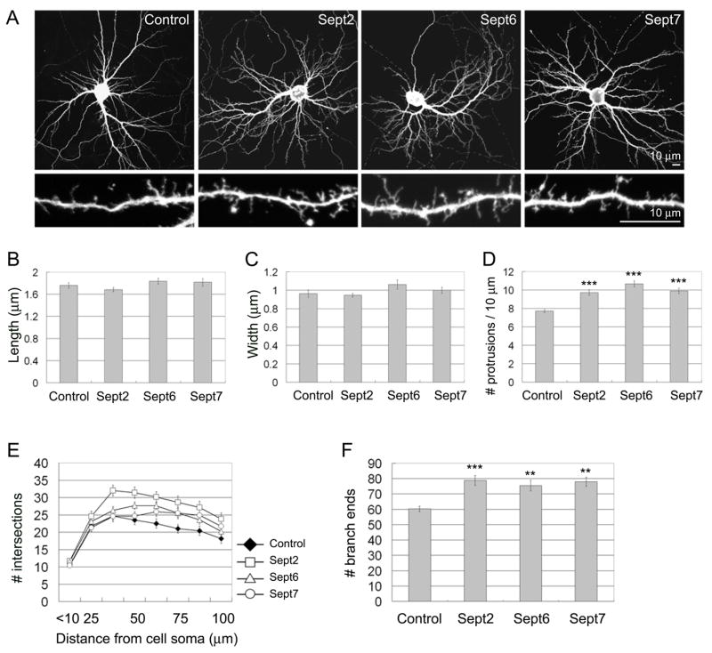
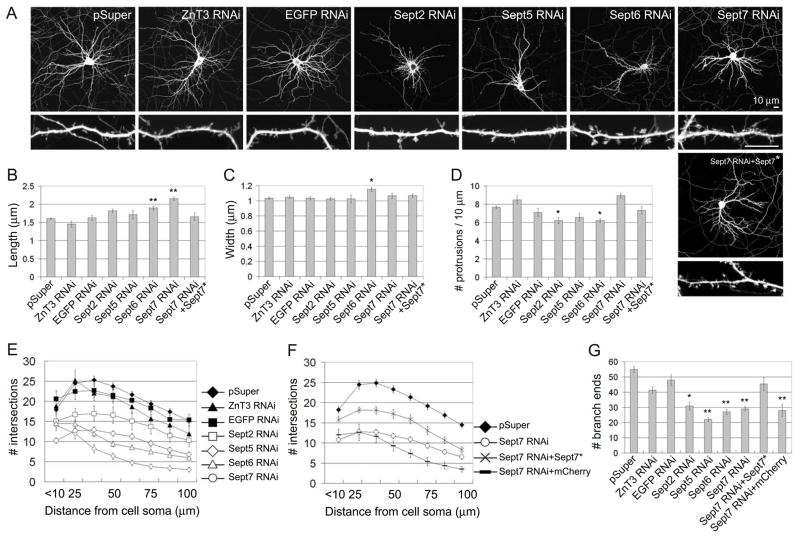
Septins are GTP-binding proteins that polymerize into heteromeric filaments and form microscopic bundles or ring structures in vitro and in vivo. Because of these properties and their ability to associate with membrane, F-actin, and microtubules, septins have been generally regarded as cytoskeletal components [1, 2]. Septins are known to play roles in cytokinesis, in membrane trafficking, and as structural scaffolds; however, their function in neurons is poorly understood. Many members of the septin family, including Septin 7 (Sept7), were found by mass-spectrometry analysis of postsynaptic density (PSD) fractions of the brain [3, 4], suggesting a possible postsynaptic function of septins in neurons. We report that Sept7 is localized at the base of dendritic protrusions and at dendritic branch points in cultured hippocampal neurons--a distribution reminiscent of septin localization in the bud neck of budding yeast. Overexpression of Sept7 increased dendrite branching and the density of dendritic protrusions, whereas RNA interference (RNAi)-mediated knockdown of Sept7 led to reduced dendrite arborization and a greater proportion of immature protrusions. These data suggest that Sept7 is critical for spine morphogenesis and dendrite development during neuronal maturation.


Figures

Figure 1
Figure 1. Expression pattern of septin family proteins in rat brain and hippocampal neuron culture
(A) Enrichment of septins in PSD preparation from adult rat forebrain. Crude synaptosomal membranes (P2) were prufied by discontinuous sucrose gradient and extracted with Triton X-100 once (PSD I), twice (PSD II), or with Triton X-100 followed by Sarkosyl (PSD III). PSD fractions (5 μg protein) were compared with 30 μg of crude synaptosomal membranes P2. (B) Regional distribution of septin proteins in the rat brain. Total homogenates (20 μg protein) from the indicated rat brain regions from P7 (postnatal day 7) and Ad (adult age; P45) animals were immunoblotted for the indicated proteins. CX: cortex, HP: hippocampus, ST: striatum, MB, midbrain, CB: cerebellum, SC: spinal cord. (C) Developmental expression pattern of septins in the rat brain. Immunoblot of total homogenates (20 μg protein) from the indicated rat brain regions at E15 (embryonic day 15), E18, P3, P5, P8 and Ad (P45). (D) Septin protein expression during development of dissociated hippocampal neurons in culture. Total extracts (15 μg) of hippocampal cultures at 1–4 weeks in vitro were immunoblotted as indicated.
Figure 2
Figure 2. Endogenous Sept7 localization at the base of dendritic protrusions and dendritic branch points in cultured hippocampal neurons
(A–D) Cultured hippocampal neurons (DIV7) were fixed and immunostained for Sept7 (red), F-actin (phalloidin staining, green) and PSD-95 (blue). Low magnification images show whole neurons (A) and higher magnification images show dendrite segments (B). Arrowheads in B mark examples of Sept7 at the base of actin-rich dendritic protrusions. (C) High magnification merged images (actin, green; Sept7, red) showing examples of Sept7 localization at the base of individual protrusions. (D) Examples of Sept7 localization at dendritic branch points (arrows) and at the base of dendritic protrusions (arrowheads). (E and F) Endogenous Sept7 (red) were immunostained together with endogenous Sept5 (blue) and F-actin (green). Low magnification images show whole neurons (E) and higher magnification images show dendrite segments (F). Arrowheads indicate examples of colocalized Sept7 with Sept5 at the base of dendritic protrusions.
Figure 3
Figure 3. Septin overexpression increases dendritic protrusions and branches
(A) Cultured hippocampal neurons (DIV7) were transfected with the empty FLAG expression vector pFLAG-CMV-2, or FLAG-tagged Sept2, 6 or 7, as indicated. Neuron morphology was visualized by cotransfected EGFP. 5 days after transfection (DIV7+5), neurons were fixed and immunostained for EGFP and FLAG. (B–D) Average length (B), head width (C) and density (D) of dendritic protrusions in neurons transfected with the indicated constructs. (E) Dendrite complexity of neurons transfected with the indicated constructs measured by Sholl analysis, which shows the number of dendrites crossing circles (vertical axis) at various radial distances from the cell soma (horizontal axis). (F) Number of dendrite branch ends in hippocampal neurons transfected with the indicated constructs. Histograms show mean ± SEM; ***p < 0.001, ** p < 0.01, One-way ANOVA. n numbers for each condition were control FLAG (41), FLAG-Sept2 (34), FLAG-Sept6 (38) and FLAG-Sept7 (29).
Figure 4
Figure 4. Effect of septin RNAi on dendritic protrusions and dendrite branching
(A) Cultured hippocampal neurons (DIV7) were transfected with empty expression vector pSuper, or with pSuper plasmids expressing ZnT3-shRNA (ZnT3 RNAi), EGFP-shRNA (EGFP RNAi), Sept2-shRNA (Sept2 RNAi), Sept5-shRNA (Sept5 RNAi), Sept6-shRNA (Sept6 RNAi), Sept7-shRNA (Sept7 RNAi), as indicated. For “rescue” experiments, Sept7-shRNA plasmid was cotransfected with RNAi-resistant Sept7 cDNA (Sept7 RNAi+Sept7*). 5 days after transfection (DIV7+5), neuron morphology was visualized by immunostaining for cotransfected β-gal. Scale bar, 10 μm (upper and lower panels). (B–D) Average length (B), head width (C) and density (D) of dendritic protrusions in neurons transfected with the indicated constructs. (E–G) Sholl analysis of dendrite branching (E and F) and branch end counts (G), as in Fig. 3. For a control of “rescue” experiments, mCherry plasmid was cotransfected with Sept7-shRNA (Sept7 RNAi+mCherry). Histograms show mean ± SEM; **p < 0.01, *p < 0.05, One-way ANOVA. n numbers for each condition were control pSuper (76), control ZnT3 RNAi (26), control EGFP RNAi (34), Sept2 RNAi (30), Sept5 RNAi (26), Sept6 RNAi (53), Sept7 RNAi (54), Sept7 RNAi+Sept7* (30), Sept7 RNAi+mCherry (30).

Comment in

References

    1. Kinoshita M, Field CM, Coughlin ML, Straight AF, Mitchison TJ. Self- and actin-templated assembly of Mammalian septins. Dev Cell. 2002;3:791–802. - PubMed
    1. Kinoshita M. Diversity of septin scaffolds. Curr Opin Cell Biol. 2006;18:54–60. - PubMed
    1. Peng J, Kim MJ, Cheng D, Duong DM, Gygi SP, Sheng M. Semiquantitative proteomic analysis of rat forebrain postsynaptic density fractions by mass spectrometry. J Biol Chem. 2004;279:21003–21011. - PubMed
    1. Collins MO, Husi H, Yu L, Brandon JM, Anderson CN, Blackstock WP, Choudhary JS, Grant SG. Molecular characterization and comparison of the components and multiprotein complexes in the postsynaptic proteome. J Neurochem 2005 - PubMed
    1. Hall PA, Jung K, Hillan KJ, Russell SE. Expression profiling the human septin gene family. J Pathol. 2005;206:269–278. - PubMed

Publication types

MeSH terms

LinkOut - more resources