. 2007 Oct;27(4):610-24.
doi: 10.1016/j.immuni.2007.08.015. Epub 2007 Oct 11.

Disruption of E-cadherin-mediated adhesion induces a functionally distinct pathway of dendritic cell maturation

Affiliations

Disruption of E-cadherin-mediated adhesion induces a functionally distinct pathway of dendritic cell maturation

Aimin Jiang et al. Immunity. 2007 Oct.

Abstract

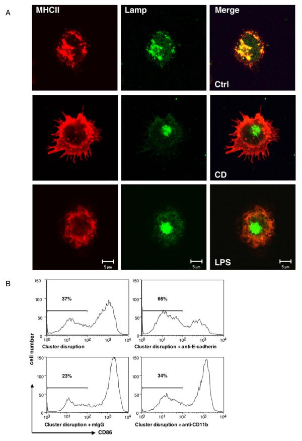
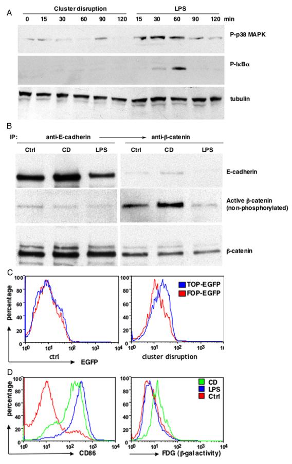
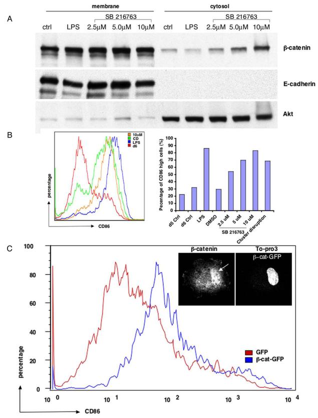
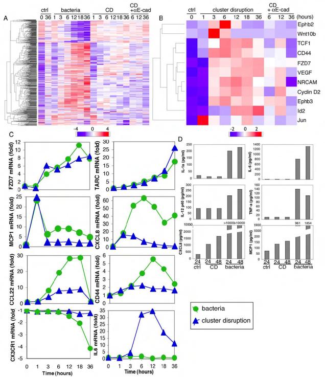
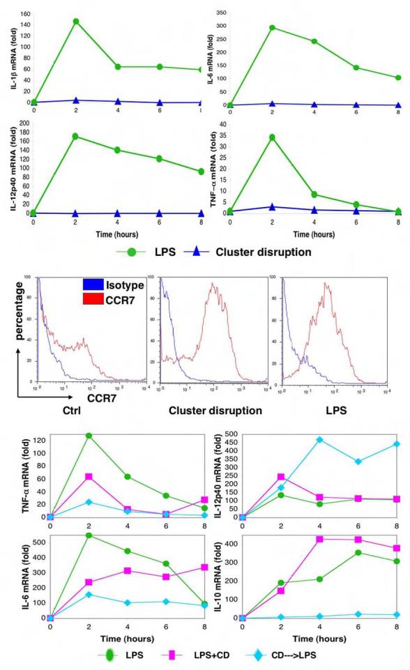
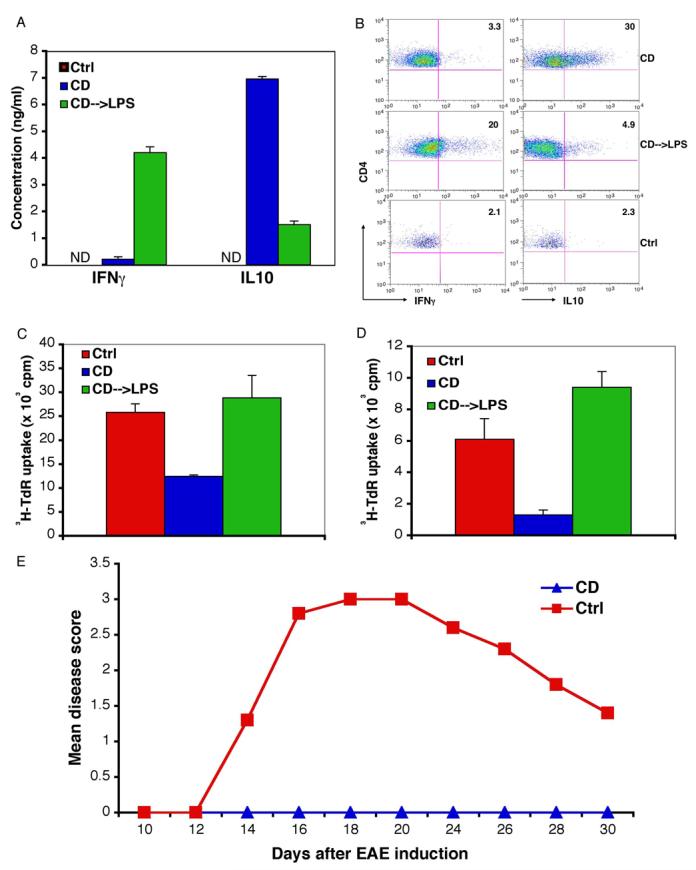
The maturation of dendritic cells (DCs) after exposure to microbial products or inflammatory mediators plays a critical role in initiating the immune response. We found that maturation can also occur under steady-state conditions, triggered by alterations in E-cadherin-mediated DC-DC adhesion. Selective disruption of these interactions induced the typical features of DC maturation including the upregulation of costimulatory molecules, MHC class II, and chemokine receptors. These events were triggered at least in part by activation of the beta-catenin pathway. However, unlike maturation induced by microbial products, E-cadherin-stimulated DCs failed to release immunostimulatory cytokines, exhibiting an entirely different transcriptional profile. As a result, E-cadherin-stimulated DCs elicited an entirely different T cell response in vivo, generating T cells with a regulatory as opposed to an effector phenotype. These DCs induced tolerance in vivo and may thus contribute to the elusive steady-state "tolerogenic DCs."


Figures

Figure 1
Figure 1. Disruption of E-cadherin-mediated clusters results in DC maturation
(Panel A) DCs matured after cluster disruption (CD) exhibited similar mophological changes as induced by LPS. DCs matured by CD or LPS were labeled for MHC II (red) and the lysosomal marker Lamp 2 (green). (Panel B) Anti-E-cadherin antibodies can block DC maturation induced by CD. BMDCs were prepared as described and CD11c+ DCs were purified at day 6 and replated at 5 × 10⁵ cells/ml. Treatment with an anti-E-cadherin mAb (Sigma) but not isotype-matched anti-CD11b mAb or mouse IgG inhibited the upregulation of CD86.
Figure 2
Figure 2. Disruption of the E-cadherin-mediated adhesion activates a distinct β-catenin/TCF signaling pathway independent of TLR signaling
(Panel A) CD did not activate NF-κB and p38 MAPK signaling pathways. Cell lysates from different treatments were analyzed by immunoblotting with anti-phospho-p38 MAPK Ab (top), phosphorylation-specific Ab against IκBα (middle) and anti-tubulin Ab (bottom). (Panel B) CD resulted in activation of β-catenin. BMDCs were either treated with LPS or CD and cell lysates from CD11c+ DCs were subject to sequential immunoprecipitation with antibodies against E-cadherin and β-catenin, followed by immunoblotting with antibodies against E-cadherin (top), active β-catenin (middle) and total β-catenin (bottom). (Panel C) CD resulted in β-catenin/TCF mediated transcription. BMDC cultures were transfected with pLTRH1 containing the TOP-EGFP or FOP-EGFP at day 2 and transfected cells were purified with magnetic columns at day 6. EGFP was measured on CD11c+ DCs immediately after purification (control) or 48 hr later (CD) by FACS. (Panel D) CD but not LPS treatment led to transactivation of TOPgal reporter. BMDCs from transgenic TOPGAL reporter mice were matured by LPS or CD, β-galactosidase activity was measured by flow cytometry using fluorescein di-β-D-galactosidase (FDG) as a substrate.
Figure 3
Figure 3
Activation of β-catenin signaling pathway induces DC maturation (Panel A) Dose-dependent accumulation of cytosolic β-catenin after treatment with GSK3β inhibitor SB216763. BMDCs were treated with either LPS or different doses of SB216763. CD11c+ DCs were then fractionated into membrane and cytosolic fractions, followed by immunoblotting with antibodies against β-catenin (top) and E-cadherin (middle). Akt was probed as a loading control (bottom). (Panel B) Inhibition of GSK3β results in DC maturation. CD11c+ DCs after different stimuli were subject to FACS analysis. The left histogram overlay shows a representative FACS profile of CD86 expression for each condition, with SB216763 at 10 μm. CD86high cells represent mature DCs on the right. (Panel C) Expression of β-catenin enhanced spontaneous DC maturation. BMDC cultures were transfected either with GFP or β-catenin-GFP and were subject to FACS analysis for CD86 expression at day 6. Expression of β-catenin-GFP but not GFP induced CD86 upregulation, although not as strongly as after CD or drug treatment. Insert: β-catenin translocates to the nucleus. 12 hr after CD of DCs expressing β-catenin-GFP, cells were fixed, labeled with a β-catenin antibody and the DNA dye TO-Pro3, and imaged by confocal microscopy. β-catenin was clearly translocated into the nucleus (arrow).
Figure 4
Figure 4. CD-matured human DCs failed to produce inflammatory cytokines
(Panel A) More than 700 genes were differentially regulated upon maturation by either CD or bacterial stimulation. Heatmap was generated as detailed in the Experimental Procedures. (Panel B) CD led to upregulation of 10 direct β-catenin/TCF target genes. Target genes were selected according to R. Nusse and colleagues and heatmap was created as described in Experimental Procedures. Wnt10b was not a target gene but was included for comparison. (Panel C) Representative gene expression profiles were plotted from the microarray data. (Panel D) Human CD34+ DCs matured by CD did not produce inflammatory cytokines. Luminex assays for multiple cytokines and chemokines were performed on supernatants from CD or bacteria-matured DCs. One of two independent experiments is shown.
Figure 5
Figure 5. CD-matured murine BMDCs upregulated CCR7 without inflammatory cytokine induction
(Panel A) CD-matured murine BMDCs did not induce inflammatory cytokines IL-1β, IL-6, IL-12p40 and TNFα. Real-time RT-PCRs were performed on total RNA isolated from DCs treated with either LPS or CD for the indicated times, the expression of each gene then was normalized to β-actin expression. (Panel B) CD-matured BMDCs express elevated level of surface CCR7. DCs untreated or matured by either CD or LPS were subjected to FACS analysis. (Panel C) Addition of LPS after cluster disruption synergistically enhances or inhibits cytokine production. Real-time RT-PCRs were performed and analyzed as described in Panel A. Cluster-disrupted DCs were stimulated with LPS simultaneously (CD+LPS) or LPS was added 14-18 hr afterwards for the indicated times (CD--->LPS). Results from one of three different sets of samples are shown.
Figure 6
Figure 6. DCs matured by CD alone generated IL10-producing CD4 T cells leading to tolerance and protection from EAE in vivo
(Panel A) Immunization with DCs matured by CD alone induced T cells that produced IL10 instead of IFN-γ. CD11c+ BMDCs were purified at day 6-7 of culture, pulsed with OVA peptide 323-339 (10 μg/ml) for 2 hr and washed extensively before resuspension in PBS. 1-2.5 × 10⁶ DCs were injected intravenously into C57BL/6 mice at day 0, 2 and 4. Splenocytes (1 × 10⁶ cells/well) were prepared at day 7 and stimulated with antigens for 3 days. The supernatants were collected and cytokines were measured with the Luminex assays. (Panel B) DC matured by CD generated IL10-producing CD4 T cells instead of IFN-γ-producing effector cells. BFA (5 μg/ml) was added for 6 hr at the end of 2-3 day restimulation, splenocytes were then stained for cytokines and analyzed by FACS. The numbers indicate the percentage of IFN-γ–or IL10-positive cells of gated CD4+ CD25+ cells. Results are representative of 4 similar experiments, each consisted of two mice for CD and CD—>LPS treatments. (Panel C) Peripheral tolerance induced by treatment with DCs matured by CD alone. Mice were injected 3 times as described in Panel A and challenged 3 days after the last injection with 200 μg OTII peptide in CFA. 5 × 10⁵ cells from pooled draining lymph nodes 3 days later were cultured with 2 × 10⁵ mitomycin C-treated splenocytes in the presence of OTII peptide and T cell proliferation was measured by 3H-thymidine incorporation. (Panel D) T cells from mice receiving CD-matured DCs showed diminished response to challenge with OTII peptide. Mice were injected 3 times with OVA peptide-pulsed DCs after adoptive transfer of naïve OTII CD4 T cells, 200 μg OTII peptide in CFA was injected subcutaneously 7 days later. 2 × 10⁵ CD4+ cells from pooled draining lymph nodes 3 days after the challenge were cultured with 1 × 10⁵ mitomycin C-treated splenocytes in the presence of OTII peptide; T cell proliferation was measured by 3H-thymidine incorporation. Similar results were obtained when the mice were injected once with DCs. (Panel E) Immunization with DCs matured by CD alone protect mice from EAE. DCs were purified as described and cultured for 12-16h before being pulsed with MOG peptide for 2h, washed extensively and 2-3 × 10⁶ cells were injected three times (at days 0, 2, 4) intravenously into 4-5 mice per group. EAE was induced 5, 7 or 9 days after the last injection and the mice were observed for paralysis. Induction of EAE on day 7 was shown.

Comment in

References

    1. Annacker O, Coombes JL, Malmstrom V, Uhlig HH, Bourne T, Johansson-Lindbom B, Agace WW, Parker CM, Powrie F. Essential role for CD103 in the T cell-mediatedregulation of experimental colitis. J Exp Med. 2005;202:1051–1061. - PMC - PubMed
    1. Banchereau J, Steinman RM. Dendritic cells and the control of immunity. Nature. 1998;392:245–252. - PubMed
    1. Barton GM, Medzhitov R. Retroviral delivery of small interfering RNA into primarycells. Proc Natl Acad Sci U S A. 2002;99:14943–14945. - PMC - PubMed
    1. Barton GM, Medzhitov R. Toll-like receptor signaling pathways. Science. 2003;300:1524–1525. - PubMed
    1. Bonifaz L, Bonnyay D, Mahnke K, Rivera M, Nussenzweig MC, Steinman RM. Efficient targeting of protein antigen to the dendritic cell receptor DEC-205 in the steady state leadsto antigen presentation on major histocompatibility complex class I products and peripheral CD8+ Tcell tolerance. J Exp Med. 2002;196:1627–1638. - PMC - PubMed

Publication types

MeSH terms

Associated data