. 2008;40(1):63-71.
doi: 10.1016/j.biocel.2007.08.014. Epub 2007 Sep 1.

A2 isoform of mammalian translation factor eEF1A displays increased tyrosine phosphorylation and ability to interact with different signalling molecules

Affiliations

A2 isoform of mammalian translation factor eEF1A displays increased tyrosine phosphorylation and ability to interact with different signalling molecules

Ganna Panasyuk et al. Int J Biochem Cell Biol. 2008.

Abstract

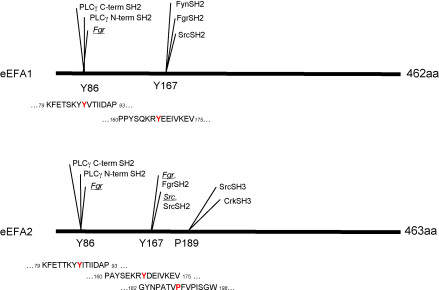
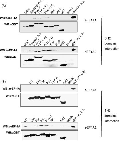
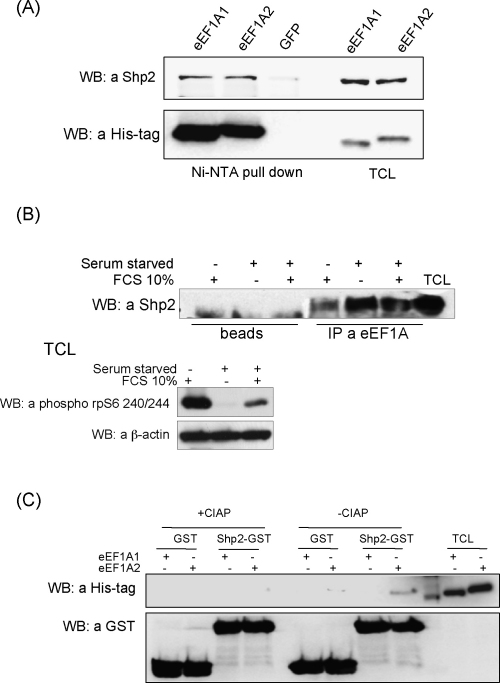
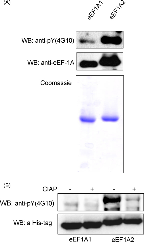
The eEF1A1 and eEF1A2 isoforms of translation elongation factor 1A have 98% similarity and perform the same protein synthesis function catalyzing codon-dependent binding of aminoacyl-tRNA to 80S ribosome. However, the isoforms apparently play different non-canonical roles in apoptosis and cancer development which are awaiting further investigations. We hypothesize that the difference in non-translational functions could be caused, in particular, by differential ability of the isoforms to be involved in phosphotyrosine-mediated signalling. The ability of eEF1A1 and eEF1A2 to interact with SH2 and SH3 domains of different signalling molecules in vitro was compared. Indeed, contrary to eEF1A1, eEF1A2 was able to interact with SH2 domains of Grb2, RasGAP, Shc and C-terminal part of Shp2 as well as with SH3 domains of Crk, Fgr, Fyn and phospholipase C-gamma1. Interestingly, the interaction of both isoforms with Shp2 in vivo was found using stable cell lines expressing eEF1A1-His or eEF1A2-His. The formation of a complex between endogenous eEF1A and Shp2 was also shown. Importantly, a higher level of tyrosine phosphorylation of eEF1A2 as compared to eEF1A1 was demonstrated in several independent experiments and its importance for interaction of eEF1A2 with Shp2 in vitro was revealed. Thus, despite the fact that both isoforms of eEF1A could be involved in the phosphotyrosine-mediated processes, eEF1A2 apparently has greater potential to participate in such signalling pathways. Since tyrosine kinases/phosphatases play a prominent role in human cancerogenesis, our observations may gave a basis for recently found oncogenicity of the eEF1A2 isoform.


Figures

Fig. 1
Fig. 1
Schematic representation of potential SH2 and SH3 interaction sites of eEF1A1 and eEF1A2. Predicted sites were identified by ScanSite search of eEF1A1 and eEF1A2 peptide sequences: core amino acids are indicated. Y: tyrosine kinase phosphorylation site; SH2: SH2 domain binding site; SH3: SH3 domain binding site.
Fig. 2
Fig. 2
Interaction of eEF1A1 and eEF1A2 with different GST-SH3 and GST-SH2 domain fusion proteins. GST-SH2 (A) or GST-SH3 (B) fusion proteins were coupled to glutathione-Sepharose beads and incubated with purified eEF1A1 (upper panels in A and B) or eEF1A2 (lower panels in A and B). The beads were washed extensively and the proteins were eluted by boiling in Laemmli sample buffer. Eluted proteins were resolved by SDS-PAGE and visualized by immunoblotting with anti-eEF1A antibodies (0.2 μg of purified eEF1A1 or eEF1A2 were loaded as controls). Amount of the GST-SH2 (A) or GST-SH3 (B) fusion proteins was controlled by immunoblotting with anti-GST antibodies. As a control, non-specific binding of eEF1A1 (upper panels in A and B) or eEF1A2 (lower panels in A and B) to GST alone or to beads alone has been analysed.
Fig. 3
Fig. 3
eEF1A1 and eEF1A2 form complexes with Shp2. (A) Complexes of His-tagged eEF1A1/eEF1A2 and Shp2 were precipitated from HEK293 stable cell lines using NiNTA sepharose. NiNTA sepharose beads were washed extensively and complexes were eluted by boiling in Laemmli sample buffer. Eluted proteins were resolved by SDS-PAGE and visualized by immunoblotting with anti-Shp2 (upper panel) or anti-His (lower panel) antibodies. NiNTA sepharose beads incubated with lysate of GFP overexpressing stable cell line were used as a control of non-specific binding of Shp2. TCL: total cell lysate. (B) Complexes were immunoprecipitated with anti-eEF1A antibodies from HEK293 cells treated under different conditions (growing, serum starved and serum starved followed by 30 min 10% serum stimulation). Protein A sepharose beads were washed extensively and the complexes were eluted by boiling in Laemmli sample buffer. Eluted proteins were resolved by SDS-PAGE and visualized by immunoblotting with anti-Shp2 antibodies. Protein A sepharose beads alone incubated with the same cell lysates were used as a control of Shp2 non-specific binding. Total cell lysates (TCL) of HEK293 cells were immunobloted with phosphospecific antibodies to phosphorylated Ser240/Thr244 in ribosomal protein S6 and β-actin to control the efficiency of treatment conditions and total protein amount used for immunoprecipitation in each sample, respectively. (C) GST or Shp2 GST-SH2 fusion proteins were coupled with glutathione-sepharose beads and incubated with phosphatase treated (10 U CIAP per 350 μg of total cell lysate) or untreated total cell lysates of stable cell lines overexpressing His-tagged eEF1A1 or eEF1A2 proteins. The beads were washed extensively and the proteins were eluted by boiling in Laemmli sample buffer. Eluted proteins were resolved by SDS-PAGE and visualized by immunoblotting with anti-His antibodies (upper panel). Amount of GST or Shp2 GST-SH2 proteins in each probe was controlled by immunoblotting with anti-GST antibodies (lower panel).
Fig. 4
Fig. 4
eEF1A1 and eEF1A2 are phosphorylated at tyrosine residues in vivo. (A) 0.5 μg of purified eEF1A1 and eEF1A2 was immunobloted with anti-phosphotyrosine-specific antibodies (4G10) followed by WB with anti-eEF1A antibodies (upper panel). SDS-PAGE of purified eEF1A1 and eEF1A2 visualized by Coomassie staining, 1 μg of each protein was loaded per line (lower panel). (B) NiNTA sepharose beads with precipitated eEF1A1/eEF1A2 proteins were divided on two samples, the first was treated with 5 U CIAP at 37 °C for 1 h, second was incubated at 37 °C for 1 h in the presence of 1 mM NaOVa. NiNTA sepharose beads were washed with the lysis buffer two times; proteins were eluted by boiling in Laemmli sample buffer and resolved by SDS-PAGE. Phosphorylated eEF1A1 and eEF1A2 were visualized by immunoblotting with anti-phosphotyrosine-specific antibodies (4G10) (upper panel) followed by WB with anti-His tag antibodies (lower panel).

References

    1. Ahmed M., Forsberg J., Bergsten P. Protein profiling of human pancreatic islets by two-dimentional gel electrophoresis and mass-spectrometry. J. Proteome Res. 2005;4(3):931–940. - PubMed
    1. Alper O., Bowden E.T. Novel insights into c-Src. Curr. Pharm. Des. 2005;11(9):1119–1130. - PubMed
    1. Amiri A., Noei F., Jeganathan S., Kulkarni G., Pinke D.E., Lee J.M. eEF1A2 activates Akt and stimulates Akt-dependent actin remodeling, invasion and migration. Oncogene. 2007;26(21):3027–3040. - PubMed
    1. Anand N., Murthy S., Amann G., Wernick M., Porter L.A., Cukier I.H. Protein elongation factor eEF1A2 is a putative oncogene in ovarian cancer. Nat. Genet. 2002;31(3):301–305. - PubMed
    1. Ann D.K., Moutsatsos I.K., Nakamura T., Lin H.H., Mao P.L., Lee M.J. Isolation and characterization of the rat chromosomal gene for a polypeptide (pS1) antigenically related to statin. J. Biol. Chem. 1991;266(16):10429–10437. - PubMed

Publication types

MeSH terms

Substances