. 2007 Nov 23;374(2):322-33.
doi: 10.1016/j.jmb.2007.09.047. Epub 2007 Sep 20.

DNA-directed DNA polymerase and strand displacement activity of the reverse transcriptase encoded by the R2 retrotransposon

Affiliations

DNA-directed DNA polymerase and strand displacement activity of the reverse transcriptase encoded by the R2 retrotransposon

Anna Kurzynska-Kokorniak et al. J Mol Biol. .

Abstract

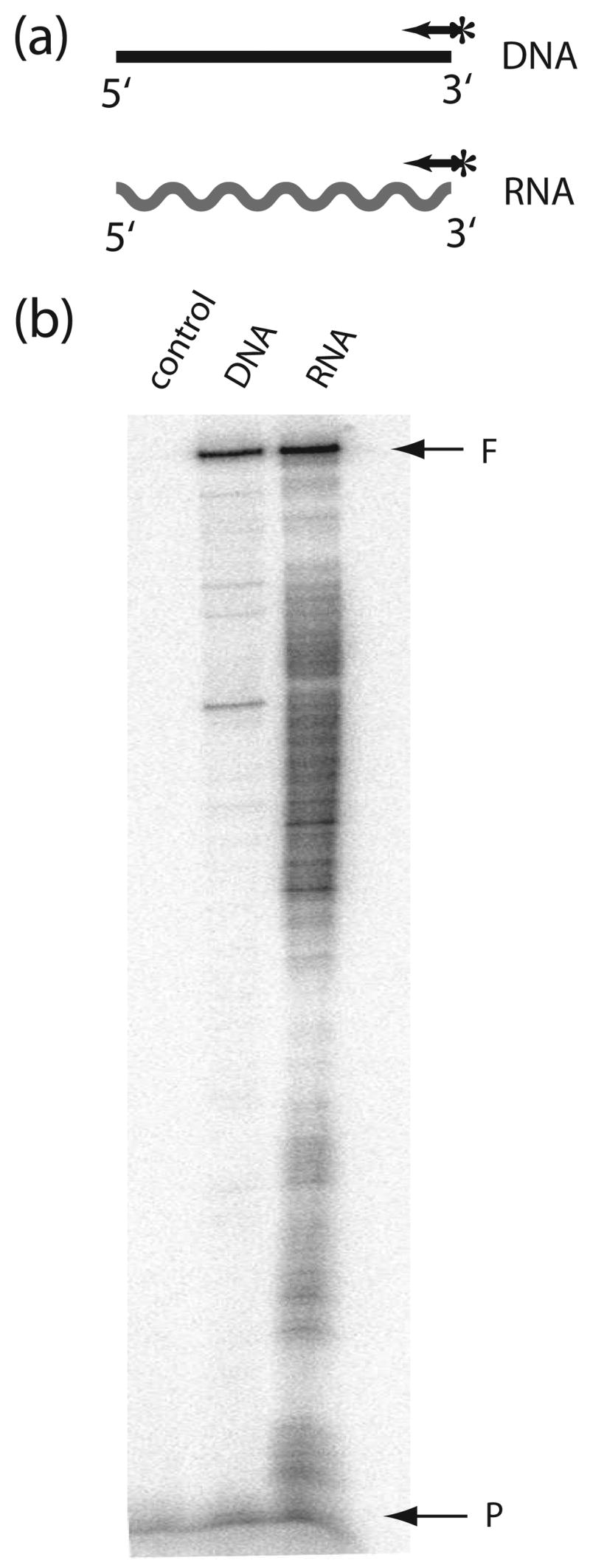
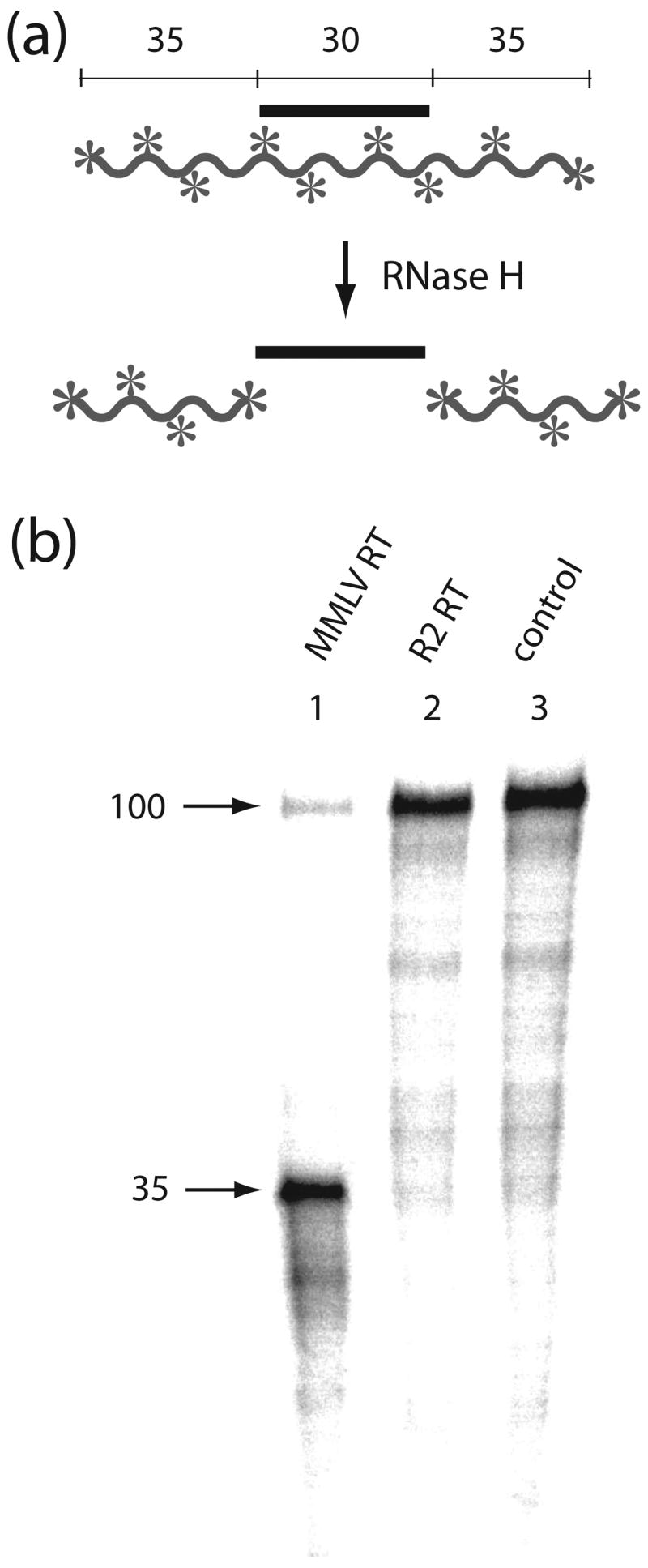
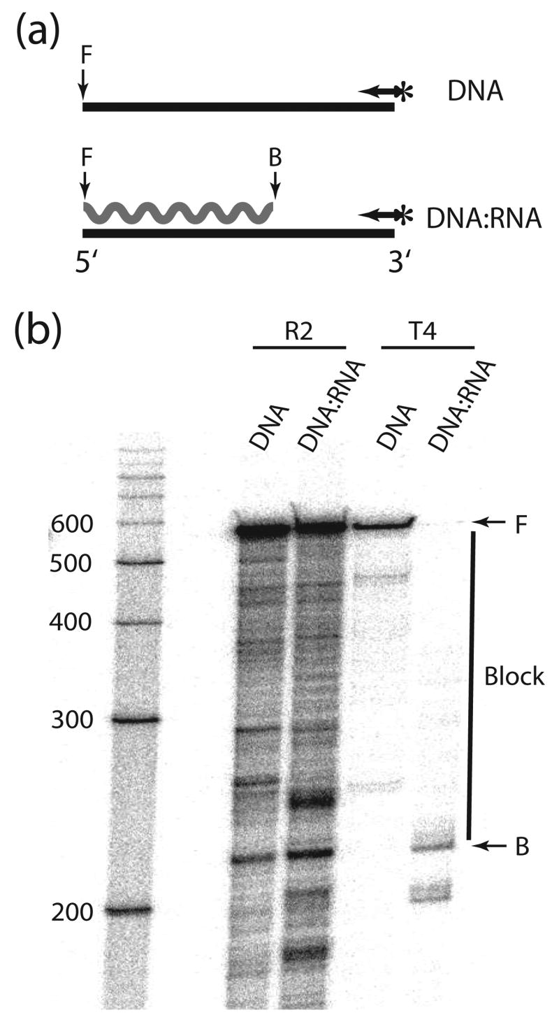
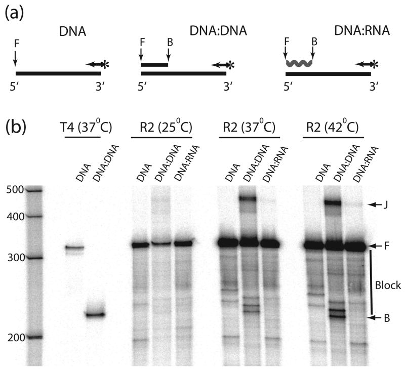
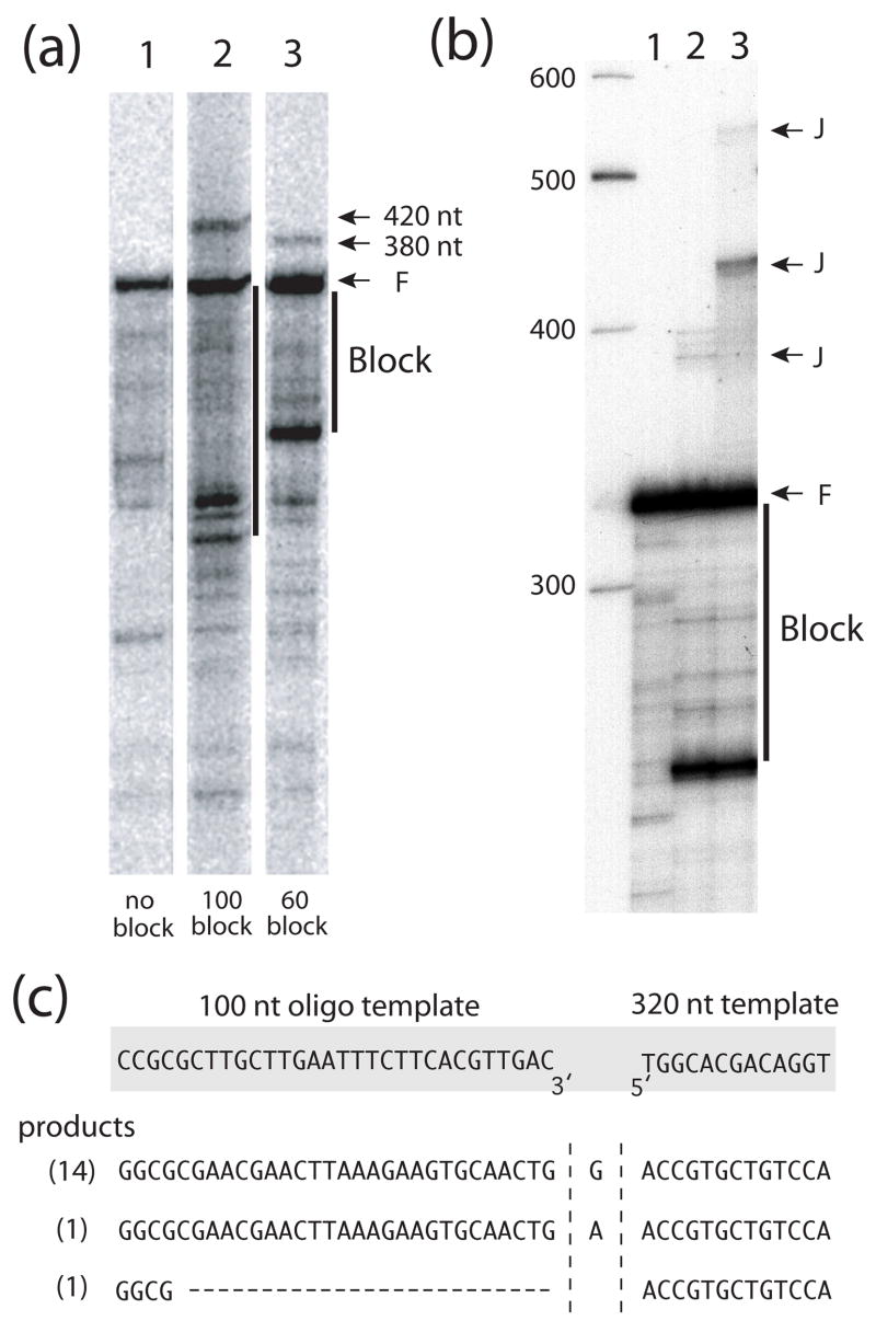
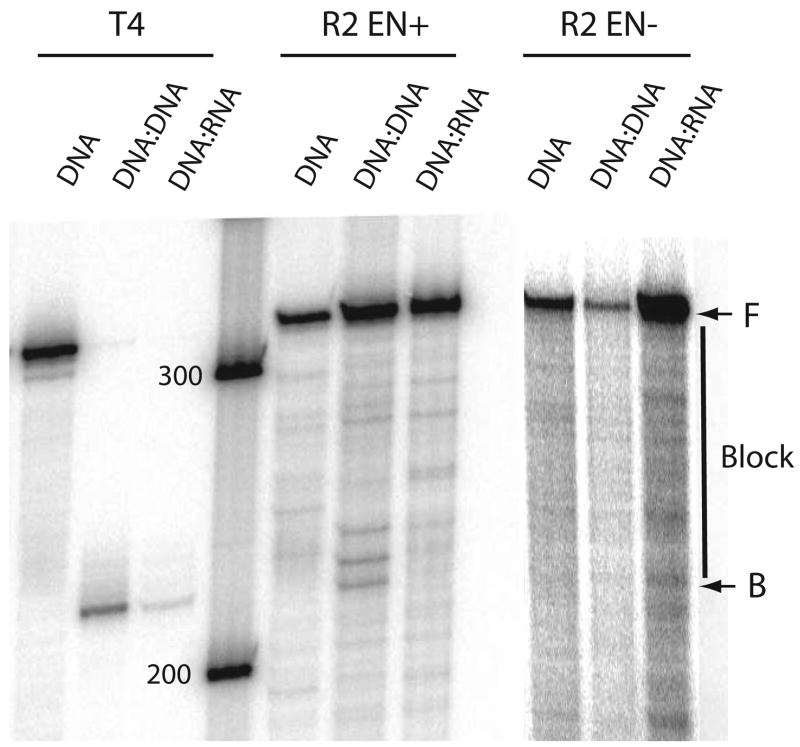
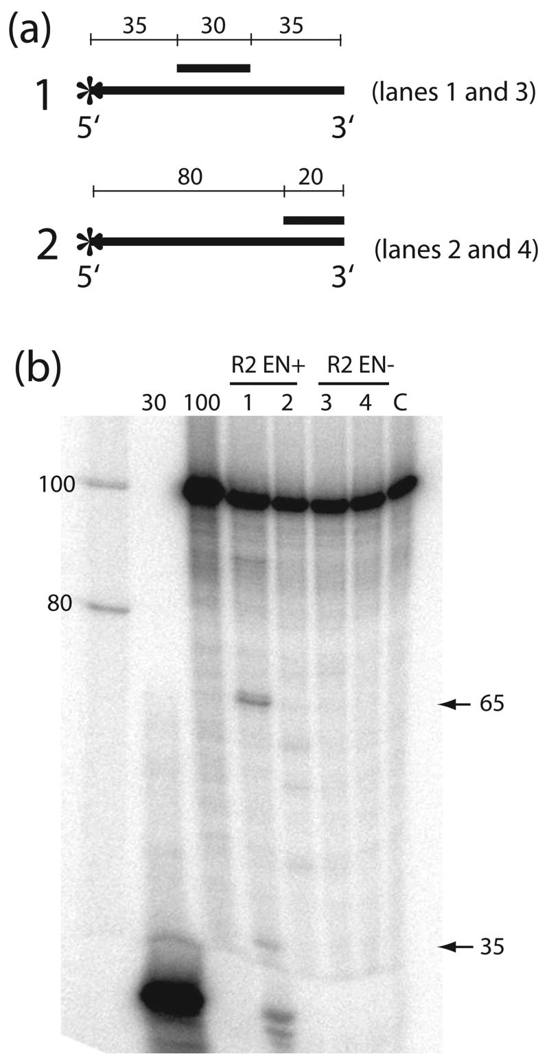
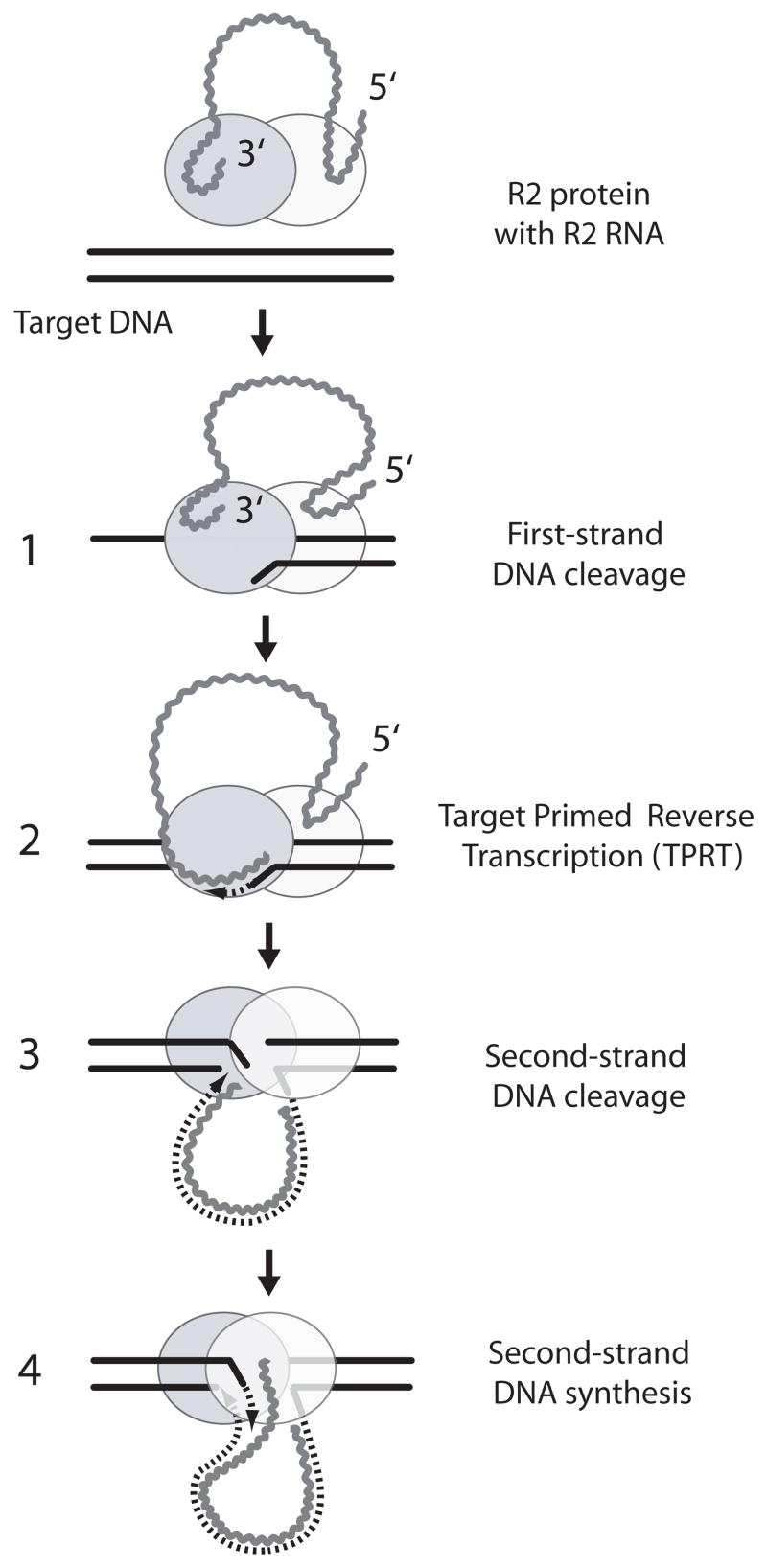
R2 elements are non-long terminal repeat (non-LTR) retrotransposons with a single open reading-frame encoding reverse transcriptase, DNA endonuclease and nucleic acid-binding domains. The elements are specialized for insertion into the 28 S rRNA genes of many animal phyla. The R2-encoded activities initiate retrotransposition by sequence-specific cleavage of the 28 S gene target site and the utilization of the released DNA 3' end to prime reverse transcription (target primed reverse transcription). The activity of the R2 polymerase on RNA templates has been shown to differ from retroviral reverse transcriptases (RTs) in a number of properties. We demonstrate that the R2-RT is capable of efficiently utilizing single-stranded DNA (ssDNA) as a template. The processivity of the enzyme on ssDNA templates is higher than its processivity on RNA templates. This finding suggests that R2-RT is also capable of synthesizing the second DNA strand during retrotransposition. However, R2-RT lacks the RNAse H activity that is typically used by retroviral and LTR-retrotransposon RTs to remove the RNA strand before the first DNA strand is used as template. Remarkably, R2-RT can displace RNA strands that are annealed to ssDNA templates with essentially no loss of processivity. Such strand displacement activity is highly unusual for a DNA polymerase. Thus the single R2 protein contains all the activities needed to make a double-stranded DNA product from an RNA transcript. Finally, during these studies we found an unexpected property of the highly sequence-specific R2 endonuclease domain. The endonuclease can non-specifically cleave ssDNA at a junction with double-stranded DNA. This activity suggests that second-strand cleavage of the target site may not be sequence specific, but rather is specified by a single-stranded region generated when the first DNA strand is used to prime reverse transcription.


Figures

Figure 1
Figure 1
The R2 polymerase can utilize both RNA and DNA templates. (a) Schematic diagrams of templates used in the experiment. DNA synthesis on a 320 nt ssDNA template (horizontal line) and a 320 nt RNA template of the same sequence (wavy line) was primed by a 5′-end labeled primer (arrow with asterisk) annealed to each template. (b) PhosphorImager scan of the extension products separated on a 6% denaturing polyacrylamide gel. Templates with annealed primers were preincubated with R2 protein for 10 min at room temperature. Reactions were started by the addition of dNTPs and a reinitiation trap [20 μg heparin and 300 ng poly(rA)/poly(dT)13–18]. Incubations were for 20 minutes at 37°C. In the control reaction the trap was added prior to the addition of the R2 protein to the template. F, full-length extension products; P, non-extended primer.
Figure 2
Figure 2
The R2 protein does not have RNase H activity. (a) Schematic diagram of the RNase H assay. In the presence of RNase H an internally 32P-labeled 100 nt RNA (waved line with asterisks) annealed with a centrally located 30 nt DNA fragment (straight line) will be cleaved to 35 nt fragments. (b) PhosphorImager scan of the products of an RNase H assay using retroviral (MMLV) RT and R2-RT. The RNA:DNA substrates were incubated for 20 min at 37°C in the presence of either MMLV RT, which has RNase H activity (lane 1), the R2 protein, which does not have RNase H activity (lane 2), or with buffer (lane 3).
Figure 3
Figure 3
R2-RT can displace RNA annealed to a DNA template. (a) Schematic diagram of the displacement assays. Polymerization on a 560 nt ssDNA template (horizontal line) was compared to polymerization on the same DNA template annealed to a 340 nt complementary RNA strand (wavy line). Polymerization was primed by a 5′-end labeled primer (arrow with asterisk) annealed to the 3′-end of the template. (b) PhosphorImager scans of the products from the displacement reactions. Reactions were conducted at 37°C as in Figure 1 but without the trap. As a control, displacement reactions were also conducted with T4 DNA polymerase. The vertical line next to the gel indicates those extension products requiring RNA displacement. T4 DNA polymerase does not have strand displacement activity, thus on the annealed template, polymerization is blocked at the beginning of the double-strand region. The RNA block has little affect on R2-RT extension. A 100 nt DNA ladder is shown at left as size markers. F, full-length extension products; B, beginning of the RNA block.
Figure 4
Figure 4
R2 polymerase displacement of DNA and RNA strands at different temperatures. (a) Schematic diagram of the primer extension reactions. Polymerization on a 320 nt ssDNA template (DNA template) was compared to polymerization on the same template annealed to a 100 nt complementary DNA (DNA:DNA template) or RNA (RNA:DNA template). Polymerization was primed by a 5′-end labeled primer (arrow with asterisk) annealed to the 3′-end of the template. (b) PhosphorImager scans of the products from the displacement reactions. Reactions were conducted as in Figure 4 at 25°C, 37°C and 42°C. Control displacement reactions with T4 DNA polymerase were also conducted as in Figure 4. The R2 protein extensions on the DNA:DNA template also generated ~420 nt products (band J). These products were generated by the R2 polymerase jumping from the 5′ end of the original template onto the 3′ end of the excess 100 nt ssDNA present in the reaction (see Figure 5 and text). F, full-length extension products; B, beginning of the RNA or DNA block; vertical line at right, those extension products requiring strand displacement.
Figure 5
Figure 5
The R2 polymerase can conduct end-to-end template jumps after strand displacement reactions. (a) PhosphoImager scans of the products from extensions reactions similar to those conducted at 37°C in Figure 4. Lane 1, 320 nt DNA template in the absence of a DNA block; lane 2, 320 nt DNA template in the presence of the 100 nt DNA block as diagramed in Figure 4a; and lane 3, 320 nt DNA template in the presence of a 60 nt DNA block. Products of the template jumps onto the excess 100 nt and 60 nt DNA molecules in the reactions appear as 420 nt and 380 nt bands, respectively. Vertical lines indicate those extension products requiring strand displacement. (b) PhospoImager scan of the products from extensions reactions as in (a): lane 1, absence of a DNA block; lane 2, presence of 60 nt DNA block; and lane 3, presence of the 60 nt DNA block and a 100 nt DNA template. The molar ratio of the 100 nt template to 60 nt block was 5:1. Extension products derived from template jumps are indicated with a J. (c) Sequences at the junctions of the template jumps from the 5′ end of 320 nt DNA template to the 3′ end of 100 nt DNA template. Shown at the top are the sequences at the ends of the DNA templates. Shown below are the junction sequences of 16 PCR amplified and cloned products from the 420 nt J band in panel b, lane 3. In parenthesis are numbers of clones obtained with each junction sequence.
Figure 6
Figure 6
Effects of an R2 endonuclease mutation on the displacement reactions. The templates and the conditions of the reactions were identical to those conducted at 37°C in Figure 4. R2 EN+, wild type R2 protein with a functional endonuclease domain; EN−, R2 protein with an amino acid substitution eliminating endonuclease activity. All DNA binding and RT activities of this mutant are similar to wild-type., The EN− mutation has similar levels of strand displacement activity to the EN+ protein, but does not show an accumulation of extension products near the beginning of the DNA:DNA block (~ 220–240 nt). F, B and vertical lines are as described in Figure 4.
Figure 7
Figure 7
Nonspecific endonuclease activity of the R2 protein. (a) Schematic diagrams of the DNA substrates used in the experiment. A 30 nt (1) or 20 nt (2) complementary oligonucleotide was annealed to a 5′-end labeled 100 nt ssDNA. DNA fragments generated by cleavage at the junctions of the duplex regions are indicated below each substrate. (b) PhosphorImager scans of the gel products of the R2 endonuclease cleavage reactions. Substrates were incubated for 20 min at 37°C in the R2 extension buffer with wild type R2 (EN+, lanes 1 and 2) or endonuclease-deficient (EN−, lanes 3 and 4) protein. Lanes 1 and 3, cleavage reactions with substrate 1; lanes 2 and 4, cleavage reactions with substrate 2; lane C, control reaction in which substrate 1 is incubated in the reaction buffer without protein. The three lanes at left contain size standards including 5′-end labeled oligonucleotides 30 and 100 nt in length.
Figure 8
Figure 8
Current model of R2 retrotransposition. The proposed mechanism involves an ordered series of cleavage and polymerization reactions carried out by two R2 protein subunits (light and dark gray ovals). Each R2 subunit binds an RNA segment from either the 3′ end or near the 5′ end of the R2 transcript (wavy lines). Binding of this RNA determines whether the protein will bind upstream or downstream of the DNA target site and thus conducts the first two or the last two steps of the integration. The endonuclease of the subunit bound upstream of the target site (darker oval binding the 3′ end of the transcript) nicks the bottom strand (step 1). The RT of this upstream subunit uses the 3′ DNA end released by the cleavage to prime reverse transcription of the R2 transcript (step 2). This step has been termed target primed reverse transcription (TPRT). When the synthesis of the first strand is completed, the second subunit which binds downstream of the target site (lighter oval binding the 5′ end of the transcript) cleaves the top DNA strand (step 3). The RT of this downstream subunit uses the 3′ DNA end released by this cleavage to prime second-strand DNA synthesis (step 4). See references 20 and 41 for a detailed description of the evidence for this model.

Similar articles

Cited by

References

    1. Ostertag EM, Kazazian HH., Jr Biology of mammalian L1 retrotransposons. Annu Rev Genet. 2001;35:501–538. - PubMed
    1. Whitcomb JM, Hughes SH. Retroviral reverse transcription and integration: progress and problems. Annu Rev Cell Biol. 1992;8:275–306. - PubMed
    1. Voytas DF, Boeke JD. Ty1 and Ty5 of Sacharomyces cerevisiae. In: Craig NL, Craigie R, Gellert M, Lambowitz AM, editors. Mobile DNA II. American Society for Microbiology; Washington DC: 2002. pp. 631–662.
    1. Luan DD, Korman MH, Jakubczak JL, Eickbush TH. Reverse transcription of R2Bm RNA is primed by a nick at the chromosomal target site: a mechanism for non-LTR retrotransposition. Cell. 1993;72:595–605. - PubMed
    1. Dewannieux M, Esnault C, Heidmann T. LINE-mediated retrotransposition of marked Alu sequences. Nat Genet. 2003;35:41–48. - PubMed

Publication types

MeSH terms

LinkOut - more resources