. 2007 Dec;89(12):1464-73.
doi: 10.1016/j.biochi.2007.09.001. Epub 2007 Sep 11.

Transforming growth factor beta mediates hepatocyte apoptosis through Smad3 generation of reactive oxygen species

Affiliations

Transforming growth factor beta mediates hepatocyte apoptosis through Smad3 generation of reactive oxygen species

Dalliah Black et al. Biochimie. 2007 Dec.

Abstract

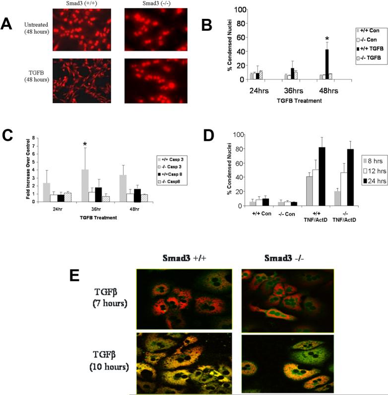
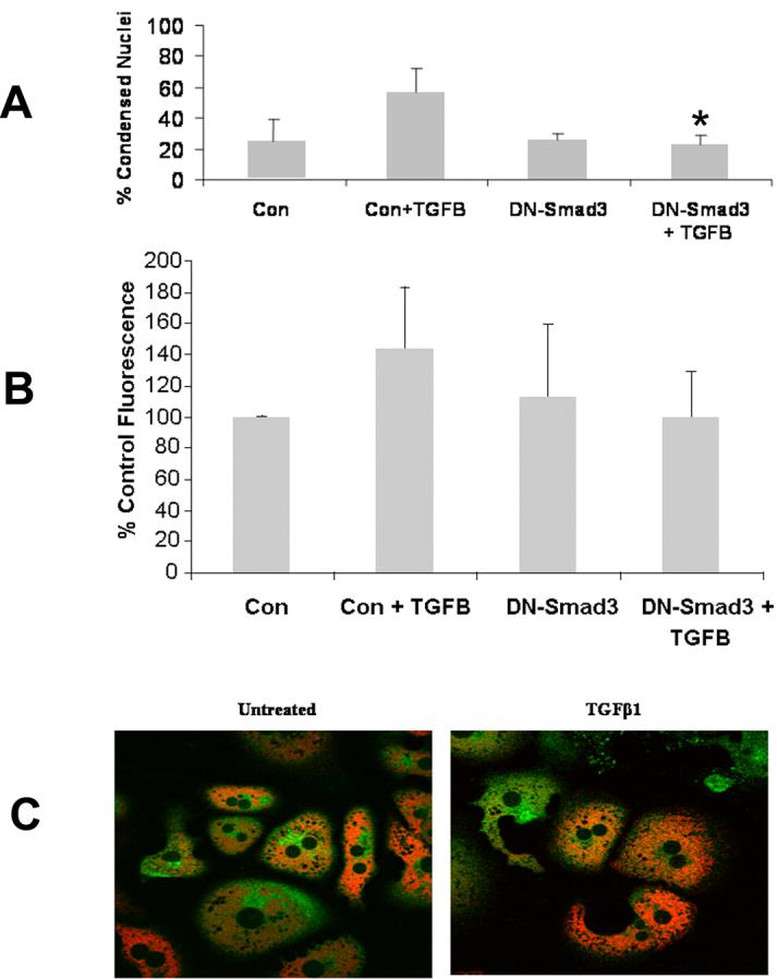
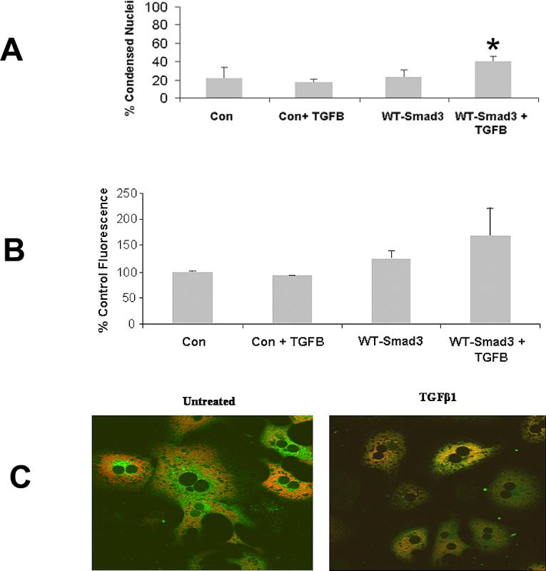
TGFbeta induces hepatocyte apoptosis via reactive oxygen species (ROS) generation, the mitochondrial permeability transition (MPT), and caspase activation. The role of the Smad pathway in these events is unknown. In this study primary hepatocytes were isolated from Smad3 wild-type (+/+) and knockout (-/-) mice, and were treated with TGFbeta (5ng/ml) and/or trolox (2mM). ROS generation, MPT, TGFbeta-dependent transcription, and apoptosis were assessed in the presence or absence of Smad3 wild-type (WT) and dominant-negative (DN) plasmids. With TGFbeta treatment, Smad3 (-/-) hepatocytes did not generate ROS activity, exhibit MPT, activate caspases, or undergo apoptosis when compared to Smad 3 (+/+) hepatocytes. Similarly, transfection of Smad3 (+/+) hepatocytes with DN-Smad3 inhibited TGFbeta-mediated transcription, ROS generation, MPT, and apoptosis. However, Smad3 (-/-) cells transfected with WT-Smad3 and treated with TGFbeta demonstrated increased transcriptional activity, the MPT, and TGFbeta-induced apoptosis. TGFbeta-mediated ROS generation occurred through an NADPH-like oxidase pathway since diphenyleneiodonium chloride inhibited ROS induction. In conclusion, TGFbeta-induced hepatocyte apoptosis occurs through Smad3 dependent activation of ROS with subsequent activation of the MPT and caspases.


Figures

Figure 1
Figure 1
Smad3 (+/+) or (−/−) hepatocytes were treated with or without TGFβ (5 ng/ml), TNFα (30 ng/ml), or actinomycin D (1 μg/ml). Apoptosis was determined morphologically by propidium iodide staining and biochemically by caspase activity. (A) Smad3 (+/+) and (−/−) hepatocytes were untreated or treated with TGFβ and morphology was assessed at 48 hours by PI staining. Smad3 (+/+) hepatocytes displayed condensed nuclei and cell shrinkage consistent with apoptosis whereas Smad3 (−/−) hepatocytes appeared similar to controls. (B) Hepatocyte apoptosis was quantified by counting the number of condensed nuclei per 5 high power fields and expressed as percent condensed nuclei over time. Smad3 (+/+) hepatocytes had significantly increased (*p<0.05) apoptosis at 48 hours of TGFβ treatment compared to Smad3 (−/−) hepatocytes. (C) Apoptosis was assessed by caspase activity assay for caspase-3 and caspase-8 activation expressed as fold increase over control at 24, 36, and 48 hours. Caspase-3 activity was increased significantly (*p<0.05) in Smad3 (+/+) hepatocytes compared to Smad (−/−) hepatocytes at 36 hours. Caspase-8 activity demonstrated a similar pattern, but was less robust. (D) To determine if inhibition of apoptosis in Smad3 (−/−) mice was specific for TGFβ, Smad3 (+/+) and (−/−) hepatocytes were treated with or without TNFα and actinomycin D (ActD) and apoptosis was determined. Both Smad3 (+/+) hepatocytes and Smad3 (−/−) hepatocytes readily underwent TNFα-induced apoptosis (no significant difference). (E) To determine if Smad3 (+/+) and (−/−) hepatocytes underwent MPT in response to TGFβ treatment; these hepatocytes were loaded with the fluorophores TMRM (red) and calcein (green). After 10 hours of treatment, Smad3 (+/+) hepatocytes demonstrated mitochondrial loss of TMRM and uptake of calcein consistent with the MPT. Smad3 (−/−) hepatocytes, however, maintained mitochondrial integrity indicating resistance to TGFβ-induced MPT.
Figure 2
Figure 2
The effect of ROS generation in Smad3 (+/+) and (−/−) was determined from 30−240 minutes following TGFβ treatment. (A) Smad3 (+/+) hepatocytes demonstrated consistently increased ROS activity compared to Smad3 (−/−) hepatocytes (*p<0.05 for the curves). A consistent burst of ROS activity was noted at 30−60 minutes after treatment, but the magnitude of the activity was variable. (B) ROS generation in Smad3 deficient JAR cells was fluorometrically determined. In contrast to Smad (+/+) hepatocytes, JAR cells did not increase ROS after TGFβ treatment (p<0.05). (C) Smad3 (+/+) hepatocytes were treated with the antioxidant, trolox, alone or in combination with TGFβ to determine if TGFβ-induced ROS activity could be inhibited. At 60 minutes, TGFβ treatment increased ROS generation significantly, and pretreatment with trolox attenuated markedly (*p<0.05) this burst of activity compared to TGFβ alone. (D) To determine the effect of inhibition of TGFβ-induced ROS on apoptosis, Smad3 (+/+) hepatocytes were treated with trolox, TGFβ, or trolox and TGFβ and apoptosis was assessed morphologically at 48 hours. Inhibition of ROS generation by trolox decreased significantly (*p<0.05) TGFβ-induced apoptosis. (E) The dependence of Smad3-induced ROS generation on transcription and translation was determined by pretreating Smad3 (+/+) hepatocytes with either actinomycin D (10 μg/ml) or cycloheximide (10 mg/ml) and measuring H2DCFDA cleavage at 60 minutes. Neither actinomycin D nor cycloheximide decreased early ROS generation suggesting that gene transcription or protein synthesis is not required for ROS activity following TGFβ treatment. (F) The involvement of NADPH oxidase-like system in ROS increase was investigated in the presence of diphenyleneiodonium chloride (DPI). Hepatocytes were treated with 1 μM of DPI for 30 min prior to TGFβ administration and ROS generation was determined by H2DCFDA fluorometry. DPI prevented TGFβ-induced increase in ROS. (G) Smad3 (+/+) and (−/−) hepatocytes were transiently transfected with a TGFβ-responsive luciferase reporter plasmid, p3TPLuc, and subsequently treated with 5 ng/ml TGFβ. In additional experiments, following transfection of the reporter plasmid, Smad3 (+/+) hepatocytes were transfected with a DN-Smad3 plasmid and Smad3 (−/−) hepatocytes transfected with a WT-Smad3 plasmid and luciferase measured after stimulation with TGFβ. Treatment of Smad3 (+/+) hepatocytes with TGFβ alone resulted in significantly increased luciferase activity compared to Smad3 (−/−) hepatocytes. However, transfection of Smad3 (+/+) hepatocytes with the DN-Smad3 followed by TGFβ treatment (light gray bar) decreased markedly transcriptional activity. Conversely, transfection of Smad3 (−/−) hepatocytes with WT-Smad3 followed by TGFβ treatment significantly increased luciferase activity (light gray bar).
Figure 3
Figure 3
Smad3 (+/+) hepatocytes were transiently transfected with control vector or the DN-Smad3 vector and subsequently treated with or without TGFβ prior to assessment of apoptosis, ROS generation, and MPT. (A) Smad3 (+/+) hepatocytes treated with 5 ng/ml TGFβ following transfection with DN-Smad3 demonstrated significantly decreased apoptosis (*p<0.05) as compared to identical cells transfected with the control vector and treated with TGFβ. (B) TGFβ-induced ROS in Smad3 (+/+) hepatocytes transfected with DN-Smad3 exhibited ROS generation similar to controls. (C) The MPT was assessed by confocal microscopy in Smad3 (+/+) hepatocytes transfected with the DN-Smad3 vector and subsequently treated with TGFβ. Transfection with DN-Smad3 inhibited the MPT following TGFβ treatment in Smad3 (+/+) hepatocytes.
Figure 4
Figure 4
Smad3 (−/−) hepatocytes were transiently transfected with either control vector or vector expressing WT-Smad3. Subsequently, apoptosis, ROS generation, and MPT were determined. (A) Smad3 (−/−) hepatocytes transiently transfected with WT-Smad3 and subsequently treated with TGFβ demonstrated significantly increased apoptosis (*p<0.05) compared to identical cells transfected with control vector and treated with TGFβ. (B) ROS was determined in Smad3 (−/−) hepatocytes transiently transfected with WT-Smad3 and subsequently treated with TGFβ. Transfection with the WT-Smad3 partially restored ROS generation in these hepatocytes. (C) Smad3 (−/−) hepatocytes transiently transfected with WT-Smad3 and treated with TGFβ showed MPT 12 hours following treatment. Control transfected Smad3 (−/−) hepatocytes treated with TGFβ failed to undergo to MPT.

References

    1. Massague J. TGF-beta signal transduction. Annu.Rev.Biochem. 1998;67:753–791. - PubMed
    1. Kim BC, Mamura M, Choi KS, Calabretta B, Kim SJ. Transforming growth factor beta 1 induces apoptosis through cleavage of BAD in a Smad3-dependent mechanism in FaO hepatoma cells. Mol.Cell Biol. 2002;22:1369–1378. - PMC - PubMed
    1. Schrum LW, Bird MA, Salcher O, Burchardt ER, Grisham JW, Brenner DA, Rippe RA, Behrns KE. Autocrine expression of activated transforming growth factor-beta(1) induces apoptosis in normal rat liver. Am.J.Physiol Gastrointest.Liver Physiol. 2001;280:G139–G148. - PubMed
    1. Shima Y, Nakao K, Nakashima T, Kawakami A, Nakata K, Hamasaki K, Kato Y, Eguchi K, Ishii N. Activation of caspase-8 in transforming growth factor-beta-induced apoptosis of human hepatoma cells. Hepatology. 1999;30:1215–1222. - PubMed
    1. Black D, Bird MA, Samson CM, Lyman S, Lange PA, Schrum LW, Qian T, Lemasters JJ, Brenner DA, Rippe RA, Behrns KE. Primary cirrhotic hepatocytes resist TGFbeta-induced apoptosis through a ROS-dependent mechanism. J.Hepatol. 2004;40:942–951. - PubMed

Publication types

MeSH terms

LinkOut - more resources