. 2007 Nov 14:1180:1-6.
doi: 10.1016/j.brainres.2007.08.039. Epub 2007 Aug 24.

Ube3a mRNA and protein expression are not decreased in Mecp2R168X mutant mice

Affiliations

Ube3a mRNA and protein expression are not decreased in Mecp2R168X mutant mice

Amy Lawson-Yuen et al. Brain Res. .

Abstract

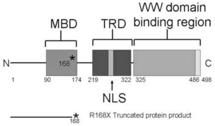
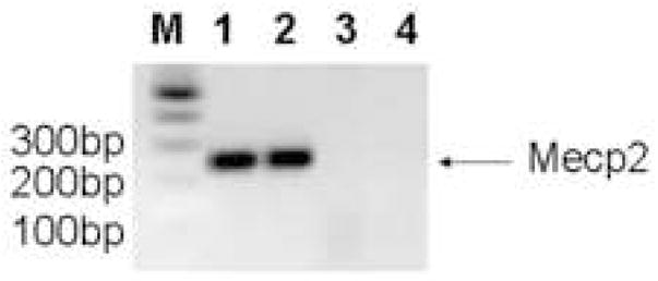
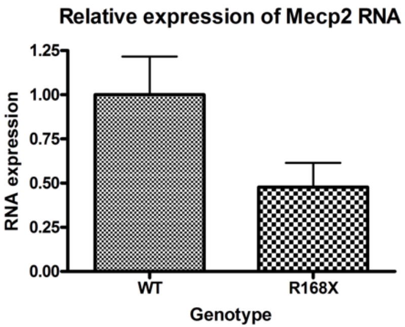
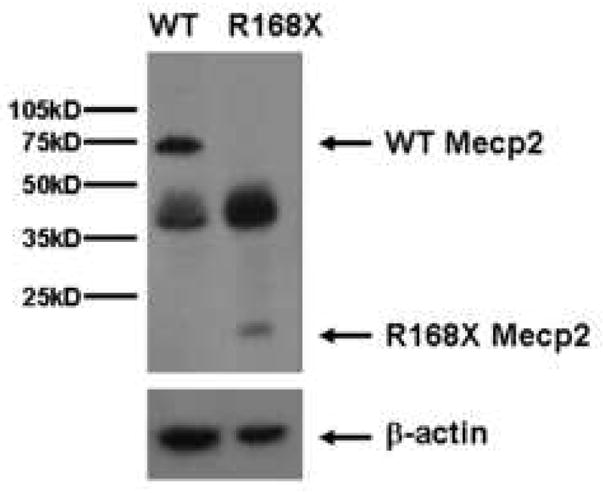
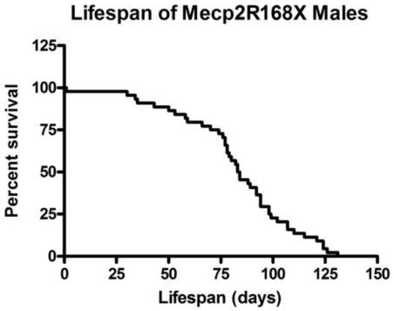
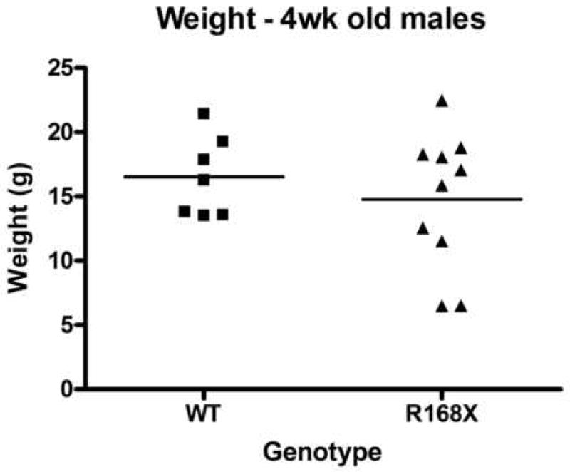
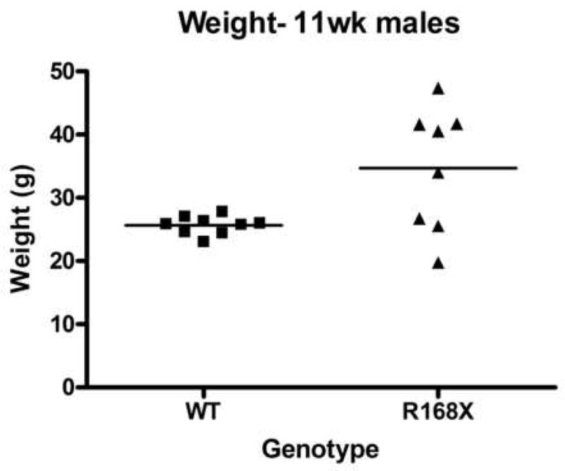
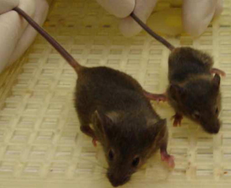
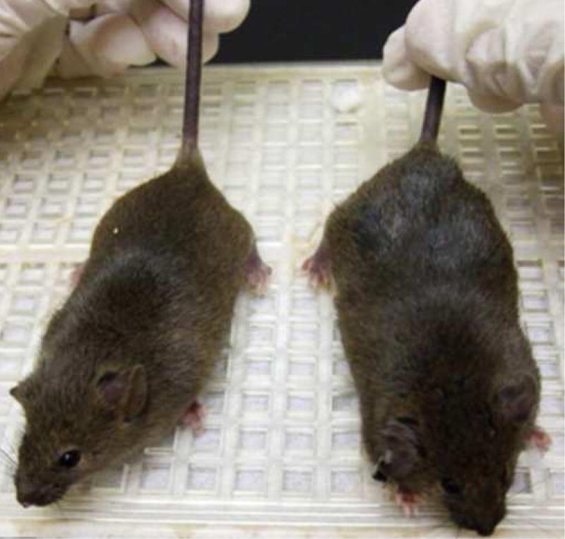
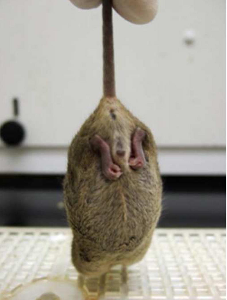
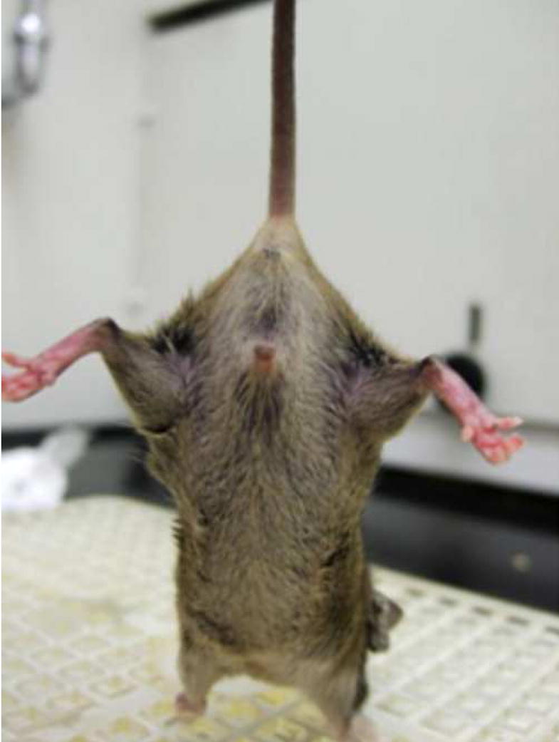
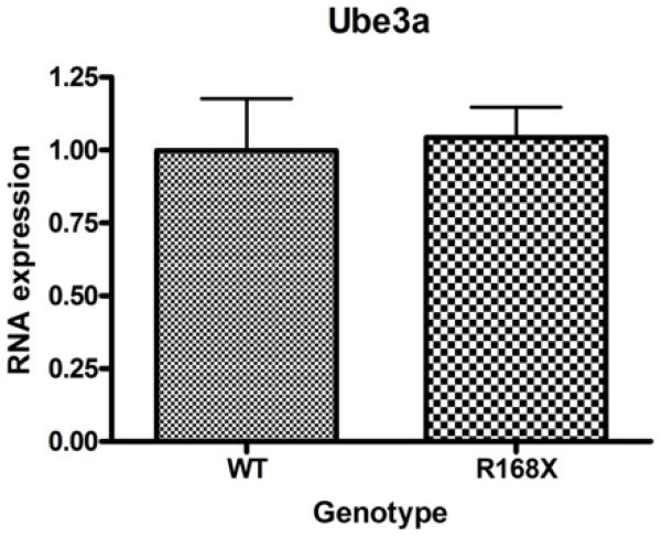
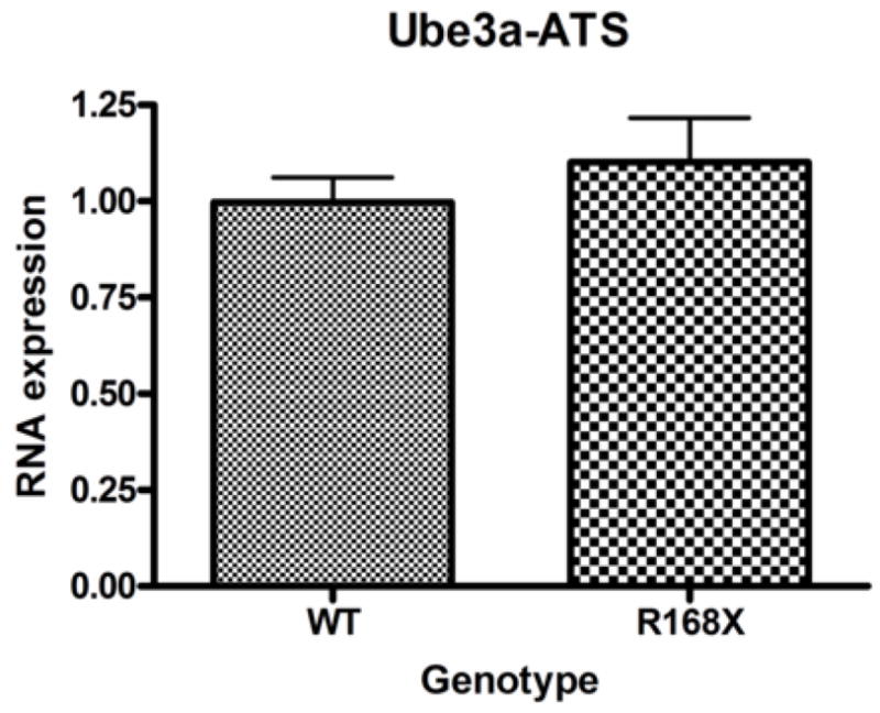
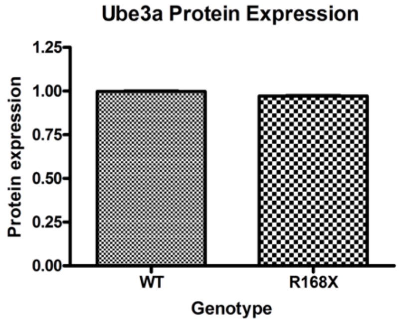
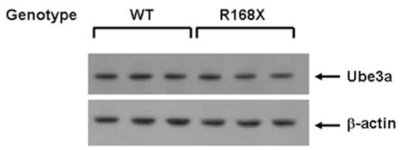
Mutations in the transcriptional repressor methyl CpG binding protein 2 (MeCP2) are responsible for most cases of Rett Syndrome (RS), a severe neurodevelopmental disorder characterized by developmental regression, minimal speech, seizures, postnatal microcephaly and hand stereotypies. Absence of the maternal copy of ubiquitin protein ligase 3A (UBE3A) results in Angelman syndrome, also a severe developmental disorder that shares some clinical features with RS. As MeCP2 regulates gene expression, this has led to the hypothesis that MeCP2 may regulate UBE3A expression; however, there are conflicting reports regarding the expression of Ube3a in MeCP2 null mutant mice. We have generated a novel MeCP2 mutant knock-in mouse with the mutation R168X, one of the most common mutations in patients with RS. These mice show features similar to RS, including hypoactivity, forelimb stereotypies, breathing irregularities, weight changes, hind limb atrophy, and scoliosis. The male mice experience early death. Analysis of Ube3a mRNA and protein levels in the Mecp2(R168X) male mice showed no significant difference in expression compared to their wild type littermates.


Figures

Figure 1
Figure 1. Mecp2R168X mutant mice
(A) An A to T point mutation was engineered to create a premature stop codon in place of an arginine at codon 168. (B) Confirmation of the mutant transcript by RTPCR. RTPCR was performed with primers flanking the mutation using cDNA reverse transcribed from WT mouse RNA (lane 1), cDNA reverse transcribed from mutant mouse RNA (lane 2), WT mouse RNA (lane 3), and mutant mouse RNA (lane 4). The expected 238bp product was detected in lanes 1 and 2 and confirmed as MeCP2 by sequencing. Larger bands are produced from genomic DNA in lanes 3 and 4 due to the presence of intron 3. (C) The Mecp2R168X mutant RNA is relatively stabled compared to the WT transcript. Error bars show standard error of the mean. (D) Mecp2R168X mutant mice (right lane) express a small protein which may be a prematurely truncated mutant MeCP2 protein, but do not express the full length WT protein. The WT mice (left lane) express the full length protein.
Figure 2
Figure 2. Characterization of Mecp2R168X mutant mice
(A) Lifespan of the Mecp2R168X mutant mice is shortened, with a median lifespan of 83.5 days (n=45). (B) At 4 weeks of age, greater variance in the weight distribution of the affected males was apparent, with numerous low weight mice. (C) In the mice surviving to 11 weeks, weight variance was also apparent, but many more mice were obese. (D) At 3 weeks, an affected hemizygous Mecp2R168X male (mouse on the right) had difficulty gaining weight while his WT littermate thrived. (E) At 7 weeks, an affected hemizygous Mecp2R168X male (mouse on the right) showed greater weight gain his WT littermate. (F) 7 week affected male showing hindlimb clasping. (G) 7 week old WT male littermate showing normal function of hindlimbs.
Figure 3
Figure 3. Ube3a and Ube3a-ATS mRNA expression
Male hemizygous Mecp2R168X mice no significant difference in Ube3a (A) or Ube3a-ATS (B) mRNA expression in cortical tissue as compared to their WT littermates when analyzed by real time qRT-PCR. Error bars show standard error of the mean.
Figure 3
Figure 3. Ube3a and Ube3a-ATS mRNA expression
Male hemizygous Mecp2R168X mice no significant difference in Ube3a (A) or Ube3a-ATS (B) mRNA expression in cortical tissue as compared to their WT littermates when analyzed by real time qRT-PCR. Error bars show standard error of the mean.
Figure 4
Figure 4. Ube3a protein expression
(A) Male hemizygous Mecp2R168X mice showed no change in Ube3a protein expression in cortical tissue as compared to their WT littermates when analyzed by Western blot. Error bars show standard error of the mean. (B) Western blot showing representative samples from 3 pairs of mice.

Similar articles

Cited by

References

    1. Amir RE, et al. Rett syndrome is caused by mutations in X-linked MECP2, encoding methyl-CpG-binding protein 2. Nat Genet. 1999;23:185–8. - PubMed
    1. Angelman H. “Puppet children.” A report of three cases. Dev Med Child Neurol. 1965;7:681–8. - PubMed
    1. Bienvenu T, Chelly J. Molecular genetics of Rett syndrome: when DNA methylation goes unrecognized. Nat Rev Genet. 2006;7:415–26. - PubMed
    1. Chamberlain SJ, Brannan CI. The Prader-Willi syndrome imprinting center activates the paternally expressed murine Ube3a antisense transcript but represses paternal Ube3a. Genomics. 2001;73:316–22. - PubMed
    1. Chen RZ, et al. Deficiency of methyl-CpG binding protein-2 in CNS neurons results in a Rett-like phenotype in mice. Nat Genet. 2001;27:327–31. - PubMed

Publication types