. 2007 Nov 15;311(2):679-90.
doi: 10.1016/j.ydbio.2007.08.036. Epub 2007 Aug 28.

Control of kidney, eye and limb expression of Bmp7 by an enhancer element highly conserved between species

Affiliations

Control of kidney, eye and limb expression of Bmp7 by an enhancer element highly conserved between species

Derek Adams et al. Dev Biol. .

Abstract

Bmp7 is expressed in numerous tissues throughout development and is required for morphogenesis of the eye, hindlimb and kidney. In this study we show that the majority if not all of the cis-regulatory sequence governing expression at these anatomical sites during development is present in approximately 20 kb surrounding exon 1. In eye, limb and kidney, multiple distinct enhancer elements drive Bmp7 expression within each organ. In the eye, the elements driving expression in the pigmented epithelium and iris are spatially separated. In the kidney, Bmp7 expression in collecting ducts and nephron progenitors is driven by separate enhancer elements. Similarly, limb mesenchyme and apical ectodermal ridge expression are governed by separate elements. Although enhancers for pigmented epithelium, nephrogenic mesenchyme and apical ectodermal ridge are distributed across the approximately 20 kb region, an element of approximately 480 base pairs within intron 1 governs expression within the developing iris, collecting duct system of the kidney and limb mesenchyme. This element is remarkably conserved both in sequence and position in the Bmp7 locus between different vertebrates, ranging from Xenopus tropicalis to Homo sapiens, demonstrating that there is strong selective pressure for Bmp7 expression at these tissue sites. Furthermore, we show that the frog enhancer functions appropriately in transgenic mice. Interestingly, the intron 1 element cannot be found in the Bmp7 genes of vertebrates such as Danio rerio and Takifugu rubripes indicating that this modification of the Bmp7 gene might have arisen during the adaptation from aquatic to terrestrial life. Mutational analysis demonstrates that the enhancer activity of the intron 1 element is entirely dependent on the presence of a 10 base pair site within the intron 1 enhancer containing a predicted binding site for the FOXD3 transcription factor.


Figures
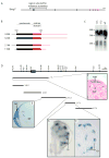
Figure 1
Figure 1. Genomic organization of the murine Bmp7 locus and screening strategy used to identify enhancer elements
A The Bmp7 gene is comprised of 7 discrete exons distributed over approximately 72 kb on mouse chromosome 2. Exon-intron structure is depicted, with black boxes corresponding to exons comprising the 5’ UTR and encoding mRNA for the prodomain of BMP7 and red boxes corresponding to exons comprising the 3’ UTR and mRNA for the mature domain of BMP7. Bar at top right represents 2.5 kb. B Predicted structures of Bmp7 transcripts based on sequencing of newborn kidney cDNA and 3’ RACE of embryonic kidney mRNA: In addition to the previously published initiation site 102 bp upstream of the first AUG (1.9 and 3.6 kb transcripts), an initiation site was identified 372 bp upstream of the AUG (2.1 and 3.8 kb transcripts). Additionally, two 3’ termini were identified: one 448 bp downstream from the UAG stop codon (1.9 and 2.1 kb transcripts), and another 1887 bp downstream from the UAG (3.6 and 3.8 kb transcripts). Red and black regions correspond to red and black exons in 1A. C Total RNA was isolated from E13.5, E17.5 and adult kidneys (13.5, 17.5 and ad respectively), and subjected to Northern hybridization with a probe corresponding to the entire Bmp7 coding sequence. Four RNA species are detected; 1.9kb, 2.1kb, 3.6kb and 3.8kb. Alternate initiation and termination sites shown in 1B account for these four distinct RNA transcripts. D Strategy used to screen for enhancer elements driving Bmp7 expression in the developing kidney. The genomic region surrounding Bmp7 exon 1 (bracketed in 1A) is shown, and lines below denote sizes of genomic subclones 142:1, 216:1, 216:2, 217:1 and 217:2. The presence of enhancer elements in each of these subclones was assayed using the Hsp68lacZpA reporter in transgenic mice. Reporter constructs drive expression at diverse anatomical sites. E The 216:2 genomic subclone drives expression in neural crest (NC) adjacent to the neural tube (NT). F Expression in nephrogenic mesenchyme (NM) of the kidney is driven by the T fragment. G, G’ Expression in collecting ducts (CD) of the kidney is driven by the 217:1 fragment.
Figure 2
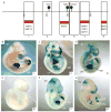
Figure 2. The intron 1 enhancer element drives expression in a significant subset of Bmp7 expression domains
Comparison of Bmp7 expression driven from the endogenous Bmp7 locus with expression driven from the intron 1 enhancer element. Heterozygous Bmp7lacZ reporter mice (Bmp7+/lacZ) and Bmp7Pst4 embryos were dissected at E8, E9.5 and E10.5 and whole-mount X-GAL stained. A At E8, endogenous Bmp7 expression is seen in head-folds (HF), heart (HT) and notochord (NT), with an expanded domain in the caudal notochord corresponding to the position of the node (ND). B The intron 1 enhancer element drives expression mainly in a diffuse region of the tail bud (TB) surrounding the node and also weakly in the heart. C At E9.5, endogenous Bmp7 expression is seen in heart, dorsal forebrain (DFB), isthmus or midbrain-hindbrain junction (IST), optic placode (OPT), otic placode (OTC), Rathke’s pouch (RP), limb bud (LB) and Wolffian duct (WD). D The intron 1 enhancer element drives expression in subset of these domains: isthmus, limb bud and Wolffian duct. Weak expression can be seen in the heart and ectopic expression is seen in the ventral forebrain (VFB). E At E10.5, additional domains of endogenous Bmp7 expression are apparent in mesonephric tubules (MN) and ureteric bud (UB). F These domains are represented in the intron 1 enhancer, where an additional ectopic domain of expression can be seen in the dorsal root ganglion (DRG).
Figure 3
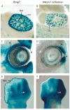
Figure 3. The intron 1 enhancer element drives expression in specific domains of the eye, limb and kidney
A Bmp7 is expressed in both the collecting duct system (CD) and nephrogenic mesenchyme (NM) of the developing kidney B The intron 1 enhancer drives expression only in collecting ducts C In the eye, Bmp7 is expressed in the pigmented epithelium (PE) and nascent iris (IR). D The intron 1 enhancer drives expression solely in the nascent iris of the eye. E Bmp7 is expressed in the apical ectodermal ridge (AER), limb mesenchyme (LM) and precartilage condensates (PC) of the developing limb. F Intron 1 enhancer driven expression is limited to limb mesenchyme.
Figure 4
Figure 4. The Bmp7 intron 1 enhancer is strongly conserved from human to frog genomes, but is not represented in the fish
A Alignment of homo sapiens (hsap), mus musculus (mmus), gallus gallus (ggal) and xenopus tropicalis (xtrop) Bmp7 loci identifies a highly homologous 480 bp region located within the murine intron 1 enhancer. This region is not represented in either zebrafish or fugu genomes. Three putative transcription factor binding sites conserved across all species were predicted using the Transfac database: OCT1, AGL3 and FOXD3. B The 480 bp region from mouse recapitulates expression in eye (E), limb (L) and Wolffian duct (WD) seen in 217XPst:Hsp68lacZpA at E10.5. C The 480 base pair region from X. tropicalis intron 1 drives Hsp68 reporter gene expression in embryonic eye (E) and limb (L), but not Wolffian duct (WD) at E11.5.
Figure 5
Figure 5. Deletion mutagenesis to localize transcription factor binding site(s) driving Bmp7 expression in limb mesenchyme (LM), Wolffian Duct (WD) and isthmus (IST)
A Schematic of Bmp7 intron 1 enhancer element showing the two different strategies used to identify putative transcription factor binding sites: i) Regions of strong homology with intronic sequence in other TGFβ superfamily ligand genes (red bars), ii) Transfac-predicted binding sites conserved in all species (black marks). Reporter genes I-VI containing deletions spanning single regions were used to generate transgenic animals which were analyzed at E10.5 for expression in isthmus (IST), limb mesenchyme (LM) and Wolffian duct (WD). BG Only deletion IV spanning the predicted FOXD3 binding site significantly affects expression in the three domains assayed.
Figure 6
Figure 6. Site directed mutagenesis of the putative FOXD3 binding site in the intron 1 enhancer and electrophoretic mobility shift assay to verify protein binding to the site in embryonic kidney and limb nuclear lysates
A The putative FOXD3 binding site of the Bmp7 intron 1 enhancer element (wild type) was altered by site directed mutagenesis to incorporate three mismatched base pairs (mutation). The mutant 480 bp intron 1 enhancer element was subsequently cloned into the Hsp68lacZ reporter. B Transgenic embryos generated using the mutagenized Bmp7 intron 1 reporter gene described in A show complete lack of expression in limb, isthmus and Wolffian duct. C A radiolabeled oligonucleotide duplex spanning the predicted FOXD3 binding site was used as a probe for a gel-shift assay with extracts of E16.5 limb and E14.5 kidney. Nucleotides removed in reporter IV (Fig. 5) of the mutational analysis are shown in bold. D Retardation of migration of the oligonucleotide by addition of crude nuclear extracts of embryonic limb or kidney was assayed on a 4% native gel in: i) the absence of cold competitor oligonucleotides (-), ii) the presence of a cold non-specific scrambled oligonucleotide (scr), and iii) the presence of a cold oligonucleotide identical in sequence to the probe (spec). The arrow marks probe retarded in the gel by the binding of protein from the extracts, and the arrowhead marks free probe.

Similar articles

Cited by

References

    1. Ahlberg PE, Milner AR. The origin and early diversification of tetrapods. Nature. 1994;368:507–514.
    1. Birney E, et al. Ensembl 2006. Nucleic Acids Res. 2006;34:D556–61. - PMC - PubMed
    1. Bush KT, et al. TGF-[beta] superfamily members modulate growth, branching, shaping, and patterning of the ureteric bud. Developmental Biology. 2004;266:285–298. - PubMed
    1. Dottori M, et al. The winged-helix transcription factor Foxd3 suppresses interneuron differentiation and promotes neural crest cell fate. Development. 2001;128:4127–38. - PubMed
    1. Dudley AT, et al. Interaction between FGF and BMP signaling pathways regulates development of metanephric mesenchyme. Genes Dev. 1999;13:1601–13. - PMC - PubMed

Publication types

MeSH terms