. 2007 Nov 15;311(2):613-22.
doi: 10.1016/j.ydbio.2007.09.004. Epub 2007 Sep 17.

Wingless activity in the precursor cells specifies neuronal migratory behavior in the Drosophila nerve cord

Affiliations

Wingless activity in the precursor cells specifies neuronal migratory behavior in the Drosophila nerve cord

Krishna Moorthi Bhat. Dev Biol. .

Abstract

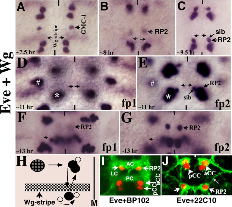
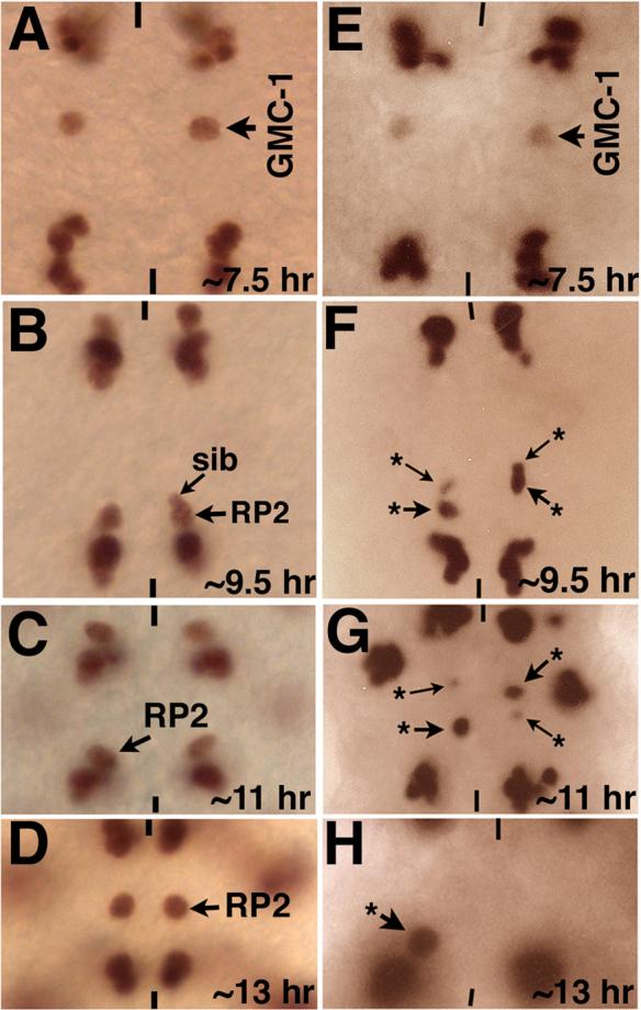
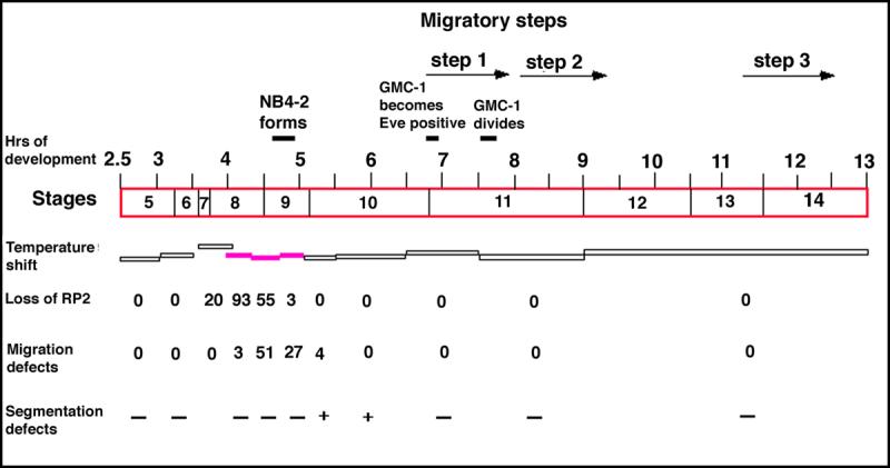
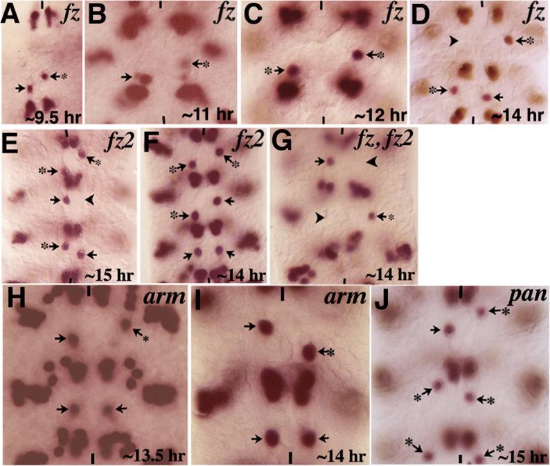
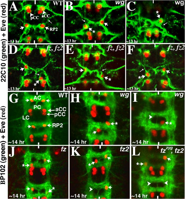
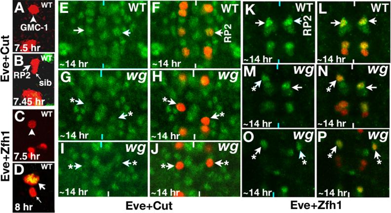
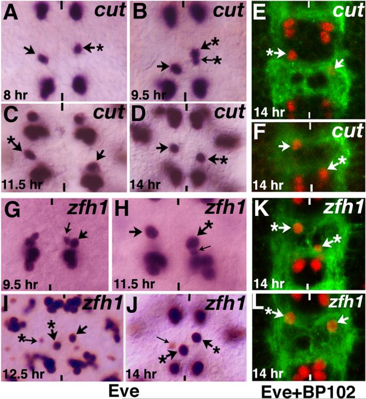
Neurons and their precursor cells are formed in different regions within the developing CNS, but they migrate and occupy very specific sites in the mature CNS. The ultimate position of neurons is crucial for establishing proper synaptic connectivity in the brain. In Drosophila, despite its extensive use as a model system to study neurogenesis, we know almost nothing about neuronal migration or its regulation. In this paper, I show that one of the most studied neuronal pairs in the Drosophila nerve cord, RP2/sib, has a complicated migratory route. Based on my studies on Wingless (Wg) signaling, I report that the neuronal migratory pattern is determined at the precursor cell stage level. The results show that Wg activity in the precursor neuroectodermal and neuroblast levels specify neuronal migratory pattern two divisions later, thus, well ahead of the actual migratory event. Moreover, at least two downstream genes, Cut and Zfh1, are involved in this process but their role is at the downstream neuronal level. The functional importance of normal neuronal migration and the requirement of Wg signaling for the process are indicated by the finding that mislocated RP2 neurons in embryos mutant for Wg-signaling fail to properly send out their axon projection.


Figures

Figure 1
Figure 1. GMC-1->RP2/sib cells follow a complex migratory path
Anterior end is up, midline is marked by vertical lines. All are wild type embryos in these panels. Panel A: The GMC-1 has already migrated several cells length towards the midline by 7.5 hours of development, though this Step 1 migration is not yet completed as the cell will migrate even further towards the midline. Panel B: The RP2/sib cells have begun their Step 2 posterior migration. The cells are slightly out of focus in this panel in order to show the Wg stripe (arrows). Panel C: The RP2/sib cells have reached the end of Step 2 migration; the Wg stripe is out of focus in this panel. Panels D and E: Two different focal panes (fp) of the same segment, in panel D, the Wg stripe is shown, whereas in panel E, the RP2/sib cells are shown. The two cells stay in this position for close to 2 hours and then they begin their Step 3 anterior migration. The asterisk marks Us/CQ neurons (just above the aCC/pCC pair) and the hash marks the EL neuron cluster. Panels F and G: Two different focal planes of the same segment; the RP2 neuron has migrated anterior to its ultimate position; the sib cell has lost its Eve expression by this time. Panel H: The migratory route of GMC-1->RP2/sib cells. The GMC-1 first moves toward the midline (M; Step 1 migration), it divides to generate an RP2 and a sib. Both RP2 and sib migrate to the posterior, parallel to the midline; they both cross the Wg stripe (and therefore the parasegmental boundary). The sib then moves toward the midline and to the anterior; it resides just about in the region of Wg stripe. The RP2 migrates anterior, it crosses the Wg stripe (the parasegmental boundary) and resides close to the original position from which it started the posterior migration. Panel I: Wild type embryo stained with Eve and BP102. AC, anterior commissure; PC, posterior commissure; LC, longitudinal connective. Panel J: Wild type embryo stained with Eve and 22C10, thin arrow indicates the ipsilateral RP2 axon projection.
Figure 2
Figure 2. Migration of GMC-1->RP2/sib cells is affected in wg mutants
Embryos in panels A-D (wild type) are stained with Eve antibody and embryos in panels E-H (wg mutant) are stained with Eve and Wg antibodies. Wild type embryos shown in panels A-D are shifted the same way as mutantembryos. Anterior end is up, midline is marked by vertical lines. Panels A-D are wild type, panels E-H are wgts mutants. The Wg activity was inactivated by shifting mutant embryos to 29°C just about NB4−2 is formed (see Materials and Methods). Panel A: The GMC-1 has migrated several cell-lengths towards the midline. Panel B: The RP2/sib cells have migrated posterior towards the aCC/pCC and U/CQ cluster. Panel C: The RP2/sib cells have completed their Step 2 migration and are located very close to the aCC/pCC and CQ cluster. Panel D: The RP2 neuron has completed its Step 3 migration and is located in its ultimate position. Panel E: The GMC-1 has migrated several cells length towards the midline, but this Step 1 migration is not yet completed. Panels F-H: The RP2/sib cells show incomplete Step 2 (panels F, G) and Step 3 (panel H) migrations. Arrow with an asterisk indicates aberrantly migrated RP2/sib cells.
Figure 3
Figure 3. Wg requirement for the proper migration of GMC-1->RP2/sib cells is in the neuroectoderm/NB4−2
Handpicked wgts mutant embryos at different developmental time points were shifted from the permissive 18°C temperature to the restrictive 29°C temperature and then shifted back to the permissive temperature. The duration at which the embryos were kept at the restrictive temperature is indicated by the horizontal bars. The filled in horizontal bars indicate sensitive period for the defect. These embryos were stained for Eve to determine the migration defects. The timings and stages correspond to developmental time/stages at 22°C; the numbers represent the percentage of hemisegments affected (number examined=220−300 per temperature-shift experiment). For example, when embryos were shifted to 29°C between 4.3−4.7 hours of developmental period, 55% of hemisegments were missing the RP2s; the percentage of migration defects indicate the defects for the remaining hemisegments where the RP2s were present. Segmentation defects were examined by cuticle preparation; at least 50 embryos were examined per temperature-shift experiment and minus symbol (−) indicates 4% or less showing the cuticle defect.
Figure 4
Figure 4. Migration of GMC-1->RP2/sib cells is affected in mutants for genes in the Wg-signaling pathway
Embryos are stained for Eve. Anterior end is up, vertical lines indicate the midline. Panels A-D: Embryos mutant for the zygotic fz showing the RP2/sib migration defects (arrows with star). Note the occasional missing RP2 (panel D, arrowhead). Panels E and F: Embryos mutant for fz2 showing the RP2/sib migration defects (arrow with star); RP2s are occasionally missing as well (arrowhead in panel E). Panel G: Embryo double mutant for zygotic fz and fz2 showing the RP2/sib migration defects (arrows with star) and missing RP2s (arrowheads). Panels H and I: Embryos mutant for zygotic arm, armY025 (panel H), and armS10C (panel I). Panels J: Embryo mutant for pan showing the RP2/sib migration defects (arrows with star). Note that consistent with our previous finding (Bhat, 1998) and contrary to a subsequent paper (Chen and Struhl, 1999), both fz and fz2 single mutants have missing RP2s in a partially penetrant manner (see also Fig. 5).
Figure 5
Figure 5. Aberrant axon projections in mislocated RP2s
Anterior end is up, vertical lines mark the midline. Embryos in panels A-F are double stained for Eve (Red) and 22C10 (Green), whereas embryos in panels G-L are doubled stained with Eve (Red) and BP102 (Green). AC, anterior commissure; PC, posterior commissure, LC, longitudinal connectives. Panel A: Wild type embryo showing the axon projection of RP2 (thin arrow); this projection fasciculates with the projection from aCC. Panels B and C: wgts mutant embryos; mislocated RP2s (arrows with star) have aberrant axon projections (thin arrows). Panels D-F: fz fz2 double mutant embryos; mislocated RP2s (arrows with star) have aberrant axon projections (thin arrows). Panel G: Wild type embryo, the RP2 is located at the inner armpit of AC. Panels H and I: wgts mutant embryos showing the mis-localization of RP2 neurons (arrow with star), note the occasional missing RP2 (arrowhead). Panels J-L: Embryos mutant for fz, fz2 or fz fz2 double mutants are shown with mislocated RP2s (and occasional missing RP2s indicated by arrowheads).
Figure 6
Figure 6. Expression of neuron-specific genes is affected in wg mutants
Anterior end is up, vertical lines mark the midline. The Wg activity was inactivated by shifting mutant embryos to 29°C just about NB4−2 is formed. Panels A and B: Embryos are double stained for Eve (Red) and Cut (Green); no detectable Cut is present in GMC-1 (panel A) or a newly generated RP2 and sib (panel B), but Cut is present at high levels in a differentiated RP2; Cut appears in an RP2 by ∼8 hours of development. Panels C and D: Embryos are double stained for Eve (Red) and Zfh1 (Green); no detectable Zfh1 is present in GMC-1 (panel C), but a newly generated RP2 has Zfh1 but not sib (yellow indicates co-localization of both Eve and Zfh1, panel D). Embryos in panels E-J are double stained for Eve (Red) and Cut (Green) and K-P are double stained for Eve (Red) and Zfh1 (Green). Panels E and F: Wild type embryo, note the levels of Cut in RP2 neurons. Panels G-J: wg mutant embryos showing mislocated RP2s (arrows with star) with reduced levels of Cut. The confocal images in Panels E-J were collected using the exact same settings. Panels K and L: Wild type embryo, note the levels of Zfh1 in RP2 neurons. Panels M-P: wg mutant embryos showing mislocated RP2s (arrows with star) with reduced levels of Zfh1. The confocal images in panels K-P were collected using the exact same settings. The aCC/pCC pairs are out of focus in panel N (left hemisegment) and we see U neuronal cluster due to some unevenness of the nerve cord in the mutant. We used the same settings for collecting images from the mutants as for collecting images from the wild type controls, and expression of Cut and Zfh1 in other cells in the nerve cord was also used as reference.
Figure 7
Figure 7. Loss of function for cut and zfh1 causes similar migration defects as loss of function for wg
Anterior end is up, vertical lines mark the midline. Arrow indicates normal RP2, arrow with star indicates RP2s with migration defects, thin arrow indicates sib. In some of the panels the sib is not visible since it is hidden under the RP2 (panels A, B, C, H) and occasionally a sib is visible with Eve even in 14 hr old embryo (cf., panel J). Panels A-F are cut mutant embryos and panels G-L are zfh1 mutant embryos showing the various RP2/sib migration defects. Embryos in panels A-D and G-J are stained with Eve; embryos in panels E, F and K, L are double stained with Eve (Red) and BP102 (Green).

References

    1. Adler PN, Vinson C, Park WJ, Conover S, Klein L. Molecular structure of frizzled, a Drosophila tissue polarity gene. Genetics. 1990;126:401–416. - PMC - PubMed
    1. Bhanot P, Brink M, Samos CH, Hsieh J-C, Wang Y, Macke JP, Andrew D, Nathans J, Nusse R. A new member of the frizzled family from Drosophila functions as a Wingless receptor. Nature. 1996;382:225–230. - PubMed
    1. Bhat KM. The patched signaling pathway mediates repression of gooseberry allowing neuroblast specification by wingless during Drosophila neurogenesis. Development. 1996;122:2911–2923. - PubMed
    1. Bhat KM. frizzled and frizzled 2 play a partially redundant role in Wingless signaling and have similar requirements to Wingless in neurogenesis. Cell. 1998;95:1027–1036. - PubMed
    1. Bhat KM. Segment polarity genes in neuroblast formation and identity specification during Drosophila neurogenesis. BioEssays. 1999;21:472–485. - PubMed

Publication types

MeSH terms

LinkOut - more resources