

Fos expression at the cerebellum following non-contact arousal and mating behavior in male rats
- PMID: 17936859
- PMCID: PMC2978247
- DOI: 10.1016/j.physbeh.2007.09.005
Fos expression at the cerebellum following non-contact arousal and mating behavior in male rats
Abstract
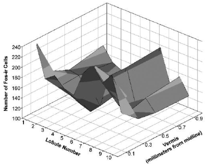
The cerebellum is considered a center underlying fine movements, cognition, memory and sexual responses. The latter feature led us to correlate sexual arousal and copulation in male rats with neural activity at the cerebellar cortex. Two behavioral paradigms were used in this investigation: the stimulation of males by distant receptive females (non-contact sexual stimulation), and the execution of up to three consecutive ejaculations. The vermis area of the cerebellum was removed following behavioral experiments, cut into sagittal sections, and analyzed with Fos immunohistochemistry to determine neuronal activation. At the mid-vermis region (sections from the midline to 0.1 mm laterally), non-contact stimulation significantly increased the activity of granule neurons. The number of activated cells increased in every lobule, but lobules 1 and 6 to 9 showed the greatest increment. In sexual behavior tests, males reaching one ejaculation had a high number of activated neurons similar to those counted after non-contact stimulation. However, two or three consecutive ejaculations showed a smaller number of Fos-ir cells. In contrast to the mid-vermis region, sections farthest from the midline (0.1 to 0.9 mm laterally) revealed that only lobule 7 expressed activated neurons. These data suggest that a well-delineated group of granule neurons have a sexual biphasic response at the cerebellar vermis, and that Fos in them is under an active degradation mechanism. Thus, they participate as a neural substrate for male rat sexual responses with an activation-deactivation process corresponding with the sensory stimulation and motor performance occurring during copulation.
Figures


Similar articles
-
Multiunit recording of the cerebellar cortex, inferior olive, and fastigial nucleus during copulation in naive and sexually experienced male rats.Cerebellum. 2010 Mar;9(1):96-102. doi: 10.1007/s12311-009-0148-y. Cerebellum. 2010. PMID: 20016964
-
Activation of the cerebellum by olfactory stimulation in sexually naive male rats.Neurologia. 2015 Jun;30(5):264-9. doi: 10.1016/j.nrl.2014.02.002. Epub 2014 Apr 4. Neurologia. 2015. PMID: 24704247 English, Spanish.
-
Olfactory stimulation induces cerebellar vermis activation during sexual learning in male rats.Neurobiol Learn Mem. 2017 Dec;146:31-36. doi: 10.1016/j.nlm.2017.11.003. Epub 2017 Nov 7. Neurobiol Learn Mem. 2017. PMID: 29104177
-
c-fos expression and (14C) 2-deoxyglucose uptake in the caudal cerebellum of the rat during motor/sensory cortex stimulation.J Comp Neurol. 1989 Jun 22;284(4):621-36. doi: 10.1002/cne.902840409. J Comp Neurol. 1989. PMID: 2504783
-
Sexual reward induces Fos in the cerebellum of female rats.Physiol Behav. 2011 Feb 1;102(2):143-8. doi: 10.1016/j.physbeh.2010.11.004. Epub 2010 Nov 6. Physiol Behav. 2011. PMID: 21059365
Cited by
-
Multiunit Recording of Cerebellar Cortex in Autistic Male Rats during Social Interaction in Enriched Environments.NeuroSci. 2023 Jul 28;4(3):178-185. doi: 10.3390/neurosci4030016. eCollection 2023 Sep. NeuroSci. 2023. PMID: 39483200 Free PMC article.
-
The role of orgasm in the development and shaping of partner preferences.Socioaffect Neurosci Psychol. 2016 Oct 25;6:31815. doi: 10.3402/snp.v6.31815. eCollection 2016. Socioaffect Neurosci Psychol. 2016. PMID: 27799080 Free PMC article.
-
Multiunit recording of the cerebellar cortex, inferior olive, and fastigial nucleus during copulation in naive and sexually experienced male rats.Cerebellum. 2010 Mar;9(1):96-102. doi: 10.1007/s12311-009-0148-y. Cerebellum. 2010. PMID: 20016964
-
Molecular mechanisms and the conflict between courtship and aggression in three-spined sticklebacks.Mol Ecol. 2016 Sep;25(17):4368-76. doi: 10.1111/mec.13766. Epub 2016 Aug 26. Mol Ecol. 2016. PMID: 27452346 Free PMC article.
-
Deletion of Endocannabinoid Synthesizing Enzyme DAGLα in Pcp2-Positive Cerebellar Purkinje Cells Decreases Depolarization-Induced Short-Term Synaptic Plasticity, Reduces Social Preference, and Heightens Anxiety.eNeuro. 2025 Jul 23;12(7):ENEURO.0400-24.2025. doi: 10.1523/ENEURO.0400-24.2025. Print 2025 Jul. eNeuro. 2025. PMID: 40523777 Free PMC article.
References
-
- Herrup K, Kuemerle B. The compartmentalization of the cerebellum. Annu Rev Neurosci. 1997;20:61–90. - PubMed
-
- Larsell O. The morphogenesis and adult pattern of the lobules and fissures of the cerebellum of the white rat. J Comp Neurol. 1952;97:281–356. - PubMed
-
- Voogd J, Glickstein M. The anatomy of the cerebellum. Trends Neurosci. 1998;21:370–375. - PubMed
-
- Ivry RB, Spencer RM, Zelaznik HN, Diedrichsen J. The cerebellum and event timing. Ann N Y Acad Sci. 2002;978:302–317. - PubMed
Publication types
MeSH terms
Substances
Grants and funding
LinkOut - more resources
Full Text Sources
