. 2007 Nov-Dec;1769(11-12):649-58.
doi: 10.1016/j.bbaexp.2007.08.004. Epub 2007 Sep 14.

Basic helix-loop-helix factors recruit nuclear factor I to enhance expression of the NaV 1.4 Na+ channel gene

Affiliations

Basic helix-loop-helix factors recruit nuclear factor I to enhance expression of the NaV 1.4 Na+ channel gene

Sadie L Hebert et al. Biochim Biophys Acta. 2007 Nov-Dec.

Abstract

We have previously shown that the basic helix-loop-helix (bHLH) transcription factors coordinate Na(V) 1.4 Na(+) channel gene expression in skeletal muscle, but the identity of the co-factors they direct is unknown. Using C2C12 muscle cells as a model system, we test the hypothesis that the bHLH factors counteract negative regulation exerted through a repressor E box (-90/-85) by recruiting positive-acting transcription factors to the nucleotides (-135/-57) surrounding the repressor E box. We used electrophoretic mobility shift assays to identify candidate factors that bound the repressor E box or these adjacent regions. Repressor E box-binding factors included the known transcription factor, ZEB/AREB6, and a novel repressor E box-binding factor designated REB. Mutations of the repressor E box that interfere with the binding of these factors prevented repression. The transcription factor, nuclear factor I (NFI), bound immediately upstream and downstream of the repressor E box. Mutation of the NFI-binding sites diminished the ability of myogenin and MRF4 to counteract repression. Based on these observations we suggest that bHLH factors recruit NFI to enhance skeletal muscle Na(+) channel expression.


Figures
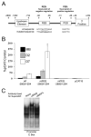
Fig. 1
Fig. 1
The repressor E box controls negative regulation of the skeletal muscle NaV 1.4 Na+ channel gene. (A) A schematic layout of several elements that regulate expression of the NaV 1.4 Na+ channel is shown, including the repressor E box (REB) and promoter E box (PEB). (B) The wild type −2800/+254 NaV 1.4 regulatory region is expressed in C2C12 muscle cells at increased levels as development proceeds from myoblasts (MB) to nascent day 2 myotubes (D2) to fully mature day 7 myotubes (D7). Mutation of the promoter E box site (mPEB) abolishes expression of the reporter gene. In contrast, mutation of the repressor E box (mREB) increases expression at all stages of development. Asterisks indicate statistical significance (p < 0.05). (C) The PEB binds both myogenin (MGN) and MRF4, as indicated by the supershift induced with antibodies to these factors in the EMSA (white asterisks indicate supershifts).
Fig. 2
Fig. 2
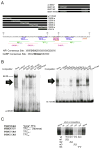
The transcription factors ZEB and REB are candidates for exerting negative regulation through the repressor E box. All EMSAs were carried out using nuclear extracts from myoblasts (MB), day 2 myotubes (D2 MT), or day 7 myotubes (D7 MT). Antibodies and competitors were added as indicated above each lane. (A) A schematic depiction of probes and competitors used in the EMSA assays is shown. (B) EMSAs were carried out with the ZEB probe. The ZEB antibody induced a supershift, as indicated by the white asterisks. The supershifted band was diminished by the wild type but not the mutant REB competitor. (C) EMSAs were carried out with the Sp1, Sp3 probe. Addition of Sp1 or Sp3 antibodies prevented formation of complexes, as indicated by the white asterisks. Competition with the wild type REB and mutant REB had little effect on the Sp1 and Sp3 complexes. (D) Using the REB probe in EMSAs, a number of complexes form. The highest complex is displaced by the wt REB but not the mREB competitors. (E) This REB factor is not supershifted by antibodies to ZEB or the Sp1 family, as shown in panel E.
Fig. 3
Fig. 3
The bHLH factors myogenin and MRF4 counteract negative regulation exerted through the repressor E box. Although there was a significant difference between the wild type and mREB reporter genes under all conditions, in the presence of myogenin and especially MRF4, expression of the wt NaV 1.4 reporter gene increased relative to the mREB, as indicated by the change in the fold difference. The asterisks indicate that the two were significantly different (p < 0.05) under each set of conditions.
Fig. 4
Fig. 4
A Transcription Factor Complex (TFC) binds sites upstream and downstream of the repressor E box. (A) Probes and competitors used in B are shown schematically above the −135 to −57 portion of the Na+ channel sequence. This portion of the sequence contains potential binding sites for nuclear factor I (NFI) and GA-Binding Protein (GABP) which are shown below the −135 to −57 sequence with their consensus sequences. (B) EMSAs with the wt 85/57 probe gave rise to two distinct bands—a high tight band indicated by the small arrow labeled 64/59, and a lower, more diffuse complex indicated by the large arrow labeled TFC (transcription factor complex). Binding sites for both the 64/59 factor and TFC are shown in A. Addition of the wt 135/95 competitor is able to displace the TFC, indicating that a common factor binds the 85/57 and 135/95 probes. To determine what sites within the 135/95 probe bound the TFC, competitions with scanning mutants indicated that competitors 135/95 c and 135/95 e were unable to displace the TFC. The TFC is also displaced by the short e and the 85/57 M2 competitors. (C) To determine the precise nucleotide involved in TFC binding scanning mutants of the short e site were used as competitors in another EMSA using the short e probe. Sequences are indicated below the EMSA. The m1 and m2 competitors were unable to displace the TFC. The “core” TFC sequences are shown to the left.
Fig. 5
Fig. 5
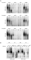
NFI is the primary component of the TFC. (A) EMSAs were carried out using the indicated probes and competitors. EMSAs with the 85/57 probe were done in the presence of the 85/57 M1 competitor (see Fig. 4A) to displace binding of all proteins except the TFC. Using nuclear extracts from the indicated stage of development, a similar developmental alteration in the mobility of the EMSAs were observed for all probes, including the NFI probe. The NFI consensus competitor, but not the C/EBP competitor, displaced the TFC from the 135/95 and 85/57 probes. (B) EMSAs were carried out with the indicated probes in the presence of the indicated antibodies. NFI antibody dissociated NFI from both the 135/95 and 85/57 probes using nuclear extracts from both myoblasts and day 7 myotubes (indicated by black asterisks). GABPα antibody induced a small supershift only in myoblasts with the 135/95 probe (indicated by the white asterisk).
Fig. 6
Fig. 6
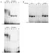
NFI is a phosphoprotein that changes appearance with development in C2C12 cells. (A) Nuclear extracts were prepared from cells at the indicated stage of development and treated with vehicle (control) or acid phosphatase (AP-treated). The appearance of NFI is diffuse and changes with development. For both probes, de-phosphorylation yielded a single “tight” band at all stages of development, although the binding to the 85/57 probe was greatly reduced. (B) Using only acid phosphatase treated nuclear extracts, EMSAs were carried out as indicated in the presence of NFI and GABPα antibodies, using the 135/95 probe. The NFI antibody interfered with binding to the 135/95 probe, as indicated by the black asterisks.
Fig. 7
Fig. 7
MGN and MRF4 recruit NFI to counteract negative regulation through the repressor E box. (A) A schematic diagram of the scanning mutations used in B are shown in the context of the full length sequence (−2800/+254). These scanning mutations correspond to those used in the EMSA analyses (Fig. 4A). (B) As shown previously in Fig. 3, expression of the wt −2800/+254 reporter gene approaches that of the mREB in the presence of the bHLH factors, especially MRF4. Individual mutations in the upstream NFI sites did not diminish expression, while the individual mutation of the downstream GABP and NFI site, M1, significantly reduced expression. Double mutation of the upstream NFI sites, c and e, significantly reduced expression, but the triple NFI mutation did not reduce expression further. Asterisks indicate a statistical significance of p < 0.05, while double asterisks indicate a statistical significance of p < 2−9.

Similar articles

Cited by

References

    1. Kallen RG, Cohen SA, Barchi RL. Structure, function and expression of voltage-dependent sodium channels. Mol Neurobiol. 1993:383–428. - PubMed
    1. Rich MM, Pinter MJ, Kraner SD, Barchi RL. Loss of electrical excitability in an animal model of acute quadriplegic myopathy. Ann Neurol. 1998:171–9. - PubMed
    1. Rich MM, Kraner SD, Barchi RL. Altered gene expression in steroid-treated denervated muscle. Neurobiol Dis. 1999:515–22. - PubMed
    1. Waxman SG. Transcriptional channelopathies: an emerging class of disorders. Nat Rev Neurosci. 2001:652–9. - PubMed
    1. Haimovich B, Schotland DL, Fieles WE, Barchi RL. Localization of sodium channel subtypes in adult rat skeletal muscle using channel-specific monoclonal antibodies. J Neurosci. 1987:2957–66. - PMC - PubMed

Publication types

MeSH terms

LinkOut - more resources