. 2007 Oct;3(10):1848-61.
doi: 10.1371/journal.pgen.0030171. Epub 2007 Aug 24.

Genome-wide expression profiling of the Arabidopsis female gametophyte identifies families of small, secreted proteins

Affiliations

Genome-wide expression profiling of the Arabidopsis female gametophyte identifies families of small, secreted proteins

Matthew W Jones-Rhoades et al. PLoS Genet. 2007 Oct.

Abstract

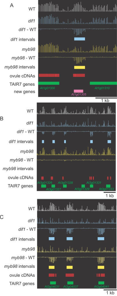
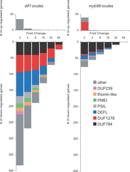
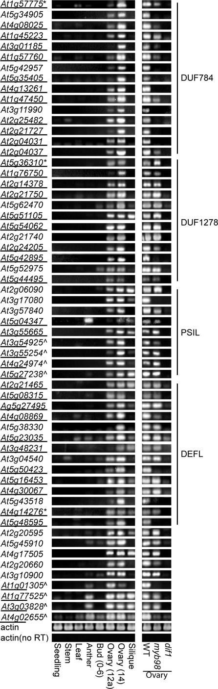
The female gametophyte of flowering plants, the embryo sac, develops within the diploid (sporophytic) tissue of the ovule. While embryo sac-expressed genes are known to be required at multiple stages of the fertilization process, the set of embryo sac-expressed genes has remained poorly defined. In particular, the set of genes responsible for mediating intracellular communication between the embryo sac and the male gametophyte, the pollen grain, is unknown. We used high-throughput cDNA sequencing and whole-genome tiling arrays to compare gene expression in wild-type ovules to that in dif1 ovules, which entirely lack embryo sacs, and myb98 ovules, which are impaired in pollen tube attraction. We identified nearly 400 genes that are downregulated in dif1 ovules. Seventy-eight percent of these embryo sac-dependent genes were predicted to encode for secreted proteins, and 60% belonged to multigenic families. Our results define a large number of candidate extracellular signaling molecules that may act during embryo sac development or fertilization; less than half of these are represented on the widely used ATH1 expression array. In particular, we found that 37 out of 40 genes encoding Domain of Unknown Function 784 (DUF784) domains require the synergid-specific transcription factor MYB98 for expression. Several DUF784 genes were transcribed in synergid cells of the embryo sac, implicating the DUF784 gene family in mediating late stages of embryo sac development or interactions with pollen tubes. The coexpression of highly similar proteins suggests a high degree of functional redundancy among embryo sac genes.


Conflict of interest statement

Competing interests. The authors have declared that no competing interests exist.

Figures

Figure 1
Figure 1. Genomic Regions Differentially Expressed between Wild-Type and dif1 or myb98 Ovules
The log2 transformed probe level expression values are plotted for wild-type (WT, white), dif1 (blue), and myb98 (yellow) ovules, as are the probe level differences in expression between wild type and mutant (dif1 − WT, myb98 − WT). Genomic intervals identified as differentially expressed in dif1 (blue) or myb98 (yellow) are plotted as colored boxes, as are the genomic alignments of ovule DNA fragments (red), previously annotated genes (green), and newly identified ORFs (purple). (A) A genomic interval between the previously annotated genes At1g01300 and At1g01310 that is downregulated in both dif1 and myb98 ovules and corresponds to a contig of ovule cDNAs is annotated as gene At1g01305. Flanking genic and intergenic probes are expressed at similar levels between all three genotypes. (B) A cluster of six DUF1278 genes, five of which are downregulated in dif1 ovules but not in myb98 ovules. (C) A cluster of three DUF784 genes that are downregulated in both dif1 and myb98 ovules.
Figure 2
Figure 2. Families of Small, Secreted Proteins Are Highly Downregulated in dif1 and myb98 Ovules
The number of genes differentially expressed between wild-type ovules and mutant ovules (p > 0.001) with changes in expression greater than the indicated cutoffs (2-fold, 4-fold, 8-fold, 16-fold, 32-fold, and 64-fold) are plotted. The numbers of genes belonging to gene families represented by at least ten members among the set of dif1 downregulated genes are indicated by color coding. (A) Genes differentially expressed between wild-type and dif1 ovules. (B) Genes differentially expressed between wild-type and myb98 ovules.
Figure 3
Figure 3. RT-PCR Analysis of Genes Downregulated in dif1 Ovules
Total RNA various ms-1 tissues, as well as from wild-type (Col) and mutant (myb98 and dif1) ovaries, was used as template for oligo-dT primed reverse transcription, and 1/100th of the first-strand cDNA was used as template per PCR. Underlined genes are not represented on the ATH1 array. Genes marked with a caret are newly annotated by this work. For genes marked with an asterisk, the PCR primers were perfectly complementary to more than one gene with highly similar sequences.
Figure 4
Figure 4. myb98 Differentially Regulated Genes Are a Subset of dif1 Differentially Regulated Genes
The overlap of the sets of gene up and downregulated in dif1 and myb98 ovules is displayed in a venn diagram.
Figure 5
Figure 5. DUF784 and DUF1278 Genes Are Transcribed in Synergid Cells
Stage 12c flowers were emasculated 24 h before ovules were stained for GUS expression. Ovules from plants transformed with promoterless GUS expression vector had no detectable GUS expression (A). Approximately 50% of ovules in T1 plants expressing GUS from one of four DUF784 promoters (B–E) or from one of four DUF1278 promoters (F–I) had a single locus of GUS expression near the micropyle, consistent with expression in the synergids, and in some cases possibly also the egg cell. Expression of GUS from a DEFL promoter (J) or one of two PSIL promoters (K, L) resulted in GUS expression within the central cell. (B) At5g35405::GUS, (C) At4g08025::GUS, (D) At5g34885::GUS, (E) At2g21727::GUS, (F) At5g54062::GUS, (G) At2g14378::GUS, (H) At5g36340::GUS, (I) At5g42895::GUS, (J) At4g14276::GUS, (K) At4g24974::GUS, and (L) At5g27238::GUS.

References

    1. Drews GN, Yadegari R. Development and function of the angiosperm female gametophyte. Annu Rev Genet. 2002;36:99–124. - PubMed
    1. Yadegari R, Drews GN. Female gametophyte development. Plant Cell. 2004;16(Suppl):S133–S141. - PMC - PubMed
    1. Christensen CA, King JK, Jordan JR, Drews GN. Megagametogenesis in Arabidopsis wild type and the Gf mutant. Sex Plant Reprod. 1997;10:49–64.
    1. Christensen CA, Subramanian S, Drews GN. Identification of gametophytic mutations affecting female gametophyte development in Arabidopsis . Dev Biol. 1998;202:136–151. - PubMed
    1. Gross-Hardt R, Kagi C, Baumann N, Moore JM, Baskar R, et al. LACHESIS restricts gametic cell fate in the female gametophyte of Arabidopsis . PLoS Biol. 2007;5:e47. doi: . - DOI - PMC - PubMed

Publication types