. 2007 Oct 15:4:75.
doi: 10.1186/1742-4690-4-75.

Requirements for the selective degradation of CD4 receptor molecules by the human immunodeficiency virus type 1 Vpu protein in the endoplasmic reticulum

Affiliations

Requirements for the selective degradation of CD4 receptor molecules by the human immunodeficiency virus type 1 Vpu protein in the endoplasmic reticulum

Julie Binette et al. Retrovirology. .

Abstract

Background: HIV-1 Vpu targets newly synthesized CD4 receptor for rapid degradation by a process reminiscent of endoplasmic reticulum (ER)-associated protein degradation (ERAD). Vpu is thought to act as an adaptor protein, connecting CD4 to the ubiquitin (Ub)-proteasome degradative system through an interaction with beta-TrCP, a component of the SCFbeta-TrCP E3 Ub ligase complex.

Results: Here, we provide direct evidence indicating that Vpu promotes trans-ubiquitination of CD4 through recruitment of SCFbeta-TrCP in human cells. To examine whether Ub conjugation occurs on the cytosolic tail of CD4, we substituted all four Ub acceptor lysine residues for arginines. Replacement of cytosolic lysine residues reduced but did not prevent Vpu-mediated CD4 degradation and ubiquitination, suggesting that Vpu-mediated CD4 degradation is not entirely dependent on the ubiquitination of cytosolic lysines and as such might also involve ubiquitination of other sites. Cell fractionation studies revealed that Vpu enhanced the levels of ubiquitinated forms of CD4 detected in association with not only the ER membrane but also the cytosol. Interestingly, significant amounts of membrane-associated ubiquitinated CD4 appeared to be fully dislocated since they could be recovered following sodium carbonate salt treatment. Finally, expression of a transdominant negative mutant of the AAA ATPase Cdc48/p97 involved in the extraction of ERAD substrates from the ER membrane inhibited Vpu-mediated CD4 degradation.

Conclusion: Taken together, these results are consistent with a model whereby HIV-1 Vpu targets CD4 for degradation by an ERAD-like process involving most likely poly-ubiquitination of the CD4 cytosolic tail by SCFbeta-TrCP prior to dislocation of receptor molecules across the ER membrane by a process that depends on the AAA ATPase Cdc48/p97.


Figures
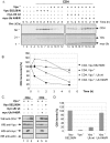
Figure 1
Figure 1
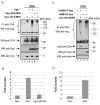
Poly-ubiquitination of CD4 is required for Vpu-mediated CD4 degradation. A. HEK 293T cells were mock-transfected or co-transfected with 1.5 μg of SVCMV CD4 wt and 8 μg of SVCMV Vpu+ (Vpu+) or the phosphorylation-defective Vpu mutant SVCMV Vpu S52,56/N (Vpu S52,56/N). In parallel, CD4/Vpu transfectants were co-transfected with 8 μg of plasmids encoding his(6)/c-myc-Ub wt (myc-Ub wt) or the TDN mutant of ubiquitin his(6)/c-myc-Ub K48/R (myc-Ub K48/R). Transfected cells were treated with BFA, pulse-labeled with [35S]methionine and [35S]cysteine and chased with complete media for the indicated time intervals. Cells were then lysed and immunoprecipitated sequentially with anti-CD4 monoclonal and polyclonal antibodies first and then with anti-Vpu and anti-myc antibodies. B. Using quantitative scanning of CD4 bands from three independent experiments, the percentage of CD4 remaining over time as compared to time 0 is plotted for each transfection. C. HEK 293T cells were mock-transfected or co-transfected as described in A. Cell transfectants were treated for two hours with BFA prior to lysis. Steady state levels of CD4, actin and tagged ubiquitin were analysed by western-blot. D. Quantitative analysis from three independent experiments showing the level of CD4 relative to CD4 expressed with Vpu S52,56/N (arbitrarily set at 100%) for each transfectant.
Figure 2
Figure 2
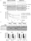
Effect of Vpu on CD4 ubiquitination. A. Vpu-mediated ubiquitination of CD4 wt when CD4 is retained in the ER through treatment with BFA. HEK 293T cells were mock-transfected or co-transfected with 1 μg of SVCMV CD4 wt, 8 μg of SVCMV Vpu+ or the phosphorylation-defective Vpu mutant SVCMV Vpu S52,56/N and 8 μg of the TDN mutant his(6)/c-myc-Ub K48/R. Samples were then treated as described in the materials and methods section. CD4 molecules were immunoprecipitated with anti-CD4 polyclonal antibodies prior to western-blot analysis with anti-myc monoclonal antibodies. (triangle) indicates the position of the heavy chains of anti-CD4 antibodies. B. Quantitative analysis of ubiquitinated CD4 conjugates. (asterisk) represents the area of the autoradiogram that was used for quantitation of CD4-Ub conjugates. The histogram shows the relative levels of ubiquitinated CD4 conjugates in presence or absence of a functional Vpu. Relative CD4-Ub conjugate levels were evaluated by quantitation of the signal detected in the area delineated on the autoradiogram relative to total CD4 as determined by quantitation of the band detected with the anti-CD4 antibodies on whole cell lysate. The relative level of ubiquitinated CD4 detected in absence of Vpu was arbitrarily set at 1. The data represent results from seven experiments. C. Vpu-mediated ubiquitination of CD4 wt in condition where CD4 is retained in the ER through binding with HIV-1 Env. HEK 293T cells were mock-transfected or co-transfected with 1 μg of pHIV CD4 wt, 10 μg of provirus encoding Vpu- (HxBH10-vpu-) or Vpu+ (HxBH10-vpu+) and 20 μg of his(6)/c-myc-Ub K48/R. Samples were then treated as in A but in absence of BFA. D. Quantitative analysis showing the relative levels of ubiquitinated CD4 detected in two independent experiments. Relative levels of ubiquitinated CD4 conjugates were determined as described in B.
Figure 3
Figure 3
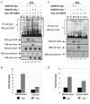
Effect of Vpu on CD4 molecules lacking lysine residues in the cytoplasmic tail. A. Analysis of CD4 wt and CD4 KRcyto turnover in presence or absence of functional Vpu by pulse-chase labeling and immunoprecipitation. HEK 293T cells were mock-transfected or co-transfected with 2 μg of pHIV CD4 wt or pHIV CD4 KRcyto and 20 μg of provirus encoding Vpu+ (HxBH10-vpu+) or phosphorylation-defective Vpu mutant (HxBH10-vpu S52,56/D). Cells were pulse-labeled with [35S]methionine and [35S]cysteine and chased in complete medium for the indicated time intervals. Cells were then lysed and immunoprecipitated sequentially with anti-CD4 antibodies first (polyclonal and monoclonal) and then with anti-Vpu antibodies. B. Using quantitative scanning of CD4 bands from two independent experiments, the percentage of CD4 remaining over time as compared to time 0 is plotted for each transfection. C. Effect of Vpu on steady-state CD4 wt and CD4 KRcyto levels. HEK 293T cells were mock-transfected or co-transfected with 1 μg of pHIV CD4 wt or pHIV CD4 KRcyto and 10 μg of proviruses encoding Vpu- or Vpu+ in addition to 25 μg of the his(6)/c-myc-Ub K48/R expressor. In the left panel (Env-), a similar experiment was performed except that HEK 293T cells were co-transfected with 10 μg of envelope-defective provirus (HxBc2-pr-, vpu-, env- or HxBH10-pr-, vpu+, env-) and treated with BFA for 2 h prior to lysis. Cell lysates were then treated as described in the materials and methods section. D. Quantitative analysis of steady-state CD4 levels. CD4 levels in presence of absence of his(6)/c-myc-Ub K48/R were arbitrarily set at 100%. The levels of CD4 in presence of Vpu are shown relative to the corresponding controls. These results are representative of the data obtained in three independent experiments for Env- and five independent experiments for Env+.
Figure 4
Figure 4
Effect of Vpu on CD4 KRcyto poly-ubiquitination. A. HEK 293T cells were mock-transfected or co-transfected with 1 μg of pHIV CD4 wt or pHIV CD4 KRcyto, 10 μg of provirus encoding Vpu- (HxBH10-vpu-) or Vpu+ (HxBH10-vpu+) and 25 μg of the TDN mutant of Ub his(6)/c-myc-Ub K48/R. Transfected cells were not treated with BFA prior to lysis. Samples were then treated as described in the materials and methods. B. Quantitative analysis of the relative levels of ubiquitinated CD4 conjugates for CD4 wt and CD4 KRcyto in two independent experiments. (asterisk) represents the area of the autoradiogram that was used for the quantitation of CD4-Ub conjugates. Relative levels of ubiquitinated CD4 conjugates were determined as described in Fig. 2B.
Figure 5
Figure 5
Vpu-mediated CD4 degradation involves dislocation of ubiquitinated CD4 conjugates from the ER membrane to the cytosol. HEK 293T cells were mock-transfected or co-transfected with 1 μg of pHIV CD4 wt, 10μg of envelope-defective provirus (HxBc2-pr-, vpu-, env- or HxBH10-pr-, vpu+, env-) and 15 μg of his(6)/c-myc-Ub K48/R expression plasmid where indicated. Cells were treated with BFA for 2 h before mechanical lysis. CD4-Ub conjugates were immunoprecipitated with anti-myc monoclonal antibodies prior to western-blot analysis with anti-CD4 polyclonal antibodies while control proteins in each fraction were revealed by western-blot. Actin and calnexin were used as cytosolic and membrane controls, respectively. A. Membrane (M) and cytosolic (C) fractions were separated and treated as described in the materials and methods section. B. Quantitative analysis of the relative amounts of ubiquitinated CD4 molecules present in each fraction relative to the amounts measured in absence of Vpu (arbitrarily set at 1). (asterisk) represents the area of the autoradiogram that was used for the quantitation of CD4-Ub conjugates. Non-specific background signal detected in lanes 7 and 8 was subtracted. Relative levels of ubiquitinated CD4 conjugates were determined as described in the legend of Fig. 2B. Error bars reflect standard deviations from duplicate independent experiments. C. Membrane (M) fractions were treated with Na2CO3 (pH 11) as described in materials and methods. Treated membrane and supernatant (S) were subsequently recovered by centrifugation. Fractions were analyzed as described above in A. D. Quantitative analysis of the relative amounts of ubiquitinated CD4 molecules (as described in the legend of Fig. 2B) present in each fraction relative to the amounts measured in absence of Vpu (arbitrarily set at 1). (asterisk) represents the area that was used for the quantitation of CD4-Ub conjugates. Non-specific background signal detected in lanes 7 and 8 was subtracted. Error bars reflect standard deviations from duplicate independent experiments.
Figure 6
Figure 6
Effect of a TDN mutant of p97 on Vpu-mediated CD4degradation. HEK 293T cells were mock-transfected or co-transfected with 1.5 μg of SVCMV CD4 wt, 12 μg of SVCMV Vpu- or Vpu+ and 1 μg of an expression plasmid encoding a FLAG-tagged version of p97 wt or the TDN mutant p97 AA. Cells were treated with BFA for 2 h prior to lysis. Cell lysates were then analyzed by western-blot as described in materials and methods. These results are representative of the data obtained in two independent experiments.

References

    1. Klatzmann D, Champagne E, Chamaret S, Gruest J, Guetard D, Hercend T, Gluckman JC, Montagnier L. T-lymphocyte T4 molecule behaves as the receptor for human retrovirus LAV. Nature. 1984;312:767–768. doi: 10.1038/312767a0. - DOI - PubMed
    1. Levesque K, Finzi A, Binette J, Cohen EA. Role of CD4 receptor down-regulation during HIV-1 infection. Curr HIV Res. 2004;2:51–59. doi: 10.2174/1570162043485086. - DOI - PubMed
    1. Levesque K, Zhao YS, Cohen EA. Vpu exerts a positive effect on HIV-1 infectivity by down-modulating CD4 receptor molecules at the surface of HIV-1-producing cells. J Biol Chem. 2003;278:28346–28353. doi: 10.1074/jbc.M300327200. - DOI - PubMed
    1. Wildum S, Schindler M, Munch J, Kirchhoff F. Contribution of Vpu, Env, and Nef to CD4 down-modulation and resistance of human immunodeficiency virus type 1-infected T cells to superinfection. J Virol. 2006;80:8047–8059. doi: 10.1128/JVI.00252-06. - DOI - PMC - PubMed
    1. Schwartz O, Dautry-Varsat A, Goud B, Marechal V, Subtil A, Heard JM, Danos O. Human immunodeficiency virus type 1 Nef induces accumulation of CD4 in early endosomes. J Virol. 1995;69:528–533. - PMC - PubMed

Publication types

MeSH terms

Substances