. 2008 Jan;52(1):98-109.
doi: 10.1128/AAC.00609-07. Epub 2007 Oct 15.

Artemisinin and a series of novel endoperoxide antimalarials exert early effects on digestive vacuole morphology

Affiliations

Artemisinin and a series of novel endoperoxide antimalarials exert early effects on digestive vacuole morphology

Maria del Pilar Crespo et al. Antimicrob Agents Chemother. 2008 Jan.

Abstract

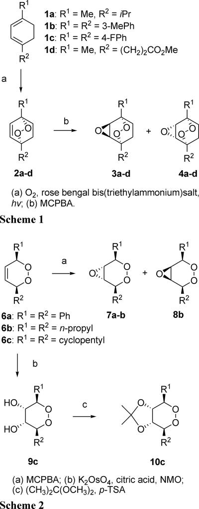
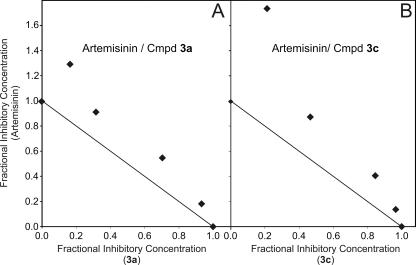
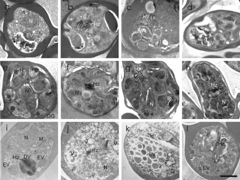
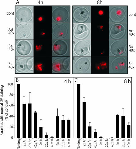
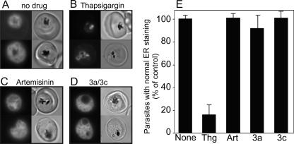
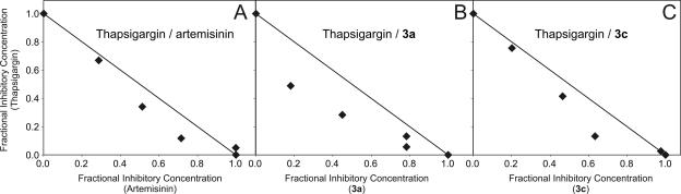
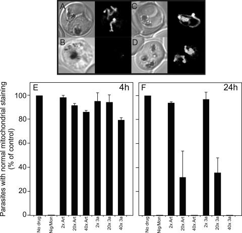
Artermisinin and its derivatives are now the mainstays of antimalarial treatment; however, their mechanism of action is only poorly understood. We report on the synthesis of a novel series of epoxy-endoperoxides that can be prepared in high yields from simple starting materials. Endoperoxides that are disubstituted with alkyl or benzyl side chains show efficient inhibition of the growth of both chloroquine-sensitive and -resistant strains of Plasmodium falciparum. A trans-epoxide with respect to the peroxide linkage increases the activity compared to that of its cis-epoxy counterpart or the parent endoperoxide. The novel endoperoxides do not show a strong interaction with artemisinin. We have compared the mechanism of action of the novel endoperoxides with that of artemisinin. Electron microscopy reveals that the novel endoperoxides cause the early accumulation of endocytic vesicles, while artemisinin causes the disruption of the digestive vacuole membrane. At longer incubation times artemisinin causes extensive loss of organellar structures, while the novel endoperoxides cause myelin body formation as well as the accumulation of endocytic vesicles. An early event following endoperoxide treatment is the redistribution of the pH-sensitive probe LysoSensor Blue from the digestive vacuole to punctate structures. By contrast, neither artemisinin nor the novel endoperoxides caused alterations in the morphology of the endoplasmic reticulum nor showed antagonistic antimalarial activity when they were used with thapsigargin. Analysis of rhodamine 123 uptake by P. falciparum suggests that disruption of the mitochondrial membrane potential occurs as a downstream effect rather than as an initiator of parasite killing. The data suggest that the digestive vacuole is an important initial site of endoperoxide antimalarial activity.


Figures

FIG. 1.
FIG. 1.
Schemes for the synthesis of endoperoxides. Me, methyl; iPr, iso-propyl; MePh, methylphenyl; FPh, fluorophenyl; MCPBA, meta-chloroperbenzoic acid; p-TSA, para-toluenesulfonic acid; NMo, N-methylmorpholine-N-oxide.
FIG. 2.
FIG. 2.
Interaction of artemisinin and two novel endoperoxides in inhibiting parasite growth. Isobolograms were constructed from the IC50 values in Table 2. For each drug combination, FICs were calculated by dividing the measured apparent IC50 values for the individual drugs in the different combinations of artemisinin with compound (Cmpd) 3a (A) or compound 3c (B) by the IC50 values obtained when the drugs were used alone.
FIG. 3.
FIG. 3.
Transmission electron microscopy analysis of endoperoxide-treated parasitized RBCs. Trophozoite-stage parasite-infected RBCs (strain D10) were incubated with no drug (a and e) or 40 times the IC50 values of artemisinin (b and f), compound 3a (c and g), or compound 3c (d and h) for 8 h. Alternatively, ring-stage parasite-infected RBCs were incubated with no drug (i) or two times the IC50 values of artemisinin (j), compound 3a (k), or compound 3c (l) for 24 h. Sections through the control trophozoite-stage parasites (a and i) show endocytic vesicles (EV) and typical hemozoin crystals (Hz) within a digestive vacuole (DV). The parasite cytoplasm is dotted with ribosomes and has a defined nucleus (N). The mitochondrion (M) is visible in some sections (i). Schizonts (e) show well-defined nuclei and developing apical organelles, rhoptries (R), and dense granules (DG). After an 8-h treatment with a high concentration of artemisinin, both trophozoite-stage parasites (b) and schizont-stage parasites (j) show the loss of digestive vacuole integrity. Many of the infected RBCs treated for 24 h with artemisinin (j) show a substantial loss of membranous features, with individual hemozoin crystals in contact with the parasite cytoplasm and the formation of vacuoles (V). Trophozoites treated for 8 h with 40 times the IC50 value of compound 3a (c) or compound 3c (d) show an accumulation of undigested endocytic vesicles in the digestive vacuole. Schizonts in the samples treated for 8 h with compound 3a (g) or compound 3c (h) show merozoites with an aberrant morphology and uneven staining of the nucleus. There is an accumulation of dense vesicles (g, asterisk) in many cells. Infected RBCs treated for 24 h with two times the IC50 value of compound 3a show an accumulation of endocytic vesicles (EV) and myelin bodies (MB) (see Fig. S1k in the supplemental material). Compound 3c causes less obvious damage (l), but development seems to be inhibited and very few schizonts are observed. Bar, 1 μm. Additional images are presented in Fig. S1 in the supplemental material.
FIG. 4.
FIG. 4.
Analysis of digestive vacuole integrity in endoperoxide-treated parasitized RBCs. Strain D10 parasites at the trophozoite stage were incubated with no additions or 2, 20, or 40 times the IC50 values of artemisinin, compound 3a, or compound 3a for 4 or 8 h. LysoSensor Blue was used to probe acidic compartments, with imaging performed by fluorescence microscopy. (A) Bright-field, fluorescence signals and overlays of control and drug-treated parasites. (B and C) The LysoSensor Blue labeling pattern in infected RBCs (300 cells) in cultures that were drug treated for 4 (B) or 8 h (C) was classified as normal or redistributed. Data are normalized relative to those for the control, to which no drug was added, and represent the means ± standard deviations of data from three separate experiments. cont, control; DV, digestive vacuole; Art, artemisinin.
FIG. 5.
FIG. 5.
Analysis of morphology of the ER in endoperoxide-treated parasitized RBCs. Strain D10 parasites at the trophozoite stage were incubated with (A) no additions or 10 times the IC50 values of (B) thapsigargin, (C) artemisinin, (D, upper panel) compound 3a, or (D, lower panel) compound 3c for 12 h. ER Tracker was used to label the ER before imaging by fluorescence microscopy. (B) Fluorescence images of infected RBCs (300 cells) were classified as normal or condensed. Data are normalized relative to those for the control, to which no drug was added, and represent the means ± standard deviations of data from three separate experiments.
FIG. 6.
FIG. 6.
Interaction of endoperoxide and thapsigarin in parasite killing. Isobolograms were constructed from the IC50 values in Table 3. For each drug combination, the FICs were calculated by dividing the measured apparent IC50 values for individual drugs in the different combinations of thapsigargin with artemisinin (A), compound 3a (B), or compound 3c (C) by the IC50 values obtained when the drugs were used alone.
FIG. 7.
FIG. 7.
Analysis of mitochondrial function in endoperoxide-treated parasitized RBCs. Strain D10 trophozoite-stage parasite-infected RBCs were incubated with (A) no additions, (B) the ionophores nigericin and monensin (20 μM each), or 40 times the IC50 values of (C) artemisinin or (D) compound 3a for 4 h. Rhodamine 123 (0.2 μM) was used to stain the mitochondrion for confocal microscopy. (E) Trophozoite-stage parasites were incubated for 4 h or (F) ring-stage infected RBCs were incubated for 24 h with no drug or with 2, 20, or 40 times the IC50 values of artemisinin or compound 3a. Infected RBCs (1,000 cells) were counted and classified as normal or rhodamine 123 depleted by fluorescence microscopy. Data are normalized relative to those for the control, to which no drug was added.

References

    1. Adovelande, J., J. Deleze, and J. Schrevel. 1998. Synergy between two calcium channel blockers, verapamil and fantofarone (SR33557), in reversing chloroquine resistance in Plasmodium falciparum. Biochem. Pharmacol. 55:433-440. - PubMed
    1. Afonso, A., P. Hunt, S. Cheesman, A. C. Alves, C. V. Cunha, V. do Rosario, and P. Cravo. 2006. Malaria parasites can develop stable resistance to artemisinin but lack mutations in candidate genes atp6 (encoding the sarcoplasmic and endoplasmic reticulum Ca2+ ATPase), tctp, mdr1, and cg10. Antimicrob. Agents Chemother. 50:480-489. - PMC - PubMed
    1. Aley, S. B., J. A. Sherwood, K. Marsh, O. Eidelman, and R. J. Howard. 1986. Identification of isolate-specific proteins on sorbitol-enriched Plasmodium falciparum infected erythrocytes from Gambian patients. Parasitology 92:511-525. - PubMed
    1. Arrow, K. J., C. B. Panosian, and H. Gelbrand. 2004. Saving lives, buying time: economics of malaria drugs in an age of resistance. The National Academies Press, Washington, DC. - PubMed
    1. Avery, M. A., W. K. M. Chong, and C. Jenning-White. 1992. Stereroselective total synthesis of (+)-artemisinin, the antimalarial constituent of Artemisia annua L. J. Am. Chem. Soc. 114:974-979.

Publication types

MeSH terms