. 2007 Dec;27(24):8748-59.
doi: 10.1128/MCB.01380-07. Epub 2007 Oct 15.

Synergistic function of DNA methyltransferases Dnmt3a and Dnmt3b in the methylation of Oct4 and Nanog

Affiliations

Synergistic function of DNA methyltransferases Dnmt3a and Dnmt3b in the methylation of Oct4 and Nanog

Jing-Yu Li et al. Mol Cell Biol. 2007 Dec.

Abstract

DNA methylation plays an important role in gene silencing in mammals. Two de novo methyltransferases, Dnmt3a and Dnmt3b, are required for the establishment of genomic methylation patterns in development. However, little is known about their coordinate function in the silencing of genes critical for embryonic development and how their activity is regulated. Here we show that Dnmt3a and Dnmt3b are the major components of a native complex purified from embryonic stem cells. The two enzymes directly interact and mutually stimulate each other both in vitro and in vivo. The stimulatory effect is independent of the catalytic activity of the enzyme. In differentiating embryonic carcinoma or embryonic stem cells and mouse postimplantation embryos, they function synergistically to methylate the promoters of the Oct4 and Nanog genes. Inadequate methylation caused by ablating Dnmt3a and Dnmt3b is associated with dysregulated expression of Oct4 and Nanog during the differentiation of pluripotent cells and mouse embryonic development. These results suggest that Dnmt3a and Dnmt3b form a complex through direct contact in living cells and cooperate in the methylation of the promoters of Oct4 and Nanog during cell differentiation. The physical and functional interaction between Dnmt3a and Dnmt3b represents a novel regulatory mechanism to ensure the proper establishment of genomic methylation patterns for gene silencing in development.


Figures

FIG. 1.
FIG. 1.
Affinity purification of Dnmt3a- and Dnmt3b-associated proteins from ES and EC cells. (A) Silver staining of copurified Dnmt3a and Dnmt3b proteins from ES cells. Affinity purification was performed with an anti-Dnmt3b monoclonal antibody, followed by protein identification by matrix-assisted laser desorption ionization (MALDI) mass spectrometry. WT, wild type. (B) Silver staining of copurified Dnmt3a and Dnmt3b proteins from P19 cells. Affinity purification was performed using a TAP-tagging procedure through stably expressed TAP-tagged Dnmt3a, followed by protein identification by MALDI mass spectrometry. The protein identification was also confirmed by Western blotting using specific antibodies (data not shown). (C) Western detection of Dnmt3a, Dnmt3b, and Dnmt3L in the fractions separated by gel filtration using a Sepharose 6 column. The input was chromatin extract prepared from ES cells. Antibodies used are shown at the right (α-Dnmt3b, -Dnmt3a2, and -Dnmt3L, anti-Dnmt3b, -Dnmt3a2, and -Dnmt3L, respectively). All three blots were from the same gel. The elution profile of the protein markers is indicated at the top.
FIG. 2.
FIG. 2.
Association between Dnmt3a and Dnmt3b in vivo. (A) FRET analysis of Dnmt3a1-YFP and Dnmt3b1-CFP fusion proteins expressed in 293T cells. Scale bar, 3 μm. The line chart to the right illustrates emission spectra of Dnmt3b1-CFP and Dnmt3a1-YFP before (black) or after (red) photobleaching. The bar chart below shows the averaged FRET efficiency for coexpression of CFP and YFP (CFP+YFP) (negative control; n = 25), Dnmt3b1-CFP and -YFP (3b-CFP+YFP) (n = 25), Dnmt3b1-CFP and Dnmt3a1-YFP (3b-CFP+3a-YFP) (n = 30), or CFP-YFP fusion protein (positive control; n = 25). Data are representative of at least three independent experiments. (B) Colocalization of endogenous Dnmt3a and Dnmt3b in the interphase nuclei of ES cells. Heterochromatin domains are brightly stained with 4′,6′-diamidino-2-phenylindole (DAPI). A similar staining pattern was observed in differentiating ES cells treated with RA for 2 days. Antibodies used did not distinguish different isoforms. wt, wild type; 3aKO, Dnmt3a knockout; 3bKO, Dnmt3b knockout; DKO, double knockout; α-Dnmt3a and -Dnmt3b, anti-Dnmt3a and -Dnmt3b antibodies. (C) Colocalization of Dnmt3a and Dnmt3b in mouse embryos. Embryos were frozen and sliced, followed by immunostaining with the antibodies indicated at the top. The squared epiblast regions are shown at higher magnification below. Scale bar, 20 μm.
FIG. 3.
FIG. 3.
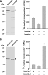
Stimulation of methyltransferase activity by Dnmt3a-Dnmt3b interaction in vitro. (A) Stimulation of methyltransferase activity between full-length enzymes. Six-His-tagged Dnmt3a1 and Dnmt3b1 purified from Escherichia coli were visualized by Coomassie blue staining in a 10% sodium dodecyl sulfate-polyacrylamide gel (left panel). Methyltransferase activity of Dnmt3a1 or Dnmt3b1 alone or in the equimolar presence of both was measured (right panel). Data for incorporation in cpm are averages from three independent experiments ± standard deviations. (B) Stimulation of methyltransferase activity between the catalytic domains. Six-His-tagged catalytic domains of Dnmt3a (Dnmt3a-C) and Dnmt3b (Dnmt3b-C) purified from E. coli were visualized by Coomassie blue staining (left panel). Their methyltransferase activities were examined alone or in equimolar combination (right panel).
FIG. 4.
FIG. 4.
Depletion of either Dnmt3a or Dnmt3b affects methylation of the Oct4 promoter in P19 cells upon differentiation. (A) Depletion of Dnmt3a and Dnmt3b in stably transfected P19 cell lines expressing small interfering RNA. The expression level of Dnmt3a and Dnmt3b in knockdown (KD) cell lines was examined by Western analysis of total cell lysates using specific antibodies (α-Dnmt3a, -Dnmt3b, and -β-actin, anti-Dnmt3a, -Dnmt3b, and -β-actin). Equal loading was verified by the detection of β-actin. (B) Schematic representation of the regulatory region upstream of Oct4 and Nanog. The distribution of CpG dinucleotides (circles), analyzed by bisulfite sequencing in panel D, is shown below. Two (filled triangles) of the 16 CpGs in the Oct4 upstream region are present in the TaqI restriction sites used for the COBRA methylation assay. DE, distal enhancer; PE, proximal enhancer; P, promoter. (C) COBRA methylation analysis of Oct4 upon cell differentiation induced with RA. Genomic DNA was isolated from wild-type (wt) and Dnmt3 knockdown cells at different days with RA treatment. The methylation status of two CpG sites was assessed by TaqI digestion of the PCR products derived from bisulfite-converted genomic DNA (left) and quantified by measuring the ratio of fully cleaved DNA to total DNA (right). The ratio from the complete digestion corresponding to full methylation at the TaqI sites was set to 1. U, uncut fragments derived from unmethylated DNA; M, cleaved fragments derived from methylated DNA. (D) Bisulfite sequencing analysis. PCR products amplified from bisulfite-treated genomic DNA were cloned and sequenced to reveal the methylation statuses of individual CpG sites. Percentages of the methylated CpG sites (filled circles) among all scored sites are indicated.
FIG. 5.
FIG. 5.
Deficiency in either Dnmt3a or Dnmt3b reduces DNA methylation at the Oct4 promoter in ES cells and embryos. (A) Confirmation of wild-type (wt) and knockout ES cell lines by Western blotting. (B) COBRA methylation analysis of the Oct4 promoter. Wild-type and knockout ES cells were induced with RA for up to 7 days, and the methylation levels of Oct4 at different time points were assessed by TaqI restriction assay of the PCR products derived from bisulfite-treated genomic DNA and quantified (bottom). Labeling of the digested fragments is as in Fig. 4C. (C) Bisulfite sequencing analysis of Oct4 and Nanog at day 3 upon RA treatment. Percentages of the methylated CpG sites (filled circles) are indicated. (D) Bisulfite sequencing analysis of Oct4 in mouse embryos. Genomic DNA was from wild-type (wt), single knockout (3aKO and 3bKO), or double knockout (DKO) embryos collected at E9.5 (three for each genotype).
FIG. 6.
FIG. 6.
Dnmt3a and Dnmt3b mutually stimulate de novo methylation at the Oct4 promoter in differentiating ES cells. (A) Expression of ectopic (with gray-shadowed background) and endogenous methyltransferases in stably established ES cell lines by Western blotting. Catalytically inactive mutant Dnmt3b1 V732G, Dnmt3a1 E752A, or wild-type (WT) counterparts were stably introduced into Dnmt3a−/−, Dnmt3b−/−, or Dnmt3a−/−3b−/− ES cells. (B) Analysis of Oct4 methylation 3 days upon RA induction by COBRA. Each bar represents an individual stable cell line.
FIG. 7.
FIG. 7.
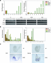
Promoter hypomethylation leads to misregulation of expression of Oct4 and Nanog in differentiating cells and developing embryos. (A) Expression of Oct4 and Nanog in P19 cells at various time points upon RA induction, measured by quantitative PCR analyses. The expression of differentiation markers Brn2 and Map2 was examined by reverse transcription-PCR (lower panels). Labels of cell lines are as in Fig. 4. (B) Expression of Oct4 and Nanog in ES cells at different time points upon RA induction measured by quantitative PCR analyses. Cell lines used are as in Fig. 5A. (C) Whole-mount in situ hybridization of Oct4 and Nanog in E9.5 wild-type and double knockout (DKO) embryos.

Similar articles

Cited by

References

    1. Aapola, U., I. Liiv, and P. Peterson. 2002. Imprinting regulator DNMT3L is a transcriptional repressor associated with histone deacetylase activity. Nucleic Acids Res. 30:3602-3608. - PMC - PubMed
    1. Bachman, K. E., M. R. Rountree, and S. B. Baylin. 2001. Dnmt3a and Dnmt3b are transcriptional repressors that exhibit unique localization properties to heterochromatin. J. Biol. Chem. 276:32282-32287. - PubMed
    1. Ben-Shushan, E., E. Pikarsky, A. Klar, and Y. Bergman. 1993. Extinction of Oct-3/4 gene expression in embryonal carcinoma x fibroblast somatic cell hybrids is accompanied by changes in the methylation status, chromatin structure, and transcriptional activity of the Oct-3/4 upstream region. Mol. Cell. Biol. 13:891-901. - PMC - PubMed
    1. Bestor, T., A. Laudano, R. Mattaliano, and V. Ingram. 1988. Cloning and sequencing of a cDNA encoding DNA methyltransferase of mouse cells. The carboxyl-terminal domain of the mammalian enzymes is related to bacterial restriction methyltransferases. J. Mol. Biol. 203:971-983. - PubMed
    1. Bestor, T. H. 2000. The DNA methyltransferases of mammals. Hum. Mol. Genet. 9:2395-2402. - PubMed

Publication types

MeSH terms