. 2007 Dec;27(24):8622-36.
doi: 10.1128/MCB.00467-07. Epub 2007 Oct 15.

c-Cbl-mediated regulation of LAT-nucleated signaling complexes

Affiliations

c-Cbl-mediated regulation of LAT-nucleated signaling complexes

Lakshmi Balagopalan et al. Mol Cell Biol. 2007 Dec.

Abstract

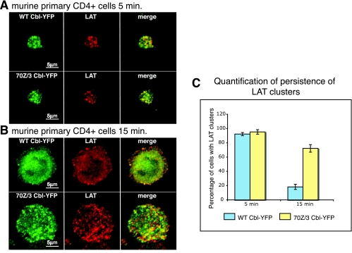
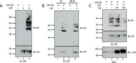
The engagement of the T-cell receptor (TCR) causes the rapid recruitment of multiple signaling molecules into clusters with the TCR. Upon receptor activation, the adapters LAT and SLP-76, visualized as chimeric proteins tagged with yellow fluorescent protein, transiently associate with and then rapidly dissociate from the TCR. Previously, we demonstrated that after recruitment into signaling clusters, SLP-76 is endocytosed in vesicles via a lipid raft-dependent pathway that requires the interaction of the endocytic machinery with ubiquitylated proteins. In this study, we focus on LAT and demonstrate that signaling clusters containing this adapter are internalized into distinct intracellular compartments and dissipate rapidly upon TCR activation. The internalization of LAT was inhibited in cells expressing versions of the ubiquitin ligase c-Cbl mutated in the RING domain and in T cells from mice lacking c-Cbl. Moreover, c-Cbl RING mutant forms suppressed LAT ubiquitylation and caused an increase in cellular LAT levels, as well as basal and TCR-induced levels of phosphorylated LAT. Collectively, these data indicate that following the rapid formation of signaling complexes upon TCR stimulation, c-Cbl activity is involved in the internalization and possible downregulation of a subset of activated signaling molecules.


Figures

FIG. 1.
FIG. 1.
LAT traffics through intracellular compartments upon stimulation of the TCR. (A) Jurkat E6.1 cells stably expressing LAT-YFP were plated onto antibody-coated coverslips as described in Materials and Methods and visualized with a spinning-disk confocal system (see Video S1 in the supplemental material). An image set made from Z stacks (five sections, 0.5 μm apart) collected after plating onto stimulatory coverslips is shown by displaying selected time points as maximum-intensity projections. (B) Primary murine CD4+ cells were transfected with LAT-YFP and plated onto stimulatory coverslips. Cells were fixed at various time points into the spreading process, as indicated, and confocal images at the coverslip were collected. (C and D) Jurkat E6.1 cells stably expressing LAT-YFP were incubated with transferrin (Tfr) (C) or CTX-B (D) at 4°C to allow binding but prevent the internalization of these markers. Cells were then plated onto stimulatory coverslips maintained at 37°C, causing the rapid internalization of these markers from the membrane, and visualized with a spinning-disk confocal system (see Videos S2 and S3 in the supplemental material). Z stacks (five stacks, 0.5 μm apart) were collected and are displayed as maximum-intensity projections at selected time points. Data are representative of a minimum of two independent experiments.
FIG. 2.
FIG. 2.
c-Cbl RING finger activity is required for the movement and dissipation of LAT-YFP clusters. (A, B, and C) E6.1 LAT-YFP cells were transiently transfected with CFP (A), WT Cbl-CFP (B), or 70Z/3 Cbl-CFP (C). Cells were plated onto stimulatory coverslips and visualized with a spinning-disk confocal system (see Videos S4, S5, and S6 in the supplemental material). Representative image sets from selected time points are shown. (A) In CFP-transfected cells, LAT-YFP clusters move for a short distance and dissipate rapidly. (B) In cells coexpressing WT Cbl-CFP, clusters containing both LAT-YFP (top panel; pseudocolored in green) and WT Cbl-CFP (bottom panel; pseudocolored in red) dissipate rapidly. Of note, the central LAT-YFP structure that persists in the cell expressing WT Cbl-CFP (top panel) is LAT-YFP localized in the Golgi compartment. (C) In cells coexpressing 70Z/3 Cbl-CFP, LAT-YFP (top panel) and 70Z/3 Cbl-CFP (bottom panel) clusters persist and do not move. (D) Average traces demonstrating the movement of LAT-YFP clusters. The y axis shows how far the clusters moved, while the x axis shows how long they were visible. (E) Traces from 10 cells expressing each construct were compared, and the average persistence of the clusters is shown in the graph. The coexpression of 70Z/3 Cbl-CFP caused a significant increase in the length of time that LAT-YFP clusters remained visible (P < 0.001), while the coexpression of WT Cbl-CFP caused a modest decrease in the persistence of clusters (P = 0.02). (F) Average traces demonstrating the movement of SLP-YFP clusters. The y axis shows how far the clusters moved, while the x axis shows how long they were visible. (G) Traces from 10 cells expressing each construct were compared, and the average persistence of the clusters is shown in the graph. The overexpression of WT Cbl-CFP caused a significant decrease in the amount of time SLP-YFP clusters were visible (P = 0.01), while the overexpression of 70Z/3 Cbl-CFP caused a significant increase in the length of time that SLP-YFP clusters remained visible (P < 0.001). Bars show the SEM.
FIG. 3.
FIG. 3.
70Z/3 Cbl inhibits endogenous LAT trafficking in primary cells. Murine primary CD4+ cells were transfected with WT Cbl-YFP or 70Z/3 Cbl-YFP as indicated. (A and B) Cells were plated onto stimulatory coverslips and fixed at 5 min (A) and 15 min (B) into the spreading process. Following fixation, cells were immunostained for LAT, and confocal images at the plane of the coverslip were collected. (C) The number of cells exhibiting LAT clusters among cells transfected with WT Cbl-YFP (blue bars) or 70Z/3 Cbl-YFP (yellow bars) was assessed at each time point. Data are representative of two independent experiments. Error bars show the SEM.
FIG. 4.
FIG. 4.
Internalization of LAT clusters in T cells lacking c-Cbl. Lymph node CD4+ cells from c-Cbl−/− or c-Cbl−/+ mice were transfected with LAT-YFP. (A and B) Cells were plated onto stimulatory coverslips and fixed at 5 min (A) and 15 min (B) into the spreading process. Following fixation, cells were immunostained for pY or pLAT and confocal images at the plane of the coverslip were collected. (C) The number of cells exhibiting LAT-YFP clusters among T cells from c-Cbl−/+ mice (blue bars) or c-Cbl−/− mice (yellow bars) was assessed at each time point. Data are presented as percentages of cells containing LAT-YFP clusters at each time point and are representative of two independent experiments. Error bars show the SEM.
FIG. 5.
FIG. 5.
70Z/3 Cbl recruitment to clusters requires the proline-rich domain of Cbl, while the persistence of 70Z/3 Cbl and LAT clusters requires the TKB domain. (A) Schematic of Cbl constructs. 4H, four-helix bundle; EF, EF hand; SH2, Src homolgy 2 domain; RF, RING finger domain; PRR, proline-rich repeat; UBA, ubiquitin-associated domain; *, point mutation in TKB domain; YYY, tyrosine residues. The table on the right summarizes the quantification of the data shown in panels B and C. Data are expressed as percentages of cells that displayed Cbl-CFP clusters (at 2 or 5 min) and LAT-YFP clusters (at 5 min). (B) Jurkat E6.1 cells were transfected with the indicated Cbl and 70Z/3 Cbl-CFP constructs, plated onto stimulatory coverslips, and fixed at 2 min into the spreading process. (C) Jurkat E6.1 cells stably expressing LAT-YFP were transfected with the indicated Cbl-CFP constructs, plated onto stimulatory coverslips, and fixed at 5 min into the spreading process. Z stacks (five stacks, 0.5 μm apart) were collected and are displayed as maximum-intensity projections. Data are representative of a minimum of three independent experiments.
FIG. 6.
FIG. 6.
Requirement of LAT and ZAP-70 binding for c-Cbl recruitment to activation clusters. Cells were dropped onto stimulatory coverslips, fixed after 2 min, and immunostained for pY, pLAT, and c-Cbl. In all cases, four panels are shown: those corresponding to pY (left; red), pLAT (second from left; blue), and c-Cbl (second from right; green) and an overlay panel emphasizing the colocalization of these proteins (right). (A) c-Cbl localization in LAT-deficient JCam2.5 cells or JCam2.5 variants reconstituted with LATY132F or LAT3YF. JCam2.5 cells reconstituted with WT LAT served as a positive control (top panel). Note the absence of pLAT staining in JCam2.5 and JCam2.5(LAT3YF) cells. c-Cbl was recruited to signaling clusters in all cell lines. (B) c-Cbl localization in ZAP-deficient P116 cells or P116 cells reconstituted with ZAP-70 Y292F. P116 cells reconstituted with WT ZAP-70 served as a positive control (top panel). c-Cbl is recruited to signaling clusters in the absence of ZAP-70 binding in the Y292F reconstituted cells. (C and D) c-Cbl localization in P116 WT ZAP-70 or P116 ZAP-70 Y292F cells transfected with siRNA for LAT (bottom panels) or control siRNA (top panels). c-Cbl is localized normally in P116 WT ZAP-70 cells depleted of LAT expression (C, bottom panel), but not in P116 ZAP-70 Y292F reconstituted cells depleted of LAT expression (D, bottom panel). (E) The numbers of cells exhibiting c-Cbl clusters among P116 WT ZAP-70 cells and P116 Y292F ZAP-70 cells transfected with control siRNA (blue bars) or LAT siRNA (yellow bars) were assessed. Data are presented as numbers of cells containing c-Cbl clusters and are representative of two independent experiments. Error bars show the SEM.
FIG. 7.
FIG. 7.
Ubiquitylation of LAT in COS-7 cells. (A) COS-7 cells were transfected with HA epitope-tagged ubiquitin (HA-Ub) alone (lane 1) or LAT alone (lane 2) or HA-Ub and LAT (lane 3). Twenty-four hours after transfection, LAT was immunoprecipitated (IP) from whole-cell lysates and the blots were immunoblotted (IB) for ubiquitin (with anti-HA) or LAT, as indicated to the right of the blots. Molecular mass standards (in kilodaltons) appear to the left of the panels. +, present; −, absent. (B) COS-7 cells were transfected with HA-Ub alone (lanes 1 and 3) or HA-Ub and LAT (lanes 2 and 4). LAT was immunoprecipitated from whole-cell lysates, and an aliquot of the immunoprecipitate was run in lanes 1 and 2. The remainder of the immunoprecipitate was boiled in a denaturing buffer to separate associated proteins and diluted in cell lysis buffer, and LAT was reimmunoprecipitated (re-IP). The precipitate from the denatured/renatured sample was run in lanes 3 and 4. The blots were immunoblotted for ubiquitin (with anti-HA) or LAT as indicated. (C) COS-7 cells were transfected with HA-Ub alone (lane 1), HA-Ub and LAT (lane 2), or HA-Ub and LAT in combination with WT Cbl (lane 3) or 70Z/3 Cbl (70Z; lane 4). Twenty-four hours after transfection, LAT was immunoprecipitated from cell lysates and blots were immunoblotted for ubiquitin (with anti-HA) or LAT as indicated. In addition, whole-cell lysates (WCL) were blotted for Cbl (bottom panel). Data are representative of a minimum of three independent experiments.
FIG. 8.
FIG. 8.
70Z/3 Cbl upregulates basal LAT levels and basal and TCR-induced levels of pLAT. Jurkat E6.1 cells stably expressing LAT-YFP were transiently transfected with CFP vector control plasmid or WT Cbl-CFP, 70Z/3 Cbl-CFP, or mutant 70Z/3 Cbl-CFP plasmids as indicated. Twenty-four hours after transfection, cells were analyzed by flow cytometry (A and B) or sorted for CFP+ YFP+ cells and analyzed by Western blotting (C and D). (A) Histogram of LAT-YFP expression in cells expressing both LAT-YFP and the CFP construct. The expression of 70Z/3 Cbl-CFP causes an increase in LAT-YFP expression. (B) Quantification of mean LAT-YFP levels in CFP+ YFP+ cells that had been transiently transfected with the indicated CFP plasmid. Data are presented as mean LAT-YFP levels (± SEM) and are normalized to the CFP vector control. Data are representative of four independent experiments. (C) Cells sorted for LAT-YFP and Cbl-CFP expression were left unstimulated or stimulated with OKT3 for various time points and lysed. Lysates were separated by electrophoresis and analyzed by Western blotting for LAT phosphorylated at tyrosine 191 (pLAT191), total LAT (LAT), GAPDH to normalize for protein expression, and HA to look at c-Cbl expression. Note that the LAT bands in panels 1 and 2 correspond to LAT-YFP. (D) pLAT levels (C, top panel) were normalized to GAPDH levels (C, third panel from top). The maximal response of 70Z/3 Cbl-CFP-expressing cells at 120 s was set to 100, and all values are presented relative to the maximal value. Data are representative of three independent experiments.

References

    1. Aguilar, R. C., and B. Wendland. 2003. Ubiquitin: not just for proteasomes anymore. Curr. Opin. Cell Biol. 15:184-190. - PubMed
    1. Alarcon, B., and M. Fresno. 1998. Transferrin receptor (CD71), p. 2389-2392. In P. J. Delves and I. M. Roitt (ed.), Encyclopedia of immunology, 2nd ed. Academic Press, London, United Kingdom.
    1. Andoniou, C. E., N. L. Lill, C. B. Thien, M. L. Lupher, Jr., S. Ota, D. D. Bowtell, R. M. Scaife, W. Y. Langdon, and H. Band. 2000. The Cbl proto-oncogene product negatively regulates the Src-family tyrosine kinase Fyn by enhancing its degradation. Mol. Cell. Biol. 20:851-867. - PMC - PubMed
    1. Andoniou, C. E., C. B. Thien, and W. Y. Langdon. 1994. Tumour induction by activated abl involves tyrosine phosphorylation of the product of the cbl oncogene. EMBO J. 13:4515-4523. - PMC - PubMed
    1. Barda-Saad, M., A. Braiman, R. Titerence, S. C. Bunnell, V. A. Barr, and L. E. Samelson. 2005. Dynamic molecular interactions linking the T cell antigen receptor to the actin cytoskeleton. Nat. Immunol. 6:80-89. - PubMed

Publication types

MeSH terms