. 2007 Dec;27(24):8683-97.
doi: 10.1128/MCB.00157-07. Epub 2007 Oct 15.

The nuclear RhoA exchange factor Net1 interacts with proteins of the Dlg family, affects their localization, and influences their tumor suppressor activity

Affiliations

The nuclear RhoA exchange factor Net1 interacts with proteins of the Dlg family, affects their localization, and influences their tumor suppressor activity

Rafael García-Mata et al. Mol Cell Biol. 2007 Dec.

Abstract

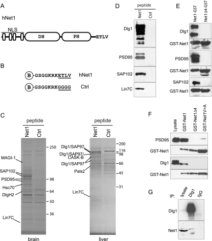
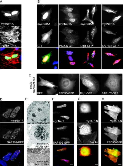
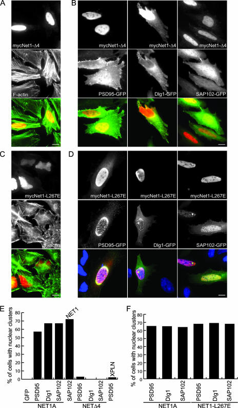
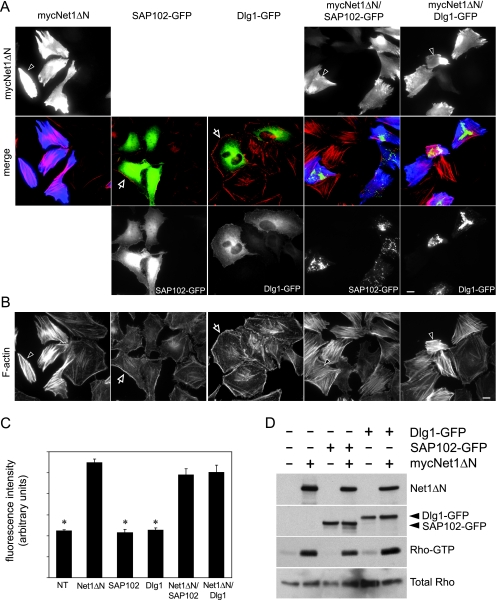
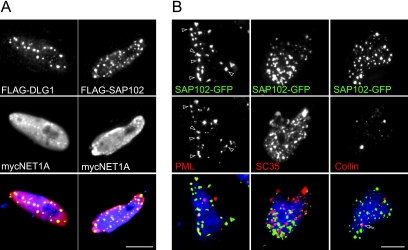
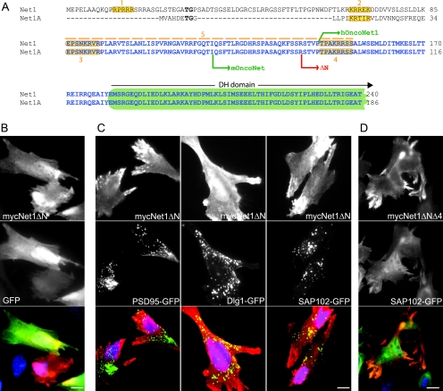
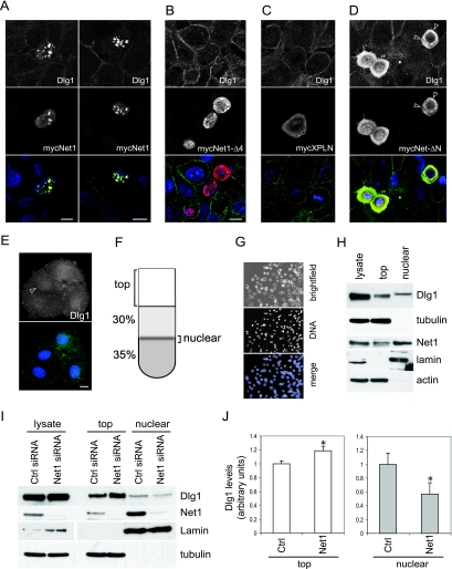
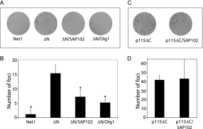
Net1 is a RhoA-specific guanine nucleotide exchange factor which localizes to the nucleus at steady state. A deletion in its N terminus redistributes the protein to the cytosol, where it activates RhoA and can promote transformation. Net1 contains a PDZ-binding motif at the C terminus which is essential for its transformation properties. Here, we found that Net1 interacts through its PDZ-binding motif with tumor suppressor proteins of the Dlg family, including Dlg1/SAP97, SAP102, and PSD95. The interaction between Net1 and its PDZ partners promotes the translocation of the PDZ proteins to nuclear subdomains associated with PML bodies. Interestingly, the oncogenic mutant of Net1 is unable to shuttle the PDZ proteins to the nucleus, although these proteins still associate as clusters in the cytosol. Our results suggest that the ability of oncogenic Net1 to transform cells may be in part related to its ability to sequester tumor suppressor proteins like Dlg1 in the cytosol, thereby interfering with their normal cellular function. In agreement with this, the transformation potential of oncogenic Net1 is reduced when it is coexpressed with Dlg1 or SAP102. Together, our results suggest that the interaction between Net1 and Dlg1 may contribute to the mechanism of Net1-mediated transformation.


Figures

FIG. 1.
FIG. 1.
Mouse brain, kidney, or liver lysates were incubated with streptavidin-Sepharose beads coupled with biotinylated peptides corresponding to the last 10 amino acids of Net1 or control (ctrl) peptides in which the last 4 amino acids were replaced by glycine (underlined). After being washed, the samples were separated by SDS-PAGE and the indicated bands were identified by MS as described in Materials and Methods. (A) Domain structure of the Net1 protein. (B) Peptide sequences used in this study. The peptides were biotinylated and contained an N-terminal glycine residue as a spacer. (C) SDS-PAGE gels from two representative experiments showing the proteins identified. Gels were stained with Coomassie blue. The bands of interest were cut from the gel and identified at the MS facility at UNC. Molecular mass markers (kDa) are indicated in each panel. (D) Protein identities were confirmed by Western blot analysis using commercially available antibodies. Peptide pulldowns were performed as described for panel C. Proteins were separated by SDS-PAGE and immunoblotted for PSD95, Dlg1, SAP102, and Lin7C. (E) Pulldown assay using brain lysates (PSD95, SAP102, and Lin7) or liver lysates (Dlg1) and GST-Net1 or a deletion mutant missing the PDZ-binding motif (GST-Net1Δ4). The precipitated proteins were immunoblotted with anti-PSD95, anti-Dlg1, and anti-SAP102 antibodies. The same blot was subsequently stripped and immunoblotted using anti-Net1 antibodies to verify the levels of GST-Net1 constructs in the assay. (F) Pulldown assay using brain lysates (PSD95) or liver lysates (Dlg1) and GST-Net1; a deletion mutant missing the PDZ-binding motif (GST-Net1Δ4) or a single-amino-acid-substitution mutant in the PDZ-binding motif (GST-Net1V→A). The precipitated proteins were immunoblotted with anti-PSD95 and anti-Dlg1 antibodies. The same blot was subsequently stripped and immunoblotted using anti-Net1 antibodies to verify the levels of GST-Net1 constructs in the assay. (G) Coprecipitation of endogenous Dlg1 and Net1 from HEK293 lysates. HEK293 lysates were incubated with anti-Dlg1 or with control antibodies, and the precipitates (IP) were immunoblotted for Net1. Net1 coprecipitated only with Dlg1, not with control antibodies.
FIG. 2.
FIG. 2.
HeLa cells were cotransfected with myc-Net1A and either PSD95-GFP, Dlg1-GFP, SAP102-GFP, or GFP alone; processed for immunofluorescence using anti-myc antibodies (red); and analyzed also for the GFP signal (green). Actin was stained with Alexa 594-phalloidin. Nuclear DNA was stained with Hoechst (blue). (A) Net1A localizes to the nucleus and to some extent to the cytosol when transfected alone. (B) Cotransfection of Net1A with any of the Dlg proteins induces a striking redistribution from a diffuse cytosolic staining (C) to a series of restricted subdomains within the nucleus. (C) The three Dlg proteins show a diffuse distribution in the cytosol in singly transfected cells. (D) Confocal sections show that both Net1 and SAP102 colocalize within the nuclear subdomains. (E) Ultrastructural analysis shows electron-dense particles in cells cotransfected with Net1A and PSD95-GFP or SAP102-GFP (arrowheads) that are not present in control cells and also severely distorted nuclei. (F) Identical subnuclear structures were formed when Net1 was cotransfected with SAP102. (G, H) The formation of the nuclear subdomains was not induced by coexpression of PSD95-GFP and XPLN, a protein homologous to Net1. Bar = 10 μm in all panels except panel E (bar = 2 μm).
FIG. 3.
FIG. 3.
HeLa cells were cotransfected with a Net1A mutant in which the last four amino acids, corresponding to the PDZ-binding motif, have been deleted (Net1Δ4) and either PSD95-GFP, Dlg1-GFP, SAP102-GFP, or GFP as a control and processed for immunofluorescence using anti-myc antibodies (red). Cells were also analyzed for the GFP signal (green). (A) Net1Δ4 localizes to the nucleus and induces actin stress fibers in a manner indistinguishable from that of wild-type Net1. (B) When Net1Δ4 is coexpressed with PSD95, Dlg1, or SAP102, the relocalization of these proteins to the nucleus and the formation of nuclear clusters are completely abolished. The three Dlg proteins also show a diffuse localization that is identical to that observed when they are expressed alone. (C) Catalytically inactive Net1 (L267E) fails to induce stress fibers when overexpressed in HeLa cells. (D) When the catalytically inactive Net1 protein is cotransfected with either PSD95, Dlg1, or SAP102, the formation of nuclear subdomains is not affected, suggesting that it is not dependent on the nucleotide exchange activity of Net1. (E, F) Experiments for Fig. 2B, 2H, 3B and 3D were quantified. At least 200 cells were counted for each condition and scored for the presence or absence of nuclear subdomains. Bar = 10 μm in all panels.
FIG. 4.
FIG. 4.
HeLa cells were transfected with mycNet1ΔN, Dlg1-GFP, or SAP102-GFP alone or in combination as indicated in the figure and processed for immunofluorescence by using monoclonal anti-myc antibodies (blue) and Alexa 594-phalloidin (red). Cells were also analyzed for the GFP signal (green). (A, B) Overexpression of Net1ΔN (the arrowhead indicates a representative cell) but not Dlg1-GFP or SAP102-GFP (arrow) induces the formation of stress fibers. Coexpression of Dlg1-GFP or SAP102-GFP with Net1ΔN does not affect Net1-mediated stress fiber formation (the arrowhead indicates a representative cell transfected with either Dlg1-GFP or SAP102-GFP with Net1ΔN). (A) A representative panel for each transfection is shown. The constructs used for each condition are indicated on top of each panel. (B) Alexa 594-phalloidin-stained images used for quantification. A representative image is shown. Arrowheads indicate cells transfected with the constructs indicated in panel A. (C) To quantify stress fiber formation, the average fluorescence intensity per cell in the red channel was measured. The fluorescence intensities in the green and blue channels were also measured to identify transfected cells. All images were acquired using the same exposure parameters. At least 25 cells were counted for each condition. Significance of difference from the control (Νet1ΔΝ) was estimated by Student's t test for nonpaired values. Bars not significantly different from the control values were left unmarked. An asterisk signifies a P value of <0.001. Bar = 10 μm in all panels. (D) HEK293 cells were transfected with mycNet1ΔN, Dlg1-GFP, or SAP102-GFP alone or in combination as indicated in the figure. Active Rho was specifically pulled down from cell lysates containing equal amounts of proteins with immobilized recombinant RBD-GST and analyzed by Western blotting using anti-RhoA antibody. Results for a representative experiment are shown.
FIG. 5.
FIG. 5.
(A) HeLa cells were cotransfected with myc-Net1A and either FLAG-Dlg1 or FLAG-SAP102 and processed for immunofluorescence using anti-FLAG (green) and anti-myc (red) antibodies. FLAG-tagged Dlg1 and SAP102 form nuclear subdomains that are identical to those formed by the GFP-tagged proteins, suggesting that GFP is not promoting the formation of these structures. (B) HeLa cells were cotransfected with myc-Net1A and either PSD95-GFP, Dlg1-GFP, or SAP102-GFP and processed for immunofluorescence using anti-PML (PML bodies), anti-SC35 (speckles), and anticoilin (Cajal bodies) antibodies (red) and analyzed also for the GFP signal (green). PML bodies showed partial colocalization with the clusters induced by Net1 (arrowheads). The arrow indicates a Cajal body colocalizing with a Net1 cluster. Bar = 10 μm in all panels.
FIG. 6.
FIG. 6.
(A) N-terminal domains of Net1 and Net1A. The oncogenic mutants and deletion mutants used in this study are indicated. NLS are shaded in orange and were previously characterized in references (regions 1 and 2), (regions 3 and 4), and (region 5). (B) Overexpression of mycNet1ΔΝ. Net1ΔΝ localizes to the nucleus and to the cytosol. (C) Single and double transfections were performed with HeLa cells, combining mycNet1ΔΝ with either GFP-PSD95, GFP-Dlg1, or GFP-SAP102. The cells were then fixed and processed for immunofluorescence using anti-myc antibodies (red) and analyzed also for the GFP signal (green). Deletion of the NLS in Net1 inhibits the formation of nuclear subdomains. The two proteins are still able to cocluster in the cytosol. (D) Deletion of the PDZ-binding motif in Net1ΔΝ (Net1ΔΝΔ4) abolishes the relocalization and clustering of the Dlg proteins in the cytosol (shown for SAP102). Bar = 10 μm in all panels.
FIG. 7.
FIG. 7.
MCF10a cells were transfected with the indicated constructs and processed for immunofluorescence, using anti-myc antibodies (red) to detect transfected cells, and analyzed also for endogenous Dlg1 by using anti-Dlg1 antibodies (green). Nuclear DNA was stained with Hoechst (blue). (A) Expression of Net1 promotes the redistribution of endogenous Dlg1 from the cell junctions to the nucleus (two representative cells are shown). (B) Deletion of the last four amino acids in Net1 (Net1Δ4) inhibits Dlg1 recruitment to the nucleus. (C) Expression of Net1's closest homologue, XPLN, has no effect in the localization of endogenous Dlg1. (D) Expression of the oncogenic Net1 protein (Net1ΔΝ) promotes the redistribution of endogenous Dlg1 from cell junctions to a diffuse cytosolic localization. Arrowheads indicate cytosolic clusters containing Dlg1 and Net1ΔN. (E) MCF7 cells were plated on coverslips and processed for immunofluorescence using anti-Dlg1 antibodies. The arrowhead indicates nuclear Dlg1. (F) Schematics of the discontinuous gradient utilized to isolate nuclei. Nucleus sediment in the 30 to 35% iodixanol interface is shown. The majority of the soluble proteins are recovered in the top fraction. (G) Representative image of a nuclear fraction. Nuclei were mixed 1:1 in mounting solution containing Hoechst to stain DNA. (H) Characterization of isolated nuclei. Proteins were separated by SDS-PAGE and immunoblotted for Dlg1, Net1, tubulin, lamin, and actin. (I) HEK293 cells were transfected with siRNA specific for Net1 or a nontargeting control siRNA. Ninety-six hours after transfection, nuclei were isolated as described in Materials and Methods. Equal amounts of protein from each fraction were then separated by SDS-PAGE and immunoblotted for Dlg1 and Net1. Membranes were stripped and reblotted for tubulin and lamin as loading controls for the top and nuclear fractions, respectively. Nuclear Dlg1 decreases when Net1 levels are reduced by siRNA treatment. (J) The amount of Dlg1 in each fraction was quantified by densitometric analysis. Values were normalized in relation to the control (ctrl) condition for each fraction. The results from three independent experiments were analyzed. Error bars represent standard errors of the means. Significance of difference from the control was estimated by Student's t test for paired values. Values that are significantly different from the control are marked with an asterisk. P = 0.0348 for the top fraction; P = 0.0033 for the nuclear fraction. Bar = 10 μm in all panels.
FIG. 8.
FIG. 8.
(A, C) Representative focus formation assay in NIH 3T3 cells. NIH 3T3 cells were transfected with the indicated constructs and analyzed for the formation of foci. (B, D) The data shown are representative of two independent experiments performed in triplicate and represent average numbers of foci generated. Error bars represent standard deviations. Significance of difference from the control (ΔΝ in panel B, p115ΔC in panel D) was estimated by Student's t test for nonpaired values. Bars not significantly different from the control values were left unmarked. An asterisk signifies a P value of <0.001.

Similar articles

Cited by

References

    1. Alberts, A. S., H. Qin, H. S. Carr, and J. A. Frost. 2005. PAK1 negatively regulates the activity of the Rho exchange factor NET1. J. Biol. Chem. 280:12152-12161. - PubMed
    1. Alberts, A. S., and R. Treisman. 1998. Activation of RhoA and SAPK/JNK signalling pathways by the RhoA-specific exchange factor mNET1. EMBO J. 17:4075-4085. - PMC - PubMed
    1. Arthur, W. T., and K. Burridge. 2001. RhoA inactivation by p190RhoGAP regulates cell spreading and migration by promoting membrane protrusion and polarity. Mol. Biol. Cell 12:2711-2720. - PMC - PubMed
    1. Arthur, W. T., S. M. Ellerbroek, C. J. Der, K. Burridge, and K. Wennerberg. 2002. XPLN, a guanine nucleotide exchange factor for RhoA and RhoB, but not RhoC. J. Biol. Chem. 277:42964-42972. - PubMed
    1. Audebert, S., C. Navarro, C. Nourry, S. Chasserot-Golaz, P. Lecine, Y. Bellaiche, J. L. Dupont, R. T. Premont, C. Sempere, J. M. Strub, A. Van Dorsselaer, N. Vitale, and J. P. Borg. 2004. Mammalian Scribble forms a tight complex with the betaPIX exchange factor. Curr. Biol. 14:987-995. - PubMed

Publication types

MeSH terms

LinkOut - more resources