. 2007 Oct 15;21(20):2629-43.
doi: 10.1101/gad.1575207.

Early pancreatic development requires the vertebrate Suppressor of Hairless (RBPJ) in the PTF1 bHLH complex

Affiliations

Early pancreatic development requires the vertebrate Suppressor of Hairless (RBPJ) in the PTF1 bHLH complex

Toshihiko Masui et al. Genes Dev. .

Abstract

PTF1a is an unusual basic helix-loop-helix (bHLH) transcription factor that is required for the development of the pancreas. We show that early in pancreatic development, active PTF1a requires interaction with RBPJ, the vertebrate Suppressor of Hairless, within a stable trimeric DNA-binding complex (PTF1). Later, as acinar cell development begins, RBPJ is swapped for RBPJL, the constitutively active, pancreas-restricted paralog of RBPJ. Moreover, the Rbpjl gene is a direct target of the PTF1 complex: At the onset of acinar cell development when the Rbpjl gene is first induced, a PTF1 complex containing RBPJ is bound to the Rbpjl promoter. As development proceeds, RBPJL gradually replaces RBPJ in the PTF1 complex bound to Rbpjl and appears on the binding sites for the complex in the promoters of other acinar-specific genes, including those for the secretory digestive enzymes. A single amino acid change in PTF1a that eliminates its ability to bind RBPJ (but does not affect its binding to RBPJL) causes pancreatic development to truncate at an immature stage, without the formation of acini or islets. These results indicate that the interaction between PTF1a and RBPJ is required for the early stage of pancreatic growth, morphogenesis, and lineage fate decisions. The defects in pancreatic development phenocopy those of Ptf1a-null embryos; thus, the first critical function of PTF1a is in the context of the PTF1 complex containing RBPJ. Action within an organ-specific transcription factor is a previously unknown function for RBPJ and is independent of its role in Notch signaling.


Figures
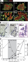
Figure 1.
Figure 1.
Ptf1a and Rbpjl expression during pancreatic development. (A) Immunofluorescent localization of PTF1a (green) at E10.5, E11.5, E12.5, and E13.5. (Red) E-cadherin. Arrowheads point to nascent acini. (B) Hybridization in situ detection of Ptf1a and Rbpjl mRNAs in early acinar cells around the periphery (solid arrowheads) compared with Ngn3 mRNA (open arrowheads) in endocrine cell precursors in the interior of the pancreatic epithelium at E14.5. Ptf1a and Rbpjl are for nearby, nonadjacent sections. (C) mRNAs were quantified by quantitative RT–PCR with RNA isolated from embryonic pancreatic rudiments and expressed relative to the levels at E18.5. The Rbpj and Rbpjl level mRNAs have a reciprocal relationship. Error bars are SEM; points without bars have SEMs equal to or smaller than the symbol.
Figure 2.
Figure 2.
Properties of the two PTF1-binding sites centered at −1341 and −92 of the mouse Rbpjl gene. (A) The nucleotide sequence of the two sites in the Rbpjl promoter are conserved. The distal site has an E-box separated from a TC-box by 21–22 bp, center-to-center; the proximal site has tandem E-boxes separated by 11 and 22 bp from a single TC-box. (B) Both Rbpjl promoter sites can bind PTF1 in nuclear extracts from adult pancreas. Binding is compared with that with the well-characterized PTF1 site from the Ela1 enhancer. Supershift analyses showed that all three sites tested bind the complete PTF1 complex (a trimer comprising PTF1a, a common E-protein [predominantly TCF12] and RBPJL) as well as a partial complex (a heterodimer of PTF1a and TCF12). (C) Comparison of the activities of the Ela1 and Rbpjl PTF1-binding sites in transfected cells. A tandem repeat of the 26-bp Ela1 or the 32-bp proximal Rbpjl PTF1 site was linked to the minimal promoter (−92 to +8) from the Ela1 gene driving a luciferase reporter gene and transfected into either the mouse 266-6 pancreatic acinar cell line or the human 293 embryonic kidney cell line. The activity of each construct was corrected for transfection efficiency and calculated relative to the activity of the same reporter, but lacking a PTF1-repeat. Error bars are standard deviations. (D) A model for the two PTF1 complexes and DNA binding that distinguishes the overlapping regions of PTF1a that interact with RBPJ or RBPJL and the basis for the differential effects of the W298A mutation. The Ws indicate the two tryptophan-containing conserved peptide motifs in the C-terminal region of PTF1a that are primary determinants for binding the RBPs. The W298A mutation is indicated by the A; based on the results of Beres et al. (2006).
Figure 3.
Figure 3.
Activity of the Rbpjl promoter in acinar cell lines. (A) Acinar cell activity is retained within the proximal 389 bp. The promoter regions extend from the distal points shown to 103 nucleotides of the 5′ untranslated region of Rbpjl and linked to the reporter vector pGL3-basic for transfection into acinar and nonpancreatic cell lines. The position of the first exon of the nearby Matrilin4 gene, transcribed in the direction opposite to Rbpjl, is shown. (B) Inactivation of the proximal PTF1-binding site by mutation of the TC-box eliminates the acinar activity; mutation of the distal PTF1 site had no discernible effect. The effects were similar for two independently derived pancreatic acinar tumor cell lines, mouse 266-6 and rat AR4-2J. The mutations, indicated by Xs, were introduced in the context of the −1380-bp promoter (hereafter, 1.4K). (C) Inactivation of the proximal PTF1-binding site by mutation of the two E-boxes eliminates the acinar activity; mutation of the distal site had no effect. For the experiments shown in A–C, the promoter alterations tested had no significant effect in the nonpancreatic 293 cell line. Luciferase reporter activity was adjusted for transfection efficiency and expressed relative to the promoterless pGL3-Basic vector. Error bars are standard deviations.
Figure 4.
Figure 4.
Activity of the Rbpjl promoter in vivo. Transgenic embryos at 17.5 d of gestation were derived from implanted, fertilized eggs injected with reporter constructs containing the 1.4-Kb promoter without alterations (A) or with the mutations described in Figure 3B of the TC-box of the proximal (B) or distal (C) PTF1-binding site. The expression of each transgene was detected by β-galactosidase activity derived from the lacZ reporter gene. The duodenum (D) has a low level of endogenous β-galactosidase activity; to verify the absence of pancreatic expression from the transgene with the mutated proximal PTF1 site, the staining was extended for B. (dP) Pancreas derived from the dorsal bud; (vP) pancreas from the ventral bud; (St) stomach; (Sp) spleen; (D) duodenum. (Bottom) The number of embryos with pancreatic β-galactosidase activity out of the total number of transgenic embryos is a measure of promoter potency.
Figure 5.
Figure 5.
PTF1 subunits reside on the Rbpjl proximal promoter region containing the PTF1 site in chromatin from adult pancreas. (A) ChIP analysis using antibodies to individual subunits showed that PTF1a and RBPJL, but not RBPJ, are present at or near the proximal PTF1 site. Results from a similar experiment showed PTF1a and RBPJL present at the distal PTF1 site as well (data not shown). Binding could not be detected using chromatin from adult liver. The schematic of the 1.4-Kb Rbpjl promoter shows the relative positions of the PTF1-binding sites and the primer pairs for PCR amplification. (B) Sequential ChIPs indicated that PTF1a and RBPJL coreside on Rbpjl promoters in adult acinar cells. The specificity of the consecutive enrichments was verified by the lack of enrichment with consecutive precipitations using an antibody to an unrelated DNA-binding protein (i.e., cMYC) and by the loss of any initial purification with anti-PTF1a or anti-RBPJL when anti-cMyc was used as the second precipitating antibody. Fold enrichment was quantified by quantitative PCR; a zero indicates that promoter DNA was not detected and a one indicates no enrichment relative to the input DNA.
Figure 6.
Figure 6.
The two E-boxes of the proximal PTF1-binding site are redundant. (A) Mutational disruption of the first (E1m) or second (E2m) E-box had no effect on the binding of the PTF1a/E12 heterodimer (cf. lanes 2,6,10), the RBPJ form of the trimer (lanes 3,7,11), or the RBPJL form of the trimer (lanes 4,8,12). (Lanes 14–16) Mutation of both E-boxes (E12m) eliminated binding of the dimer and both forms of the trimer. The positions of the monomer RBPJ (1), the PTF1a/E12 heterodimer (2), and the trimers (3) are indicated. The PTF1 subunits were synthesized by cell-free translation. (B) The E-box mutations shown in A were incorporated into the −389 RbpL-Luc plasmid to test whether both E-boxes were also required for activity of the Rbpjl promoter in transfected acinar or kidney cells. Mutational inactivation of either E-box had no effect on acinar activity, whereas inactivation of both nearly eliminated it. None of the mutations affected activity of the promoter in 293 kidney cells. (C) A proper stereochemical relationship between the TC-box and an E-box is required for binding the complete PTF1 trimer in EMSAs. To simplify the complexity of two E-boxes, we mutated the proximal one (which had no discernible effect on complex binding or activity) and tested the effects of altered spacing between the TC-box and the remaining E-box by inserting 6 bp (E2m + 6 bp) or deleting 6 bp (E2m − 6 bp) (see the diagram in D). Either alteration decreased binding of the PTF1 trimer, but not the PTF1a/E12 heterodimer. (D) Proper spacing between the TC-box and an E-box is required for the acinar activity of the promoter. The spacing changes described for C were incorporated into −389 RbpL-Luc for transfection into 266-6 acinar cells. Either alteration decreased acinar activity by at least 90%. Error bars are standard deviations.
Figure 7.
Figure 7.
The activity of the proximal PTF1 site can be reconstituted by ectopic expression of the PTF1 subunits. (A) Coexpression of PTF1a, HEB, and RBPJ or RBPJL in 293 kidney cells induces transcription from a synthetic promoter containing three copies of the proximal PTF1 site from the Rbpjl promoter (described in Fig. 2C). The activity in response to PTF1a alone was due to the presence of substantial RBPJ and E-proteins in 293 kidney cells. The activity was enhanced by the overexpression of HEB/TCF12 or RBPJL (which is absent in 293 cells). (B) The activities of the PTF1 sites from the Rbpjl and Ela1 promoters differ quantitatively and qualitatively. The Rbpjl site is many-fold more active than the Ela1 site in response to either the J or the JL forms of the reconstituted PTF1 (note the different scales). Moreover, the difference in the ability of the J and JL forms to activate the Ela1 site (approximately sixfold) is nearly absent for the Rbpjl site (approximately twofold). Error bars are standard deviations.
Figure 8.
Figure 8.
RBPJ is exchanged for RBPJL on the Rbpjl promoter during pancreatic development. ChIP analysis of PTF1a, RBPJL, and RBPJ at the proximal promoter region of Rbpjl (A) and the proximal Cpa1 promoter (B). An antibody to rabbit IgG was used to estimate background precipitation of Rbpjl promoter DNA. Arrows flanking the PTF1-binding sites in the promoters indicate the positions of the PCR primers. Error bars are SEM for three ChIPs.
Figure 9.
Figure 9.
Defective pancreatic development for the W298A mutant of Ptf1a. Pancreatic tissue derived from Ptf1a-expressing endoderm is stained for β-galactosidase activity induced by Ptf1a-cre activation of the R26R locus. (A–C) Normal pancreatic development of heterozygous Ptf1a+/cre embryos at E11.5, E12.5, and E17.5. (C) The normal perinatal pancreas is composed of two lobes derived from the dorsal pancreatic bud (dP) and one from the ventral pancreatic bud (vP), which lies within the first loop of the duodenum (D). A includes a view of the truncated pancreatic development of a homozygous Ptf1acre/cre embryo for comparison. (A′–C′) Abnormal pancreatic development of Ptf1aWA/cre embryos. One Ptf1acre allele is necessary to follow the fate of the mutant pancreatic cells; homozygous Ptf1aWA/WA embryos have an identical mutant phenotype (Supplementary Fig. S2). (A′) Altered pancreatic development is evident at E11.5, and is indistinguishable from that of homozygous-deficient Ptf1acre/cre embryos. (B′) Compared with normal development at E12.5, growth of the dorsal pancreatic epithelium is curtailed, and a ventral epithelium has not escaped the duodenal mucosa. (C′) The dorsal pancreas forms a truncated, undeveloped ductal structure (dP) without islet or acinar tissues. A ventral pancreas does not form; rather, the mutant, β-galactosidase-positive cells incorporate into the gut mucosa and acquire intestinal phenotypes (vP). (Inset) Cells within the normal duodenal epithelium stain for β-galactosidase activity and occasionally costain red with PAS, indicative of goblet cells. (dP) Dorsal pancreas; (vP) ventral pancreas; (D) duodenum; (St) stomach; (Sp) spleen.
Figure 10.
Figure 10.
Loss of pancreatic development markers in homozygous W298A mutant embryos. (Top) For Ptf1aWA/WA embryos at E10.5 (A′–D′), epithelial morphogenesis (A,A′) of the dorsal pancreas and the levels of PTF1a (B,B′) and PDX1 (C,C′) were indistinguishable from those for normal development of heterozygous Ptf1a+/WA (A–D) and wild-type embryos (data not shown). By E14.5 in mutant embryos, morphogenesis was defective (E,E′), PTF1a did not appear in newly forming acini (F,F′,H,H′; acini), and cells with high levels of PDX1 were absent (G,G′). (A,A′,E,E′) E-cadherin delineates the pancreatic epithelium. (Bottom) Neither islets nor acini form for Ptf1aWA/WA embryos. At E14.5, CPA1, an early marker of acinar cell differentiation (I; arrow), was absent from mutant embryonic pancreas (I′). Endocrine cell clusters in the mutant pancreas coexpress prohormone convertase 1/3 (PC1/3) (e.g., arrow, J′) and glucagon (arrow, K′). Insulin-expressing islet β-cells are normally present at high numbers (inset of L; insulin, blue), but are rare in the mutant (inset of L′; insulin, blue). Bars, 50 μm.

Similar articles

Cited by

References

    1. Ahlgren U., Jonsson J., Edlund H., Jonsson J., Edlund H., Edlund H. The morphogenesis of the pancreatic mesenchyme is uncoupled from that of the pancreatic epithelium in IPF1/PDX1-deficient mice. Development. 1996;122:1409–1416. - PubMed
    1. Apelqvist A., Li H., Sommer L., Beatus P., Anderson D.J., Honjo T., de Hrabe Angelis M., Lendahl U., Edlund H., Li H., Sommer L., Beatus P., Anderson D.J., Honjo T., de Hrabe Angelis M., Lendahl U., Edlund H., Sommer L., Beatus P., Anderson D.J., Honjo T., de Hrabe Angelis M., Lendahl U., Edlund H., Beatus P., Anderson D.J., Honjo T., de Hrabe Angelis M., Lendahl U., Edlund H., Anderson D.J., Honjo T., de Hrabe Angelis M., Lendahl U., Edlund H., Honjo T., de Hrabe Angelis M., Lendahl U., Edlund H., de Hrabe Angelis M., Lendahl U., Edlund H., Lendahl U., Edlund H., Edlund H. Notch signalling controls pancreatic cell differentiation. Nature. 1999;400:877–881. - PubMed
    1. Beres T.M., Masui T., Swift G.H., Shi L., Henke R.M., MacDonald R.J., Masui T., Swift G.H., Shi L., Henke R.M., MacDonald R.J., Swift G.H., Shi L., Henke R.M., MacDonald R.J., Shi L., Henke R.M., MacDonald R.J., Henke R.M., MacDonald R.J., MacDonald R.J. PTF1 is an organ-specific and Notch-independent basic helix–loop–helix complex containing the mammalian Suppressor of Hairless (RBP-J) or its paralogue, RBP-L. Mol. Cell. Biol. 2006;26:117–130. - PMC - PubMed
    1. Birren S.J., Lo L., Anderson D.J., Lo L., Anderson D.J., Anderson D.J. Sympathetic neuroblasts undergo a developmental switch in trophic dependence. Development. 1993;119:597–610. - PubMed
    1. Brinster R.L., Chen H.Y., Trumbauer M.E., Yagle M.K., Palmiter R.D., Chen H.Y., Trumbauer M.E., Yagle M.K., Palmiter R.D., Trumbauer M.E., Yagle M.K., Palmiter R.D., Yagle M.K., Palmiter R.D., Palmiter R.D. Factors affecting the efficiency of introducing foreign DNA into mice by microinjecting eggs. Proc. Natl. Acad. Sci. 1985;82:4438–4442. - PMC - PubMed

Publication types

MeSH terms