. 2007 Oct 15;21(20):2659-70.
doi: 10.1101/gad.445307.

Regulation of the sigmaE stress response by DegS: how the PDZ domain keeps the protease inactive in the resting state and allows integration of different OMP-derived stress signals upon folding stress

Affiliations

Regulation of the sigmaE stress response by DegS: how the PDZ domain keeps the protease inactive in the resting state and allows integration of different OMP-derived stress signals upon folding stress

Hanna Hasselblatt et al. Genes Dev. .

Abstract

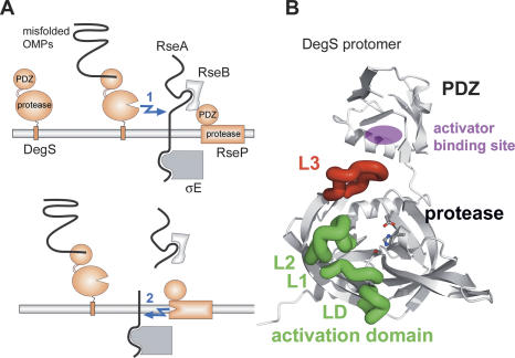
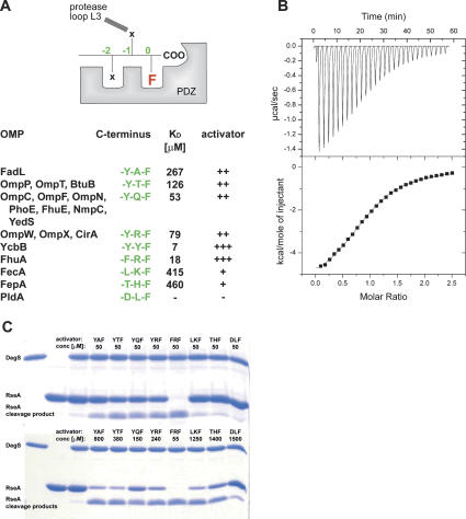
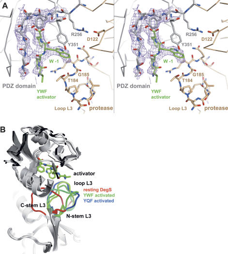
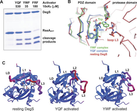
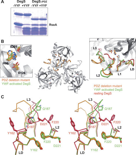
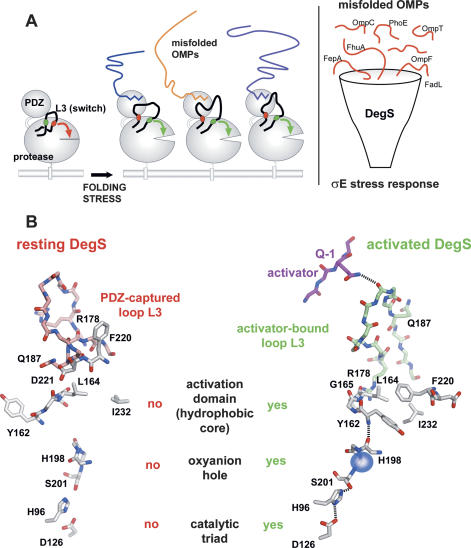
The unfolded protein response of Escherichia coli is triggered by the accumulation of unassembled outer membrane proteins (OMPs) in the cellular envelope. The PDZ-protease DegS recognizes these mislocalized OMPs and initiates a proteolytic cascade that ultimately leads to the sigmaE-driven expression of a variety of factors dealing with folding stress in the periplasm and OMP assembly. The general features of how OMPs activate the protease function of DegS have not yet been systematically addressed. Furthermore, it is unknown how the PDZ domain keeps the protease inactive in the resting state, which is of crucial importance for the functioning of the entire sigmaE stress response. Here we show in atomic detail how DegS is able to integrate the information of distinct stress signals that originate from different OMPs containing a -x-Phe C-terminal motif. A dedicated loop of the protease domain, loop L3, serves as a versatile sensor for allosteric ligands. L3 is capable of interacting differently with ligands but reorients in a conserved manner to activate DegS. Our data also indicate that the PDZ domain directly inhibits protease function in the absence of stress signals by wedging loop L3 in a conformation that ultimately disrupts the proteolytic site. Thus, the PDZ domain and loop L3 of DegS define a novel molecular switch allowing strict regulation of the sigmaE stress response system.


Figures

Figure 1.
Figure 1.
Function of DegS in the σE stress response. (A) Schematic presentation of the σE stress response. (B) Ribbon presentation of one subunit of the DegS trimer highlighting loop L3 (red) that mediates communication between the PDZ and protease domains, and the activation domain (loops L1, L2, and LD, green) that allows formation of a functional catalytic triad comprising residues His96, Asp126, and Ser201, which are shown in ball-and-stick mode.
Figure 2.
Figure 2.
Binding and activation potential of different OMP-derived C termini. (A) Activating peptides used in this study. In the top panel, the general binding mode to the PDZ domain is indicated together with the nomenclature used here. For the ligand, the C-terminal phenylalanine present in all OMPs is highlighted. The table shown in the bottom panel summarizes the C termini of the analyzed peptides (green), the corresponding OMPs in E. coli, and the individual dissociation constants and activation capabilities. (B) ITC measurement of the binding of the FRF-peptide to the DegS wild-type protein. (Top panel) Ten-microliter aliquots of the FRF peptide (1 mM) were injected into the sample cell containing 85 μM DegS. (Bottom panel) The area under each peak was integrated and plotted against the molar ratio peptide/DegS inside the sample cell. The black line represents the fit to a binding isotherm, assuming one binding site per protomer. (C) SDS-PAGE of RseA cleavage by DegS. The periplasmic domain of RseA was present in 30 μM, DegS was present in 10 μM, and the activating peptides were present in the indicated concentrations. The reactions were stopped after 18 h. In the control reactions (first three lanes), the assay was conducted without activating peptide.
Figure 3.
Figure 3.
Structure of the complex between DegS and YWF. (A) Stereo presentation of the omit density of the bound YWF activator and of the main-chain-binding segment of the PDZ domain. The 3Fo − 2Fc electron-density map was calculated at 2.5 Å resolution (contoured at 1.0 σ.) after omitting the PDZ domain and the YWF activator from the refined model. Only the four C-terminal residues of the activator were defined by electron density. The YWF peptide (green)and the DegS residues that interact with the tryptophan in the −1 position are shown as a stick model. Furthermore, the Arg256:Asp122 salt bridge, which mainly defines the orientation of protease (brown) and the PDZ domain (gray), is indicated. (B) Ribbon plot of the superimposed structures of resting and activated DegS. The bound YWF peptide is shown as a stick model to indicate the position of the activator-binding site. Loop L3 is highlighted by color (resting DegS in red, YQF-activated in blue, and YWF-activated in green). Although the central segment of loop L3 differs in all activated forms, the stem regions show a mostly conserved conformation.
Figure 4.
Figure 4.
Differences in the YQF- and YWF-activated DegS. (A) SDS-PAGE of the cleavage of the periplasmic domain of RseA (30 μM) by DegS (10 μM), applying saturating activator concentrations. For each activator, the 10-fold concentration of the individual KD value was applied. The reactions were carried out at 30°C and stopped after 3 h. The control reaction was carried out without activating peptide. (B) Comparison of YQF- and YWF-binding mode. Alignment of the PDZ domains with bound activator (YQF, blue; YWF, green) onto the resting DegS structure (red) illustrates the different binding positions of the −1 glutamine and tryptophan side chains, respectively. In contrast to the YQF peptide, binding of the indol ring of the YWF activator leads to a sterical clash with loop L3 of the resting protease. (C) Ribbon plot showing the protease domain of DegS with mapped thermal motion factors: (blue) rigid parts; (red) flexible parts. The relevant active site loops are labeled. For both activated complexes, the active site loops L1, L2, and LD are better defined as in resting DegS. However, loop L2 was more flexible in the DegS/YQF structure than in the DegS/YWF structure. The differences between the average thermal motion factors of loops LD/L1/L2 and the protease domain are as follows: 46.1, 29.7, and n.d. for resting DegS; −10.3, −9.9, and 49.7 for YQF-activated DegS; and −12.9, −9.7, and 36.3 Å2 for YWF-activated DegS.
Figure 5.
Figure 5.
Structure–function analysis of the PDZ deletion mutant DegSΔPDZ. (A) SDS-PAGE of RseA cleavage by DegS. The periplasmic domain of RseA was present in 30 μM, DegS and DegSΔPDZ were present in 10 μM, and the YYF peptide was present in 100 μM. The reaction was stopped after 5 h. (B) Overall fold of the DegSΔPDZ trimer. Mechanistic important loops L1, L2, L3, and LD of one monomer are colored orange. To indicate the position of the proteolytic site, the individual catalytic triads are shown as a stick model. (Left inset) Superposition of the catalytic triad of activated DegS (green) and DegSΔPDZ (orange). For the PDZ deletion mutant, the triad was observed in a different configuration in each subunit of the trimer, illustrating its flexibility. (Right inset) Superposition of active site loops of DegSΔPDZ (orange), resting DegS (red), and activated DegS (green). Notably, the loops of the activation domain (L1, L2, LD) of DegSΔPDZ and activated DegS fit nicely to each other. (C) Stereoview of the superposition of DegSΔPDZ (orange) with YWF-activated (green) and resting DegS (red), highlighting the different conformations of loop L3. The presence of the PDZ domain as well as the binding of activating peptides reorient loop L3 in a distinct but specific manner. Key residues for the interaction between loop L3 and loops L2 and LD of the activation domain are labeled and shown as a stick model. The superposition also illustrates the inherent flexibility of the central part of loop L3, which obtains a different conformation in each structure.
Figure 6.
Figure 6.
Model for the integration of different OMP-derived stress signals by DegS. (A) The left panel illustrates activation of DegS by different OMP C termini. Loop L3 is highlighted with its inhibitory (red) and activating (green) structural elements. Molecular details of both inhibitory and activating processes are given in B. In latent DegS, loop L3 directly inhibits protease function by disrupting the activation domain. Binding of the allosteric activator to the PDZ domain triggers a switch of this loop into its active position, where it now supports the setup of a functional proteolytic site. The right panel illustrates the cellular function of DegS acting as a mechanistic funnel to integrate the information from different mislocalized OMPs into the σE stress response. (B) Working model for how DegS switches from the resting to the activated state. Key residues that are important for regulation and for signal propagation are labeled. Loop L3 of the resting DegS is drawn in red, loop L3 of the active DegS is in green. Details of the signal transduction through the DegS molecule leading to the functional protease are described in the text.

Similar articles

Cited by

References

    1. Ades S.E., Connolly L.E., Alba B.M., Gross C.A., Connolly L.E., Alba B.M., Gross C.A., Alba B.M., Gross C.A., Gross C.A. The Escherichia coli σ(E)-dependent extracytoplasmic stress response is controlled by the regulated proteolysis of an anti-σ factor. Genes & Dev. 1999;13:2449–2461. - PMC - PubMed
    1. Akiyama Y., Kanehara K., Ito K., Kanehara K., Ito K., Ito K. RseP (YaeL), an Escherichia coli RIP protease, cleaves transmembrane sequences. EMBO J. 2004;23:4434–4442. - PMC - PubMed
    1. Alba B.M., Leeds J.A., Onufryk C., Lu C.Z., Gross C.A., Leeds J.A., Onufryk C., Lu C.Z., Gross C.A., Onufryk C., Lu C.Z., Gross C.A., Lu C.Z., Gross C.A., Gross C.A. DegS and YaeL participate sequentially in the cleavage of RseA to activate the σ(E)-dependent extracytoplasmic stress response. Genes & Dev. 2002;16:2156–2168. - PMC - PubMed
    1. Brown M.S., Ye J., Rawson R.B., Goldstein J.L., Ye J., Rawson R.B., Goldstein J.L., Rawson R.B., Goldstein J.L., Goldstein J.L. Regulated intramembrane proteolysis: A control mechanism conserved from bacteria to humans. Cell. 2000;100:391–398. - PubMed
    1. Brunger A.T., Adams P.D., Clore G.M., DeLano W.L., Gros P., Grosse-Kunstleve R.W., Jiang J.S., Kuszewski J., Nilges M., Pannu N.S., Adams P.D., Clore G.M., DeLano W.L., Gros P., Grosse-Kunstleve R.W., Jiang J.S., Kuszewski J., Nilges M., Pannu N.S., Clore G.M., DeLano W.L., Gros P., Grosse-Kunstleve R.W., Jiang J.S., Kuszewski J., Nilges M., Pannu N.S., DeLano W.L., Gros P., Grosse-Kunstleve R.W., Jiang J.S., Kuszewski J., Nilges M., Pannu N.S., Gros P., Grosse-Kunstleve R.W., Jiang J.S., Kuszewski J., Nilges M., Pannu N.S., Grosse-Kunstleve R.W., Jiang J.S., Kuszewski J., Nilges M., Pannu N.S., Jiang J.S., Kuszewski J., Nilges M., Pannu N.S., Kuszewski J., Nilges M., Pannu N.S., Nilges M., Pannu N.S., Pannu N.S., et al. Crystallography & NMR system: A new software suite for macromolecular structure determination. Acta Crystallogr. D Biol. Crystallogr. 1998;54:905–921. - PubMed

Publication types

MeSH terms

Substances

LinkOut - more resources