A mechanism of polarized light sensitivity in cone photoreceptors of the goldfish Carassius auratus
- PMID: 17938422
- PMCID: PMC2025664
- DOI: 10.1529/biophysj.107.112292
A mechanism of polarized light sensitivity in cone photoreceptors of the goldfish Carassius auratus
Abstract
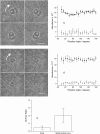
An integrated laser tweezer and microphotometry device has been used to characterize in detail how individual, axially orientated goldfish photoreceptors absorb linearly polarized light. This work demonstrates that the mid-wavelength sensitive members of double cone photoreceptors display axial differential polarization sensitivity. The polarization contrast was measured to be 9.2 +/- 0.4%. By comparison, rod photoreceptors only exhibit isotropic absorbance. These data, combined with the square cone mosaic of double cones in the retina, suggest that intrinsic axial dichroism forms part of the underlying biophysical detection mechanism for polarization vision in this species.
Figures


Similar articles
-
Ultraviolet polarization vision and visually guided behavior in fishes.Brain Behav Evol. 2010;75(3):186-94. doi: 10.1159/000314275. Epub 2010 Aug 20. Brain Behav Evol. 2010. PMID: 20733294 Review.
-
No evidence of UV cone input to mono- and biphasic horizontal cells in the goldfish retina.J Comp Physiol A Neuroethol Sens Neural Behav Physiol. 2010 Dec;196(12):913-25. doi: 10.1007/s00359-010-0574-9. Epub 2010 Aug 24. J Comp Physiol A Neuroethol Sens Neural Behav Physiol. 2010. PMID: 20734051
-
Temporal expression of rod and cone opsins in embryonic goldfish retina predicts the spatial organization of the cone mosaic.Invest Ophthalmol Vis Sci. 1996 Feb;37(2):363-76. Invest Ophthalmol Vis Sci. 1996. PMID: 8603841
-
Biochemistry and physiology of zebrafish photoreceptors.Pflugers Arch. 2021 Sep;473(9):1569-1585. doi: 10.1007/s00424-021-02528-z. Epub 2021 Feb 17. Pflugers Arch. 2021. PMID: 33598728 Free PMC article. Review.
-
Cones perform a non-linear transformation on natural stimuli.J Physiol. 2010 Feb 1;588(Pt 3):435-46. doi: 10.1113/jphysiol.2009.179036. Epub 2009 Dec 14. J Physiol. 2010. PMID: 20008463 Free PMC article.
Cited by
-
Behavioural and physiological mechanisms of polarized light sensitivity in birds.Philos Trans R Soc Lond B Biol Sci. 2011 Mar 12;366(1565):763-71. doi: 10.1098/rstb.2010.0196. Philos Trans R Soc Lond B Biol Sci. 2011. PMID: 21282180 Free PMC article. Review.
-
A vertebrate retina with segregated colour and polarization sensitivity.Proc Biol Sci. 2017 Sep 13;284(1862):20170759. doi: 10.1098/rspb.2017.0759. Proc Biol Sci. 2017. PMID: 28878058 Free PMC article.
-
Retinal region of polarization sensitivity switches during ontogeny of rainbow trout.J Neurosci. 2013 Apr 24;33(17):7428-38. doi: 10.1523/JNEUROSCI.5815-12.2013. J Neurosci. 2013. PMID: 23616549 Free PMC article.
-
Primary processes in sensory cells: current advances.J Comp Physiol A Neuroethol Sens Neural Behav Physiol. 2009 Jan;195(1):1-19. doi: 10.1007/s00359-008-0389-0. Epub 2008 Nov 15. J Comp Physiol A Neuroethol Sens Neural Behav Physiol. 2009. PMID: 19011871 Review.
-
Inversion by P4: polarization-picture post-processing.Philos Trans R Soc Lond B Biol Sci. 2011 Mar 12;366(1565):638-48. doi: 10.1098/rstb.2010.0205. Philos Trans R Soc Lond B Biol Sci. 2011. PMID: 21282167 Free PMC article. Review.
References
-
- Horváth, G., and D. Varjú. 2004. Polarized Light in Animal Vision. Springer, Berlin, Heidelberg.
-
- Wehner, R. 2001. Polarization vision—a uniform sensory capacity? J. Exp. Biol. 204:2589–2596. - PubMed
-
- Waterman, T. H. 1981. Polarization sensitivity. In Handbook of Sensory Physiology, Vol. VII/6B. Springer-Verlag, Berlin, Heidelberg, New York.
-
- Bernard, G. D., and R. Wehner. 1977. Functional similarities between polarization vision and color vision. Vision Res. 17:1019–1028. - PubMed
-
- Coughlin, D. J., and C. W. Hawryshyn. 1995. A cellular basis for polarized-light vision in rainbow trout. J. Comp. Physiol. [A]. 176:261–272.
Publication types
MeSH terms
LinkOut - more resources
Full Text Sources

