

Unusual presentation of cystic lymphangioma of the gallbladder
- PMID: 17939338
- PMCID: PMC2687694
- DOI: 10.3904/kjim.2007.22.3.197
Unusual presentation of cystic lymphangioma of the gallbladder
Abstract
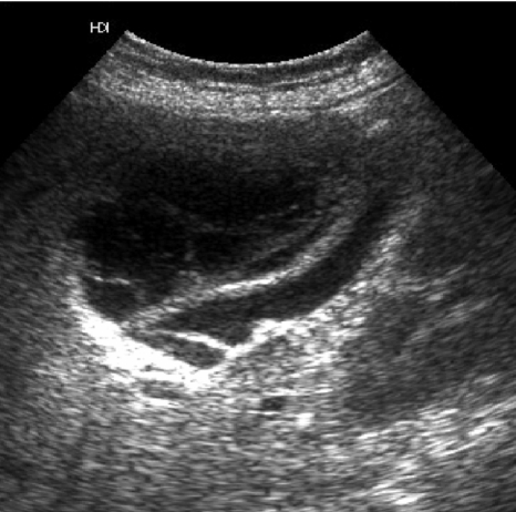
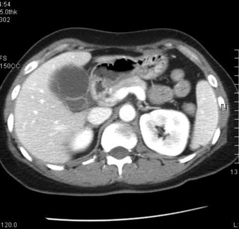
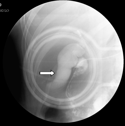
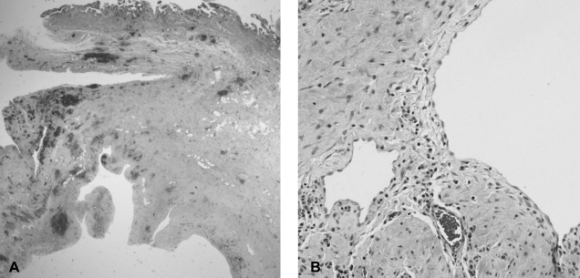
Cystic lymphangioma of the gallbladder is quite a rare tumor with only a few cases having been reported in the literature. We describe here a rare case of cystic lymphangioma of the gallbladder, which was unusual in that the patient presented with biliary pain and an abnormal liver test. Ultrasonography and computed tomography of the abdomen showed a multi-septated cystic mass in the gallbladder fossa and an adjacent compressed gallbladder. Endoscopic retrograde cholangiography showed there was no communication between the bile tract and the lesion, and there were no other abnormal findings with the exception of a laterally compressed gallbladder. After performing endoscopic sphincterotomy, a small amount of sludge was released from the bile duct. The histological findings were consistent with a cystic lymphangioma originating from the subserosal layer of the gallbladder. This unusual clinical presentation of a gallbladder cystic lymphangioma was attributed to biliary sludge, and this was induced by gallbladder dysfunction that was possibly from compression of the gallbladder due to the mass.
Figures
Similar articles
-
Cystic lymphangioma of the gall-bladder: a case report.J Gastroenterol Hepatol. 1995 Nov-Dec;10(6):693-6. doi: 10.1111/j.1440-1746.1995.tb01373.x. J Gastroenterol Hepatol. 1995. PMID: 8580416
-
Cystic lymphangioma of the gallbladder: report of a case.Surg Today. 2008;38(1):81-4. doi: 10.1007/s00595-007-3564-y. Epub 2007 Dec 24. Surg Today. 2008. PMID: 18085372
-
Gallbladder lymphangioma: a case report and review of the literature.World J Gastroenterol. 2007 Jan 14;13(2):320-3. doi: 10.3748/wjg.v13.i2.320. World J Gastroenterol. 2007. PMID: 17226918 Free PMC article. Review.
-
Gall-bladder and hepatoduodenal ligament lymphangioma - case report and literature review.Pol Przegl Chir. 2013 Jan;85(1):39-43. doi: 10.2478/pjs-2013-0007. Pol Przegl Chir. 2013. PMID: 23509201 Review.
-
Gallbladder lymphangioma: MR findings.Abdom Imaging. 2002 Jan-Feb;27(1):54-7. doi: 10.1007/s00261-001-0051-6. Abdom Imaging. 2002. PMID: 11740609
Cited by
-
Mesenteric cystic lymphangioma in an adolescent male; a diagnostic dilemma: A case report.Int J Surg Case Rep. 2023 Dec;113:109042. doi: 10.1016/j.ijscr.2023.109042. Epub 2023 Nov 14. Int J Surg Case Rep. 2023. PMID: 37984261 Free PMC article.
-
Gallbladder fossa lymphangioma encasing the common bile duct: a case report and review of the literature.Ann Med Surg (Lond). 2024 Apr 24;86(6):3702-3707. doi: 10.1097/MS9.0000000000002083. eCollection 2024 Jun. Ann Med Surg (Lond). 2024. PMID: 38846820 Free PMC article.
-
Is laparoscopic cholecystectomy safe for lymphangioma of the gallbladder? A complicated case mimicking subhepatic abscess.Updates Surg. 2012 Mar;64(1):73-6. doi: 10.1007/s13304-011-0080-9. Epub 2011 May 27. Updates Surg. 2012. PMID: 21618037
-
Surgery of multiple lymphangioma in small bowel: a rare case report of chronic gastrointestinal bleeding.Ann Surg Treat Res. 2018 Jan;94(1):52-56. doi: 10.4174/astr.2018.94.1.52. Epub 2017 Dec 28. Ann Surg Treat Res. 2018. PMID: 29333427 Free PMC article.
-
Hemolymphangioma of the transverse mesocolon: a case report and literature review.Transl Cancer Res. 2021 Aug;10(8):3849-3855. doi: 10.21037/tcr-21-176. Transl Cancer Res. 2021. PMID: 35116683 Free PMC article.
References
-
- Enzinger FM, Weiss SW. Tumors of lymph vessels. In: Enzinger FM, Weiss SW, editors. Soft tissue tumors. 3rd ed. St Louis: Mosby; 1995. pp. 679–699.
-
- Roisman I, Manny J, Field S, Shiloni E. Intra-abdominal lymphangioma. Br J Surg. 1989;76:485–489. - PubMed
-
- Takiff H, Calabria R, Yin L, Stabile BE. Mesenteric cysts and intraabdominal cystic lymphangiomas. Arch Surg. 1985;120:1266–1269. - PubMed
-
- Yang HR, Jan YY, Huang SF, Yeh TS, Tseng JH, Chen MF. Laparoscopic cholecystectomy for gallbladder lymphangiomas. Surg Endosc. 2003;17:1676. - PubMed
Publication types
MeSH terms
LinkOut - more resources
Full Text Sources
Medical