. 2007 Oct 23;104(43):16934-9.
doi: 10.1073/pnas.0704860104. Epub 2007 Oct 15.

Chromosome mobility during meiotic prophase in Saccharomyces cerevisiae

Affiliations

Chromosome mobility during meiotic prophase in Saccharomyces cerevisiae

Harry Scherthan et al. Proc Natl Acad Sci U S A. .

Abstract

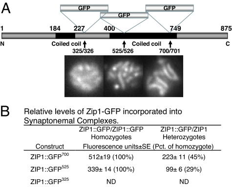
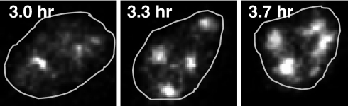
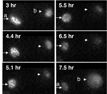
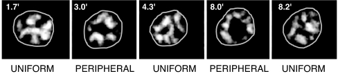
In many organisms, a synaptonemal complex (SC) intimately connects each pair of homologous chromosomes during much of the first meiotic prophase and is thought to play a role in regulating recombination. In the yeast Saccharomyces cerevisiae, the central element of each SC contains Zip1, a protein orthologous to mammalian SYCP1. To study the dynamics of SCs in living meiotic cells, a functional ZIP1::GFP fusion was introduced into yeast and analyzed by fluorescence video microscopy. During pachytene, SCs exhibited dramatic and continuous movement throughout the nucleus, traversing relatively large distances while twisting, folding, and unfolding. Chromosomal movements were accompanied by changes in the shape of the nucleus, and all movements were reversibly inhibited by the actin antagonist Latrunculin B. Normal movement required the NDJ1 gene, which encodes a meiosis-specific telomere protein needed for the attachment of telomeres to the nuclear periphery and for normal kinetics of recombination and meiosis. These results show that SC movements involve telomere attachment to the nuclear periphery and are actin-dependent and suggest these movements could facilitate completion of meiotic recombination.


Conflict of interest statement

The authors declare no conflict of interest.

Figures

Fig. 1.
Fig. 1.
Structure of ZIP1::GFP constructs and relative efficiency of their incorporation into SCs. (A) ZIP1 gene showing location of GFP inserts. Constructs were introduced into S. cerevisiae as described in Materials and Methods. Representative fluorescent images of pachytene nuclei for each construct are shown below. (B) Relative efficiency of incorporation into SCs in heterozygotes and homozyotes. Strains HW122, HW123, EW102, and EW103 were incubated in sporulation medium and analyzed when the percentage of cells in pachytene was highest. Zip1 fluorescence was quantitated as described in Materials and Methods. ND, not determined.
Fig. 2.
Fig. 2.
Meiotic reciprocal recombination in strains containing different ZIP1::GFP constructs. The percent recombination (cM ± SE) was calculated from the number of parental ditype (PD), nonparental ditype (NPD), and tetratype (TT) asci, as described in Materials and Methods. Pct. WT, total recombination relative to WT. iG418 is YCL056C::kanmx, and iURA is an insertion of URA3 in YCL040W.
Fig. 3.
Fig. 3.
Kinetics of SC formation and meiosis in ZIP1::GFP strains. (Upper) Initiation and completion of SCs were monitored by determining the percentage of cells showing punctate (Zyg-zygotene) and mature worm-like (Pach-full-length pachytene) ZIP1::GFP fluorescent structures in strains EW102 (GFP525) and HW122 (GFP700). (Lower) Meiotic divisions were monitored in strains EW102 (GFP525), HW122 (GFP700), and NKY279 (WT nonfluorescent control) by DAPI staining samples taken at the times noted from cultures incubated in liquid sporulation medium and scoring the number of cells containing one (not shown), two (MI, meiosis I), and four nuclei (MII, meiosis II). At least 200 cells were scored per time point sample.
Fig. 4.
Fig. 4.
Zip1-GFP700 containing nucleus undergoing transition from zygotene into pachytene. Time elapsed after introduction into sporulation medium and the outline of the nucleus is displayed.
Fig. 5.
Fig. 5.
Zip1-GFP700 containing nuclei undergoing transitions during imaging. Images were captured at the noted times. a, Rearrangements of pachytene SCs (3–5.1 h) followed by diplotene SC disassembly (5.5–6.5 h) and loss of most fluorescence (7.5 h). b, Early diplotene nucleus with last remnants of SC (3–4.4 h) followed by loss of most fluorescence (5.1 h). This nucleus began to autofluoresce (7.5 h) and formed a four-spored ascus ≈1 h after recording was stopped (not shown).
Fig. 6.
Fig. 6.
Consecutive images showing rapid mobility of Zip-GFP-labeled SCs in WT strains and impaired mobility in ndj1 mutants. (Upper) WT nucleus (strain HW122), Arrowhead points to a single SC that remains immobile providing a reference point near fixed line. Upper Left arrow points to a single more elongated SC that comes into the plane of focus, extends, and then slides along the nuclear periphery. Arrows on Lower Right point to three U-shaped SCs that change shape. (Lower) An ndj1 mutant (strain EW105) nucleus where all SCs appear almost immobile and relatively extended except one which slides into the image plane (arrowhead). Elapsed time is shown at top. Nuclei are outlined.
Fig. 7.
Fig. 7.
Quantitation of chromosomal movement and nuclear deformations. (A) Paths of the ends of randomly chosen individual fluorescent SCs were followed in WT and ndj1 mutant nuclei as described in Materials and Methods by using images captured every 0.5 sec. The effects of LatB and formaldehyde (Formald) addition are shown. (B) Changes in nuclear shape (Δ[l/w]) were quantitated as described in Materials and Methods by using images captured every 5 sec. Error bars denote SEM.
Fig. 8.
Fig. 8.
Zip1-GFP containing nuclei showing extensive chromosome movement and nuclear deformations in WT and impaired movement in LatB-treated and ndj1 mutant cells. Video frames captured at the noted intervals with nuclei outlined. (A) WT. Arrows point to deforming nucleus and movement of “maverick” chromosomes out of and then back to the bulk of the chromosomal mass. (B) WT + LatB. WT nucleus treated with LatB where all SC appear virtually immobile. (C) Mutant ndj1 where SCs and nucleus show limited mobility and arrow points to a stationary reference chromosome.
Fig. 9.
Fig. 9.
SCs shuttling between the peripheral and uniformly distributed arrays. Frames from videos taken at the noted intervals after 3 h of incubation in sporulation medium. Nuclei are outlined.

References

    1. Baker BS, Carpenter AT, Esposito MS, Esposito RE, Sandler L. Annu Rev Genet. 1976;10:53–134. - PubMed
    1. Trelles-Stricken E, Loidl J, Scherthan H. J Cell Sci. 1999;112:651–658. - PubMed
    1. Zickler D, Kleckner N. Annu Rev Genet. 1999;33:603–754. - PubMed
    1. Esponda P, Gimenez-Martin G. Chromosoma. 1972;38:405–417. - PubMed
    1. Byers B, Goetsch L. Proc Natl Acad Sci USA. 1975;72:5056–5060. - PMC - PubMed

Publication types

MeSH terms

LinkOut - more resources