. 2007 Oct 23;104(43):17216-21.
doi: 10.1073/pnas.0704680104. Epub 2007 Oct 16.

Galactolipid synthesis in chloroplast inner envelope is essential for proper thylakoid biogenesis, photosynthesis, and embryogenesis

Affiliations

Galactolipid synthesis in chloroplast inner envelope is essential for proper thylakoid biogenesis, photosynthesis, and embryogenesis

Koichi Kobayashi et al. Proc Natl Acad Sci U S A. .

Abstract

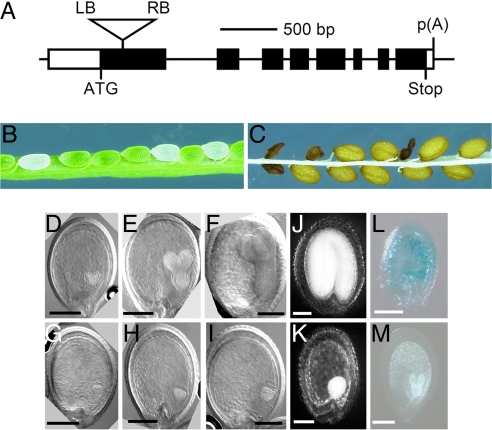
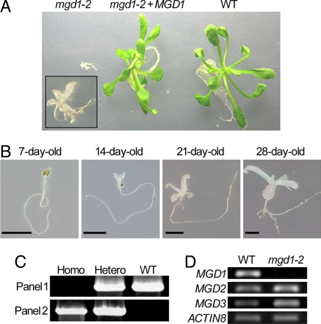
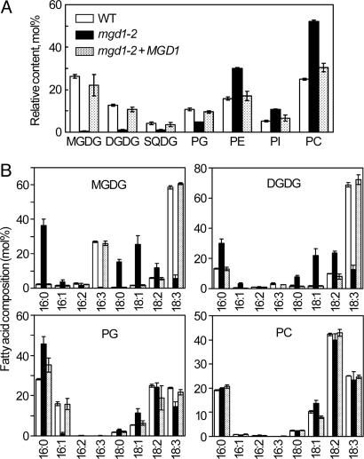
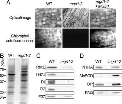
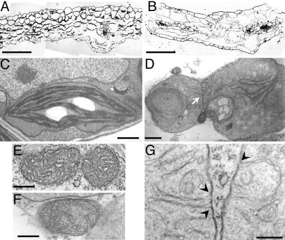
The biogenesis of thylakoid membranes, an indispensable event for the photoautotrophic growth of plants, requires a significant increase in the level of the unique thylakoid membrane lipid monogalactosyldiacylglycerol (MGDG), which constitutes the bulk of membrane lipids in chloroplasts. The final step in MGDG biosynthesis occurs in the plastid envelope and is catalyzed by MGDG synthase. Here we report the identification and characterization of an Arabidopsis mutant showing a complete defect in MGDG synthase 1. The mutant seeds germinated as small albinos only in the presence of sucrose. The seedlings lacked galactolipids and had disrupted photosynthetic membranes, leading to the complete impairment of photosynthetic ability and photoautotrophic growth. Moreover, invagination of the inner envelope, which is not seen in mature WT chloroplasts, was observed in the mutant, supporting an old hypothesis that envelope invagination is a major event in early chloroplast biogenesis. In addition to the defective seedling phenotype, embryo development was arrested in the mutant, although seeds with impaired embryos could germinate heterotrophically. These results demonstrate the importance of galactolipids not only in photosynthetic growth but also in embryogenesis.


Conflict of interest statement

The authors declare no conflict of interest.

Figures

Fig. 1.
Fig. 1.
Identification of the MGD1 knockout mutant mgd1-2. (A) Schematic diagram showing the T-DNA insertion site in mgd1-2. Exons and untranslated regions are represented by filled and open boxes, respectively. Lines between the boxes correspond to introns. The T-DNA insertion site is represented by the inverted triangle in the first exon. LB, left border of the T-DNA; RB, right border of the T-DNA; ATG, translation initiation codon; Stop, translation termination codon; p(A), polyadenylation site. (B and C) Light microscopic images of the dissected green silique (B) and mature seeds (C) of heterozygous mgd1-2 plants. (D–I) Nomarski microscopic images of green (D–F) and white (G–I) seeds removed from self-pollinated siliques of heterozygous mgd1-2 plants. The embryos are observed at heart (D and G), torpedo (E and H) and bent cotyledon (F and I) stages. (J and K) Light microscopic images of the normal (J) and aborted (K) seeds removed from dehydrated siliques of heterozygous mgd1-2 plants. (L and M) Histochemical analysis of the expression of MGD1::GUS (β-glucuronidase) constructs at the globular (L) and heart (M) stages. Developing immature seeds of MGD1::GUS transformants (10) were used for GUS analysis. (Scale bars: 100 μm.)
Fig. 2.
Fig. 2.
Characterization of mgd1-2 seedlings germinated from aborted seeds. (A) mgd1-2, mgd1-2 + MGD1, and WT seedlings (21 days old) on sucrose-containing medium. (Inset) Higher magnification of the mgd1-2 seedling. (B) Growth phenotypes of the mgd1-2 mutant. Homozygous mgd1-2 plants were grown in rotated liquid medium for the indicated times. (Scale bars: 2.5 mm.) (C) PCR analysis of genomic DNA from WT, heterozygous (Hetero), and homozygous (Homo) lines of mgd1-2. (Upper) Amplified bands representing the sequence between the translation initiation site and the second exon of MGD1. (Lower) Amplified bands representing the sequence between the right end of the T-DNA and the second exon of MGD1. (D) Semiquantitative RT-PCR analysis of three atMGD genes in WT and mgd1-2 homozygous plants. ACTIN8 transcripts were analyzed as a loading control.
Fig. 3.
Fig. 3.
Lipid analyses of the mgd1-2 mutant. (A) Composition of polar glycerolipids in WT, mgd1-2, and mgd1-2 + MGD1 plants. SQDG, sulfoquinovosyldiacylglycerol; PG, phosphatidylglycerol; PE, phosphatidylethanolamine; PI, phosphatidylinositol; PC, phosphatidylcholine. Values are mean ± SE from three independent measurements. (B) Fatty acid composition of each lipid extracted from WT (open bars), mgd1-2 (filled bars), and mgd1-2 + MGD1 (stippled bars) plants. Values are mean ± SE from six independent measurements for mgd1-2 and three measurements for WT and mgd1-2 + MGD1 plants.
Fig. 4.
Fig. 4.
Impairment of the photosynthetic system in mgd1-2. (A) Fluorescence microscopic analysis of WT, mgd1-2, and mgd1-2 + MGD1 plants. Bright fluorescence represents chlorophyll accumulation in the chloroplasts. mgd1-2 plants showed no fluorescence. (B) Coomassie brilliant blue staining of an SDS/PAGE of total proteins extracted from WT and mgd1-2 plants. (C and D) Immunoblot analysis of plastidic (C) and extraplastidic (D) proteins from WT and mgd1-2 plants. RbcL, large subunits of ribulose-1,5-bisphosphate carboxylase/oxygenase; NTRA, NADPH-dependent thioredoxin reductase A; MnSOD, manganese-containing superoxide dismutase; PAQ, plasma membrane aquaporin.
Fig. 5.
Fig. 5.
Leaf morphology and ultrastructure of mgd1-2 plastids. (A and B) Light microscopy of WT (A) and mgd1-2 mutant (B) leaf sections. (Scale bars: 100 μm.) (C and D) Electron micrographs of plastids from WT (C) and mgd1-2 (D) leaves. (Scale bars: 1.0 μm.) (E and F) Electron micrographs of mitochondria from WT (E) and mgd1-2 (F). (Scale bars: 0.2 μm.) (G) A close-up of the membrane structures indicated by an arrow in D. Arrowheads indicate the sites of inner envelope invagination. (Scale bar: 0.2 μm.)

Similar articles

Cited by

References

    1. Block MA, Dorne AJ, Joyard J, Douce R. J Biol Chem. 1983;258:13281–13286. - PubMed
    1. Dörmann P, Benning C. Trends Plant Sci. 2002;7:112–118. - PubMed
    1. Lee AG. Curr Biol. 2000;10:R377–R380. - PubMed
    1. Jordan P, Fromme P, Witt HT, Klukas O, Saenger W, Krauss N. Nature. 2001;411:909–917. - PubMed
    1. Loll B, Kern J, Saenger W, Zouni A, Biesiadka J. Nature. 2005;438:1040–1044. - PubMed

Publication types