

Germ cells are essential for sexual dimorphism in the medaka gonad
- PMID: 17940041
- PMCID: PMC2040408
- DOI: 10.1073/pnas.0609932104
Germ cells are essential for sexual dimorphism in the medaka gonad
Abstract
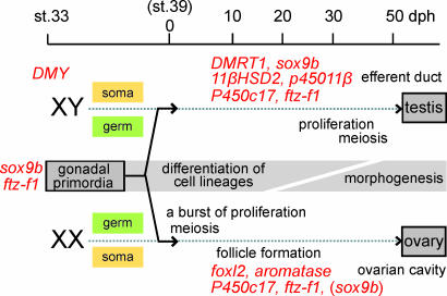
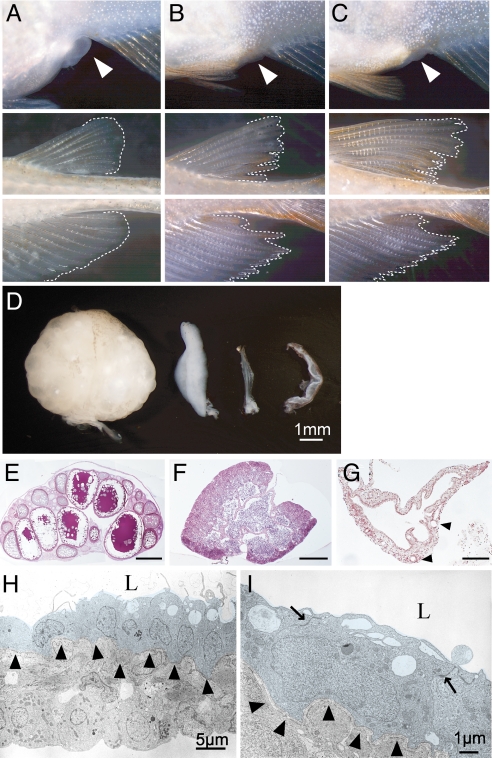
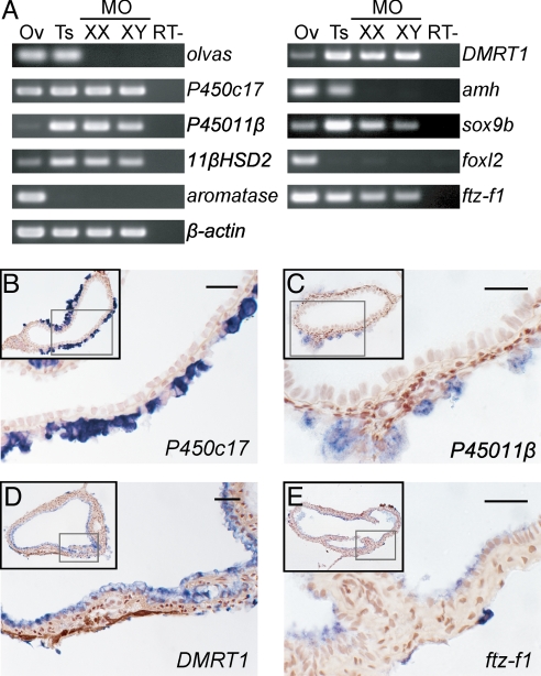
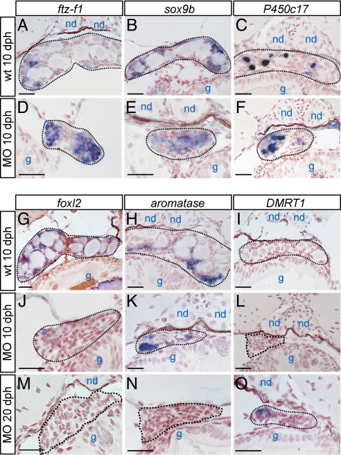
To further elucidate the roles of germ cells in the sex differentiation of gonads, we have used the medaka, a teleost fish, to generate mutants that lack germ cells from the onset of gonadogenesis by the morpholino-mediated knockdown of cxcr4. The resulting germ-cell-deficient medaka show female-to-male sex reversal of their secondary sex characteristics, accompanied by increased levels of androgen and reduced levels of estrogen. A failure to maintain granulosa cells or estrogen-producing cells also occurs at early stages of sex differentiation in the cxcr4 morphants, before the initiation of gonadal morphogenesis. In contrast, androgen-producing cells are unaffected in germ-cell-deficient medaka of either sex. In addition, a single tube-like gonad that expresses male-specific genes is formed in these mutants irrespective of the genetic sex. Significantly, each of these mutant phenotypes occurs in a somatic cell-autonomous manner, suggesting that gonadal somatic cells are predisposed toward male development in the absence of germ cells. This highlights the importance of germ cells in the sexual dimorphism of the gonads.
Conflict of interest statement
The authors declare no conflict of interest.
Figures
Similar articles
-
Estrogen Receptor β2 Oversees Germ Cell Maintenance and Gonadal Sex Differentiation in Medaka, Oryzias latipes.Stem Cell Reports. 2019 Aug 13;13(2):419-433. doi: 10.1016/j.stemcr.2019.07.013. Stem Cell Reports. 2019. PMID: 31412286 Free PMC article.
-
Cross talk between germ cells and gonadal somatic cells is critical for sex differentiation of the gonads in the teleost fish, medaka (Oryzias latipes).Dev Growth Differ. 2008 May;50(4):273-8. doi: 10.1111/j.1440-169X.2008.01015.x. Dev Growth Differ. 2008. PMID: 18366386 Review.
-
Ultrastructure of fetal human gonad before sexual differentiation and during early testicular and ovarian development.J Submicrosc Cytol Pathol. 1990 Jul;22(3):389-400. J Submicrosc Cytol Pathol. 1990. PMID: 2390761
-
Gsdf is not indispensable for male differentiation in the medaka species Oryzias hubbsi.Biochem Biophys Res Commun. 2024 Sep 10;724:150227. doi: 10.1016/j.bbrc.2024.150227. Epub 2024 Jun 6. Biochem Biophys Res Commun. 2024. PMID: 38870865
-
Comparative aspects of gonadal sex differentiation in medaka: a conserved role of developing oocytes in sexual canalization.Sex Dev. 2009;3(2-3):99-107. doi: 10.1159/000223075. Epub 2009 Aug 10. Sex Dev. 2009. PMID: 19684455 Review.
Cited by
-
Figla Favors Ovarian Differentiation by Antagonizing Spermatogenesis in a Teleosts, Nile Tilapia (Oreochromis niloticus).PLoS One. 2015 Apr 20;10(4):e0123900. doi: 10.1371/journal.pone.0123900. eCollection 2015. PLoS One. 2015. PMID: 25894586 Free PMC article.
-
Gonadal Transcriptome Analysis and Sequence Characterization of Sex-Related Genes in Cranoglanis bouderius.Int J Mol Sci. 2022 Dec 13;23(24):15840. doi: 10.3390/ijms232415840. Int J Mol Sci. 2022. PMID: 36555482 Free PMC article.
-
Gonad morphogenesis in vertebrates: divergent means to a convergent end.Annu Rev Cell Dev Biol. 2009;25:457-82. doi: 10.1146/annurev.cellbio.042308.13350. Annu Rev Cell Dev Biol. 2009. PMID: 19807280 Free PMC article. Review.
-
Abundance of Early Embryonic Primordial Germ Cells Promotes Zebrafish Female Differentiation as Revealed by Lifetime Labeling of Germline.Mar Biotechnol (NY). 2019 Apr;21(2):217-228. doi: 10.1007/s10126-019-09874-1. Epub 2019 Jan 22. Mar Biotechnol (NY). 2019. PMID: 30671659 Free PMC article.
-
Induced autoimmunity against gonadal proteins affects gonadal development in juvenile zebrafish.PLoS One. 2014 Dec 1;9(12):e114209. doi: 10.1371/journal.pone.0114209. eCollection 2014. PLoS One. 2014. PMID: 25436775 Free PMC article.
References
-
- Koopman P, Munsterberg A, Capel B, Vivian N, Lovell-Badge R. Nature. 1990;348:450–452. - PubMed
-
- Sinclair AH, Berta P, Palmer MS, Hawkins JR, Griffiths BL, Smith MJ, Foster JW, Frischauf AM, Lovell-Badge R, Goodfellow PN. Nature. 1990;346:240–244. - PubMed
-
- Koopman P, Gubbay J, Vivian N, Goodfellow P, Lovell-Badge R. Nature. 1991;351:117–121. - PubMed
-
- Matsuda M, Nagahama Y, Shinomiya A, Sato T, Matsuda C, Kobayashi T, Morrey CE, Shibata N, Asakawa S, Shimizu N, et al. Nature. 2002;417:559–563. - PubMed
Publication types
MeSH terms
Substances
Associated data
- Actions
- Actions
LinkOut - more resources
Full Text Sources
Research Materials
