. 2007 Oct 16;104(42):16486-91.
doi: 10.1073/pnas.0706847104. Epub 2007 Oct 10.

Polyphosphate kinase 1, a conserved bacterial enzyme, in a eukaryote, Dictyostelium discoideum, with a role in cytokinesis

Affiliations

Polyphosphate kinase 1, a conserved bacterial enzyme, in a eukaryote, Dictyostelium discoideum, with a role in cytokinesis

Haiyu Zhang et al. Proc Natl Acad Sci U S A. .

Abstract

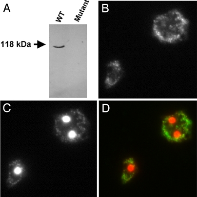
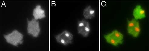
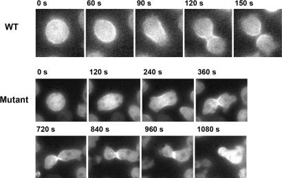
Polyphosphate kinase 1 (PPK1), the principal enzyme responsible for reversible synthesis of polyphosphate (poly P) from the terminal phosphate of ATP, is highly conserved in bacteria and archaea. Dictyostelium discoideum, a social slime mold, is one of a few eukaryotes known to possess a PPK1 homolog (DdPPK1). Compared with PPK1 of Escherichia coli, DdPPK1 contains the conserved residues for ATP binding and autophosphorylation, but has an N-terminal extension of 370 aa, lacking homology with any known protein. Polyphosphate or ATP promote oligomerization of the enzyme in vitro. The DdPPK1 products are heterogeneous in chain length and shorter than those of E. coli. The unique DdPPK1 N-terminal domain was shown to be necessary for its enzymatic activity, cellular localization, and physiological functions. Mutants of DdPPK1, as previously reported, are defective in development, sporulation, and predation, and as shown here, in late stages of cytokinesis and cell division.


Conflict of interest statement

The authors declare no conflict of interest.

Figures

Fig. 1.
Fig. 1.
SDS/PAGE (12%) analysis of purified DdPPK1. Samples were precipitated with 0.015% deoxycholate/5% trichloroacetic acid. Proteins were visualized by Coomassie blue staining. Crude lysate (≈50 μg) and purified fraction (≈3 μg) (Mono Q column) were applied as indicated. Mm, protein molecular mass markers (Bio-Rad).
Fig. 2.
Fig. 2.
Size-exclusion chromatography of DdPPK1. Lysates of D. discoideum DdPPK1 overexpressing cells were prepared and incubated in elution buffer only (A), elution buffer with 10 mM poly P (type 15) (B), or elution buffer with 2 mM ATP (C). After incubating at 0°C for 30 min, 200-ml lysates were applied onto a Tosoh G3000SWXL HPLC column and eluted with the same incubation buffer. Enzyme activity was measured in each fraction. Molecular mass standards were thyroglobulin (670 kDa), bovine gamma globulin (158 kDa), and chicken ovalbumin (44 kDa).
Fig. 3.
Fig. 3.
Poly P products of DdPPK1. Poly P synthesis with 40 ng of DdPPK1 was carried out with [γ-32P]ATP under the optimal conditions for 30 min. Products were purified, treated with exopolyphosphatase (PPX) (as described in Materials and Methods) and separated by 20% PAGE containing 7 M urea, and visualized by PhosphorImager (Molecular Dynamics). P750 is a purified product of EcPPK1, which has a chain length of ≈750 residues. P300, P50, and P30 indicate the position of poly P with different chain lengths. PPi, pyrophosphate. (A) Poly P products before (−) and after (+) PPX-treatment. (B) The region between poly P750 and PPi from gel A is magnified to highlight the chain lengths of poly P.
Fig. 4.
Fig. 4.
Growth of different strains of D. discoideum on a K. aerogenes lawn. D. discoideum cells (20–50) were mixed with K. aerogenes and plated on SM5 agar. Plaque sizes were measured over a 7-day period.
Fig. 5.
Fig. 5.
Cellular localization of DdPPK1. (A) Western blot of anti-EcPPK1 against D. discoideum WT and DdPPK1 mutant (AX2 Ddppk1::bsr) soluble extracts. (B) Immunofluorescent detection of DdPPK1 in WT cells using anti-EcPPK1 antibody. (C) DAPI staining image of the same field as B. (D) The merged image of B and C with anti-EcPPK1 signal in green and DAPI in red. Preimmune serum was tested and was found to be negative.
Fig. 6.
Fig. 6.
Cellular location of N-terminal truncated DdPPK1. Log-phase mutant cells containing pTX-gfp-373-Ddppk1 were fixed on cover slips, stained with DAPI, and visualized under microscope. (A) GFP signal of N-terminal-truncated DdPPK1. (B) DAPI staining image of the same field as A. (C) Merged image of A and B with GFP in green and DAPI staining in red.
Fig. 7.
Fig. 7.
Cytokinesis of mutant cells (AX2 Ddppk1::bsr) expressing GFP-myosin II. Time-lapse images show the localization of GFP-myosin II. The WT cell underwent cytokinesis in 150 s. The mutant cell showed normal GFP-myosin II localization and initiation of cleavage furrow ingression. However, it did not complete cleavage furrow ingression and cytokinesis in 18 min (observed in 16 of 39 cells imaged).

Similar articles

Cited by

References

    1. Kornberg A, Rao NN, Ault-Riché D. Annu Rev Biochem. 1999;68:89–125. - PubMed
    1. Rao NN, Kornberg A. J Bacteriol. 1996;178:1394–1400. - PMC - PubMed
    1. Kuroda A, Nomura K, Ohtomo R, Kato J, Ikeda T, Takiguchi N, Ohtake H, Kornberg A. Science. 2001;293:705–708. - PubMed
    1. Rashid MH, Rao NN, Kornberg A. J Bacteriol. 2000;182:225–227. - PMC - PubMed
    1. Rashid MH, Kornberg A. Proc Natl Acad Sci USA. 2000;97:4885–4890. - PMC - PubMed

Publication types

MeSH terms

Substances

Associated data