. 2007 Dec;13(12):2300-11.
doi: 10.1261/rna.626207. Epub 2007 Oct 16.

Protein-free spliceosomal snRNAs catalyze a reaction that resembles the first step of splicing

Affiliations

Protein-free spliceosomal snRNAs catalyze a reaction that resembles the first step of splicing

Saba Valadkhan et al. RNA. 2007 Dec.

Abstract

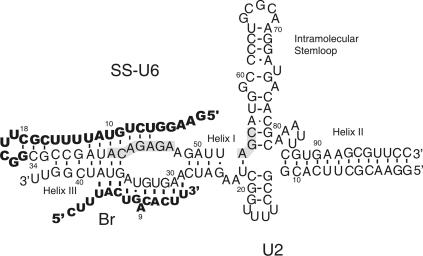
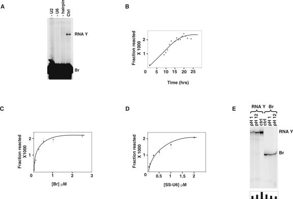
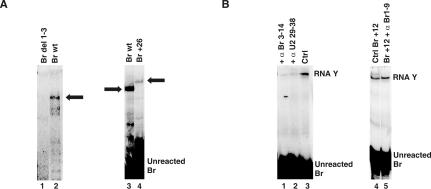
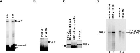
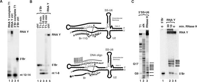
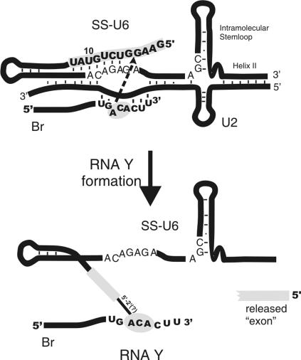
Splicing of introns from mRNA precursors is a two-step reaction performed by the spliceosome, an immense cellular machine consisting of over 200 different proteins and five small RNAs (snRNAs). We previously demonstrated that fragments of two of these RNAs, U6 and U2, can catalyze by themselves a splicing-related reaction, involving one of the two substrates of the first step of splicing, the branch site substrate. Here we show that these same RNAs can catalyze a reaction between RNA sequences that resemble the 5' splice site and the branch site, the two reactants of the first step of splicing. The reaction is dependent on the sequence of the 5' splice site consensus sequence and the catalytically essential domains of U6, and thus it resembles the authentic splicing reaction. Our results demonstrate the ability of protein-free snRNAs to recognize the sequences involved in the first splicing step and to perform splicing-related catalysis between these two pre-mRNA-like substrates.


Figures

FIGURE 1.
FIGURE 1.
U6/U2 base-paired complex and splicing substrates. The splicing substrates are shown in bold letters. Base-pairing interactions between Br and U2 and the bulged A are shown. SS is covalently attached to the 5′ end of U6 via a linker sequence and a hairpin (nt 11–25 of the bold region of SS-U6). The positions of the U6 intramolecular stem–loop and U6/U2 helices I, II, and III are shown. The highlighted areas mark the invariant domains of U6. The numbering of the U6 part of SS-U6 and U2 refers to human numbering. Base-pairing interactions are shown by black bars. The mutually exclusive base-pairing interactions shown between U6, U2, and SS in helix III region do not form concurrently.
FIGURE 2.
FIGURE 2.
Characterization of RNA Y formation. (A) Requirements for RNA Y formation. Position of RNA Y and Br is shown to the right. The reactions in lanes marked “−U2” and “−U6” lack U2 and U6, respectively. Lane marked “−hairpin” contains a SS-U6 in which the hyperstable hairpin structure linking SS to U6 is replaced by a random sequence. Lane marked “Ctrl” contains a typical RNA Y formation reaction. (B) Time course of RNA Y formation. (C) Dependence of RNA Y formation on Br concentration. (D) Dependence of RNA Y formation on the concentration of SS-U6. (E) Effect of alkaline and acidic pH on formed RNA Y. Locations of RNA Y and Br are shown to the right. The pH of the buffer used is shown on top. The amount of RNA Y in each lane is shown in the graph at the bottom of the gel.
FIGURE 3.
FIGURE 3.
Sequence and ionic requirements for RNA Y formation. (A) Requirement of SS for RNA Y formation. The position of RNA Y and unreacted Br is shown to the right (Wt, wild type). (B) Effect of point mutations in SS on RNA Y formation (wt, wild type). Values represent the average of at least three independent experiments. The diagram illustrates the potential base-pairing interactions between SS and ACAGAGA in SS-U6. (C) Cationic requirement for RNA Y formation. Position of RNA Y and unreacted Br is shown to the side of gel panels. Identity and concentration of the sole cation used in the reaction are shown above each lane. Lane labeled “MgCl2+EDTA+Co(NH3)6” was initially folded in 60 mM MgCl2 and later received 0.5 M NaCl, 5 mM Co(NH3)6, and 40 mM EDTA. (D) Effect of mutations in U6 on RNA Y formation (wt, wild type). Values represent the average of three independent experiments. (E) Mutational study of the role of ACAGAGA in RNA Y formation. Position of RNA Y is shown to the right. RNA species used in each reaction is shown above each lane (Ctrl, control).
FIGURE 4.
FIGURE 4.
Br requirements in RNA Y formation. (A) Effect of changing the length of Br on RNA Y formation. The positions of RNA Y made with wild type (wt) and a Br species that has 26 extra nucleotides added to the 5′ end (Br+26) is marked by arrows. Location of unreacted Br is indicated. (B) Effect of blocking the base-pairing interaction between Br and U2 on RNA Y formation. Positions of RNA Y and unreacted Br are indicated to the right (Ctrl, control).
FIGURE 5.
FIGURE 5.
Determining the RNA sequences present in RNA Y. (A) 5′ or 3′ end-labeled Br in RNA Y formation. Positions of RNA Y and unreacted Br are shown to the right. (B) 5′ or 3′ end-labeled SS-U6 in RNA Y formation; bracket indicates the position of the gel piece that was cut to analyze for the presence of RNA Y. Position of RNA Y is shown to the right. (C) Relabeling of the RNAs eluted from the region marked by bracket in (B). Position of RNA Y is shown to the right. The SS-U6 used is shown above each lane. Arrows point to unreacted 5′-labeled SS-U6 that was co-purified with the unlabeled sample. (D) RNA Y formation with SS-U6 containing a 17-nt extension. Position of wild type (wt) SS-U6 and the SS-U6 containing a 17-nt extension at the 5′ end are shown to the right. Position of RNA Y formed is shown to the left.
FIGURE 6.
FIGURE 6.
Defining the sites of linkage in Br and SS in RNA Y. (A) Complete RNase T1 digestion of RNA Y made with 3′-labeled Br. Locations of 3′Br, RNA Y, and the nucleotides 12–14 fragment resulting from complete T1 digestion are shown to the right. (B) Complete RNase T1 digestion of RNA Y made with 5′-labeled Br. Positions of RNA Y, 5′Br, and the nucleotides 1–8 fragment resulting from T1 digestion are shown to the right (min, minutes of RNase T1 digestion). The identity of the RNA species is shown on top. Top diagram depicts the result of the complete RNA digestions. Arrowheads point to the sites of T1 digestion. Fragments released after complete T1 digestion are highlighted in gray. (C) RNase H digestion of 5′-labeled SS-U6 (left panel) and RNA Y (right panel) with a DNA oligo complementary to the first 18 nt of SS (T1, partial RNase T1 digestion; alk, partial alkali hydrolysis ladder). Positions of fragments released by RNase T1 are shown to the left. Positions of RNA Y and 5′Br are shown to the right. Fragments released by RNase H treatment are marked by arrows (min, minutes of RNase H digestion). The bottom diagram illustrates the outcome of RNase H digestion of RNA Y. The DNA oligonucleotide is shown in gray. The arrows indicate the 7 most downstream sites of RNase H digestion (see text). The location of the DNA oligo, SS-U6, U2 and Br is shown. The asterisk marks the location of the radioactive label. The dotted arrow shows the likely site of the linkage between the two molecules.
FIGURE 7.
FIGURE 7.
RNA Y. Locations of Br, SS-U6, U2, U6 intramolecular stem–loop, and U6/U2 helix II are shown. Gray areas mark the regions that contain the site of linkage between Br and SS. Arrow indicates the direction of the nucleophilic reaction between putative reacting nucleotides involved in RNA Y formation.

Similar articles

Cited by

References

    1. Anderson, P., Monforte, J., Tritz, R., Nesbitt, S., Hearst, J., Hampel, A. Mutagenesis of the hairpin ribozyme. Nucleic Acids Res. 1994;22:1096–1100. doi: 10.1093/nar/22.6.1096. - DOI - PMC - PubMed
    1. Brow, D.A. Allosteric cascade of spliceosome activation. Annu. Rev. Genet. 2002;36:333–360. - PubMed
    1. Burke, J.M., Butcher, S.E., Sargueil, B. Structural analysis and modification of the hairpin ribozyme. In: Eckstein F., Lilley D.M.J., editors. Nucleic acids and molecular biology. Springer; Berlin, Germany: 1996. pp. 129–143.
    1. Collins, C.A., Guthrie, C. The question remains: Is the spliceosome a ribozyme? Nat. Struct. Biol. 2000;7:850–854. - PubMed
    1. Cowan, J.A. Metallobiochemistry of RNA. Co(NH3)6 3+ as a probe for Mg2+(aq) binding sites. J. Inorg. Biochem. 1993;49:171–175. - PubMed

Publication types

LinkOut - more resources