. 2007 Dec;13(12):2287-99.
doi: 10.1261/rna.562907. Epub 2007 Oct 16.

Alternately spliced WT1 antisense transcripts interact with WT1 sense RNA and show epigenetic and splicing defects in cancer

Affiliations

Alternately spliced WT1 antisense transcripts interact with WT1 sense RNA and show epigenetic and splicing defects in cancer

Anthony R Dallosso et al. RNA. 2007 Dec.

Abstract

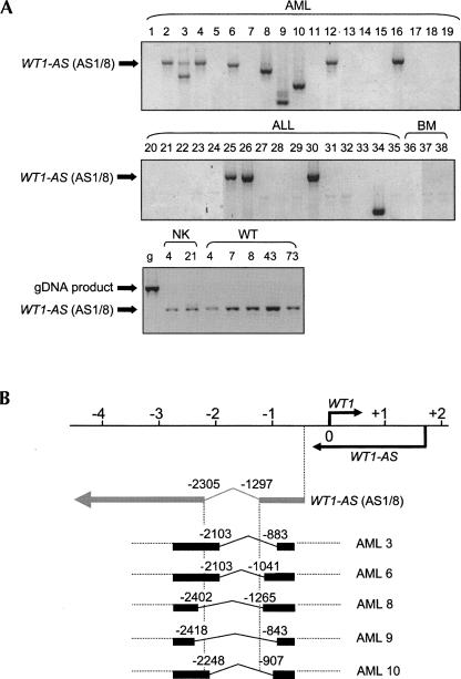
Many mammalian genes contain overlapping antisense RNAs, but the functions and mechanisms of action of these transcripts are mostly unknown. WT1 is a well-characterized developmental gene that is mutated in Wilms' tumor (WT) and acute myeloid leukaemia (AML) and has an antisense transcript (WT1-AS), which we have previously found to regulate WT1 protein levels. In this study, we show that WT1-AS is present in multiple spliceoforms that are usually expressed in parallel with WT1 RNA in human and mouse tissues. We demonstrate that the expression of WT1-AS correlates with methylation of the antisense regulatory region (ARR) in WT1 intron 1, displaying imprinted monoallelic expression in normal kidney and loss of imprinting in WT. However, we find no evidence for imprinting of mouse Wt1-as. WT1-AS transcripts are exported into the cytoplasm and form heteroduplexes with WT1 mRNA in the overlapping region in WT1 exon 1. In AML, there is often abnormal splicing of WT1-AS, which may play a role in the development of this malignancy. These results show that WT1 encodes conserved antisense RNAs that may have an important regulatory role in WT1 expression via RNA:RNA interactions, and which can become deregulated by a variety of mechanisms in cancer.


Figures

FIGURE 1.
FIGURE 1.
Antisense transcripts at the human and mouse WT1 loci. (A) Human WT1 antisense RNAs. The top scale is numbered in kilobase pairs relative to the major WT1 transcriptional start site (Hofmann et al. 1993), which is indicated by the right-angled black arrow, along with the furthest upstream start site identified by Fraizer et al. (1994) (right-angled dashed arrow) and the WT1-AS transcriptional start site (Malik et al. 1995) (right-angled arrow below line). Below are shown the structures of previously published WT1-AS RNAs (double lines), WT1-AS transcripts identified by cDNA library screening (AS1, AS8, AS9; this study; gray lines) and human ESTs representing WT1-AS RNAs (black lines). (B) Mouse Wt1 antisense RNAs. The top scale is numbered in kilobase pairs relative to the major Wt1 transcriptional start site. Below are shown the structures of three mouse ESTs representing Wt1-as RNAs. Transcripts are annotated with GenBank accession number and tissue source. Vertical dotted lines indicate shared donor and acceptor splice sites and alignment with WT1/Wt1 transcriptional start sites.
FIGURE 2.
FIGURE 2.
Expression of human WT1-AS in normal and malignant tissues. (A) Ribonuclease protection assay. Total RNA was hybridized to a probe corresponding to sequence −127 to +83 of WT1 (shaded box, left), synthesized to detect sense or antisense transcripts (shown above autoradiograph of gel of protected fragments, right). (P) Probe; (P+) probe plus RNase; (1) WT1-expressing cell line 7.92; (2) WT1 nonexpressing line 17.94. Positions of molecular weight markers are shown on the right. Full-length protected probe in antisense lane 1 indicates expression of WT1-AS from exon 1. (B) Strand–specific RT–PCR for human WT1-AS. Fetal kidney cDNA was synthesized using primers specific for WT1 antisense RNA (WT1AScsyn) or WT1 sense RNA (WT1csyn) and then amplified using the following primer pairs: (I) Primers across the AS1/8 splice (WITKBF1 and WITKBR1), (II) Primers within WT1 exon 1 (WT15 and WT8), (III) Primers spanning WT1 exon 1 and intron 1 (WT15 and WTEX1AS). (+, –) Reactions with and without reverse transcriptase, respectively. Specific signal was seen for cDNA made in the antisense (AS) direction in all cases, but only in exon 1 for sense cDNA (S). (C) Expression of WT1-AS in human tissues and Wilms’ tumor. Real-time RT–PCR of cDNA from human term placenta (P), fetal spleen (S), fetal liver (L), fetal brain (B), and fetal kidney (K), kidney adjacent to Wilms’ tumors 28 and 53 (K28 and K53) and Wilms’ tumors (T28, T53, T43, T57, and T62), with primers specific for WT1 (WTRQF and WTRQR) and WT1-AS spliceoforms AS1/AS8 (WT1-ASRQF and WT1-ASRQR), AS9 (AS9x2rnaF and AS9x3rnaR), AI648530 (AI648530rnaF and AI648530rnaR). Results were normalized relative to the housekeeping gene TBP (TBPRQF and TBPRQR). Error bars show the range of duplicate measurements; results are representative of two separate experiments. WT1-AS expression paralleled WT1 in all tissues except term placenta, where WT1 was expressed at the lowest detectable level. Primer sequences are shown in Table 1.
FIGURE 3.
FIGURE 3.
Expression of mouse Wt1-as. (A) Mapping expression of mouse Wt1-as. Strand-specific RT–PCR for mouse Wt1-as using fetal kidney cDNA synthesized using: (I) Primers in the first 2 exons of mouse Wt1-as (552314S and 552314AS); (II) Primers spanning Wt1 exon 1 and intron 1 (WT1 and MWT1); (III) Primers within Wt1 intron 1 (MWT3A and MWT4A). In each case, specific signal was only seen for cDNA made in the antisense (AS) direction and not the sense (S). (+, –) Reactions with and without reverse transcriptase, respectively. (B) Expression of Wt1-as in mouse tissues. Real-time RT–PCR expression analysis using cDNA from mouse fetal tissues (E17.5 d); placenta (P), spleen (S), brain (B), liver (L), and kidney (K), and postnatal (P3, 7, 22, and 65 d) kidney (K), with primers specific for Wt1-as (MASPLICE1RQF and MASPLICE1RQR) and Wt1 (MWTRQF2 and MWTRQR2), normalized relative to the housekeeping gene Tbp (MTBPRQF and MTBPRQR). Error bars show the range of duplicate measurements; results are representative of two separate experiments. Wt1-as was coexpressed with Wt1 in fetal placenta, spleen, and kidney. Primer sequences are shown in Table 1.
FIGURE 4.
FIGURE 4.
Epigenetic regulation of WT1-AS. (A) Allelic expression and methylation of WT1-AS in Wilms’ tumor. (Left) Allelic expression of the WT1-AS AS9 transcript (AS9-FOR and AS9-REV) in paired normal kidney (NK) and Wilms’ tumor (WT), using RT–PCR across a DdeI restriction site polymorphism. DdeI digestion gives two allelic bands (A1 and A2) and one constant band (arrowed). (+, –) Reactions with and without reverse transcriptase, respectively. DNA lane shows the alleles amplified from corresponding genomic DNA. WT1-AS AS9 was monoallelically expressed in NK and biallelically expressed in WT. (Right) Methylation of the ARR in the NK and WT assayed by methylation-sensitive Southern blotting of corresponding genomic DNAs, showing differential allelic methylation in NK (731 and 542 bp bands) and complete hypomethylation in WT (542 bp band only). (B) Azacytidine—mediated modulation of transcription at the human WT1 locus. (Left) Agarose gel (HPRT; HPRT5′ and HPRT3′) or autoradiograph (WT1-AS spliceoforms AS1/8; WITKBF1 and WITKBR1 and AS9; AS9-FOR and AS9-REV) of RT–PCR reactions using total RNA extracted from the Wilms’ tumor cell line 17.94, treated with (+AZA) or without (−AZA) 5- azacytidine for 4 d. (+, –) Reactions with and without reverse transcriptase, respectively. AZA treatment induced expression of both WT1-AS spliceoforms. (Right) Methylation-sensitive Southern blot of corresponding 17.94 cell genomic DNAs showed partial demethylation at the Antisense Regulatory Region (ARR) in WT1 intron 1 after 4 d of AZA treatment, as demonstrated by the appearance of unmethyated bands at 731 and 542 bp. (C) Wt1 (MWTRQF2 and MWTRQR2) and Wt1-as (MASPLICE1RQF and MASPLICE1RQR) expression in Pax6 Sey-H mice. RNA expression was assayed by real-time PCR of normal kidney cDNA from wild-type mice (WT) and mice heterozygous for the Pax6 Sey-H deletion (which includes Wt1), inherited from father (Pat KO) or mother (Mat KO). Expression levels are shown as fold differences compared with wild-type controls, normalized relative to the housekeeping gene Tbp (MTBPRQF and MTBPRQR). Each mouse kidney cDNA was assayed twice and the results show the mean expression values +/− SEM for five WT, five Pat KO, and two Mat KO mice. Wt1-as expression was maintained in both Pat and Mat KO mice, indicating no imprinting of Wt1-as in mice. Primer sequences are shown in Table 1.
FIGURE 5.
FIGURE 5.
Subcellular localization of WT1-AS transcripts and RNA duplex analysis. (A) Subcellular localization. The 7.92 cells were fractionated into cytoplasmic (C) and nuclear (N) extracts as described in Materials and Methods. (Left) Agarose gel of total RNA, demonstrating unprocessed pre-rRNA in the nuclear fraction. (Right) Table showing distribution of WT1 antisense RNAs, protein-coding RNAs, and other noncoding RNAs in the cytoplasmic fraction, as assessed by real-time RT–PCR. Results show that WT1-AS is transported into the cytoplasm. (B) RNA duplex analysis. Total RNA was made from 7.92 cells using nondenaturing conditions, to extract intact RNA:RNA duplexes. Panels I to V show agarose gel electrophoresis of RT–PCR products from the WT1 locus made from RNA incubated in the presence (lanes 2,3) or absence (lane 1) of RNase A, followed by first-strand cDNA synthesis in the presence (lanes 1,3) or absence (lane 2) of reverse transcriptase. I: Upstream (WITKBR1 + WITKBF2), II: Exon 1 (WT14 + WT16), III: Exon 1a (CPG-USTR + CPG-AS), IV: Exons 6–10 (WT2 + WT4), V: Exon 10 UTR (WT6 +WT7). Above is a schematic of WT1, WT1-AS, and AWT1 transcripts with the positions of the primers shown as arrowheads. Amplification products are only visible in lane 3 if duplex formation spanning the primer pair has “protected” the amplicon from RNase A digestion. The only protected product is in panel II, suggesting RNA:RNA duplex formation within the WT1 exon 1 region. Primer sequences are shown in Table 1.
FIGURE 6.
FIGURE 6.
Expression of aberrant WT1-AS spliceoforms in human leukaemia. (A) Agarose gel of RT–PCR for WT1-AS expression in samples from acute myeloid leukaemia (AML; 1–19), acute lymphoblastic leukaemia (ALL; 20–35), normal bone marrow (BM; 36–38), normal kidney (NK; 4 and 21), and Wilms’ tumor (WT; 4, 7, 8, 43, 73). Nested RT–PCR was performed with primers WITKBR1 and WITKBF3A, followed by WT18 and WITKBF3B, which produced the expected 793-bp product in most tissues for AS1/8 (arrowed), but demonstrated abnormal smaller products in AMLs 3, 6, 8, 9, 10, and ALL 34. No abnormal-sized products were detected in Wilms’ tumors. The larger sized PCR product from genomic DNA (g; first lane, bottom) shows that all cDNA products derived from spliced WT1-AS RNAs. Primer sequences are shown in Table 1. (B) The structure of the normal AS1/8 transcripts are shown relative to the WT1 transcriptional start site, and underneath the structures of the new spliceoforms isolated from AMLs 3, 6, 8, 9, and 10, with the product sizes and positions of their splices indicated. None of the splice junctions coincided with those of the major WT1-AS RNAs found in normal tissues.

Similar articles

Cited by

References

    1. Brannan, C.I., Dees, E.C., Ingram, R.S., Tilghman, S.M. The product of the H19 gene may function as an RNA. Mol. Cell. Biol. 1990;10:28–36. - PMC - PubMed
    1. Brightwell, G., Poirier, V., Cole, E., Ivins, S., Brown, K.W. Serum-dependent and cell cycle-dependent expression from a cytomegalovirus-based mammalian expression vector. Gene. 1997;194:115–123. - PubMed
    1. Brown, K.W., Watson, J.E., Poirier, V., Mott, M.G., Berry, P.J., Maitland, N.J. Inactivation of the remaining allele of the WT1 gene in a Wilms’ tumour from a WAGR patient. Oncogene. 1992;7:763–768. - PubMed
    1. Brown, K.W., Wilmore, H.P., Watson, J.E., Mott, M.G., Berry, P.J., Maitland, N.J. Low frequency of mutations in the WT1 coding region in Wilms’ tumor. Genes Chromosomes Cancer. 1993;8:74–79. - PubMed
    1. Caballero, O.L., Souza, S.J., Brentani, R.R., Simpson, A.J. Alternative spliced transcripts as cancer markers. Dis. Markers. 2001;17:67–75. - PMC - PubMed

Publication types