Review
. 2007 Jul-Aug;18(4):248-59.

Magnetic resonance imaging of the coronary arteries

Affiliations
Review

Magnetic resonance imaging of the coronary arteries

S C Gerretsen et al. Cardiovasc J Afr. 2007 Jul-Aug.

Abstract

Despite progress in prevention and early diagnosis, coronary artery disease (CAD) remains one of the leading causes of mortality in the world. For many years, invasive X-ray coronary angiography has been the method of choice for the diagnosis of significant CAD. However, up to 40% of patients referred for elective X-ray coronary angiography have no clinically significant stenoses. These patients still remain subjected to the potential risks of X-ray angiography. As an alternative, magnetic resonance imaging (MRI) is currently one of the most promising techniques for noninvasive imaging of the coronary arteries. Over the past two decades, many technical developments have been implemented that have led to major improvements in coronary MRI. Nowadays, both anatomical and functional information can be obtained with high temporal and spatial resolution and good image quality. In this review we will discuss the technical foundations and current status of clinical coronary MRI, and some potential future applications.


Figures

Fig. 1.
Fig. 1.
Three-tesla images of the right coronary artery (RCA). (A) Three-dimensional gradient echo sequence of the RCA using a T2 pre-pulse and fat saturation. (B) Dual inversion recovery (DIR) and fat-saturated gradient echo sequence of the RCA vessel wall. Both images show a high signal-to-noise ratio (SNR). The coronary vessel wall is clearly visible in B (arrows). Ao: aorta. (Courtesy M Stuber, Johns Hopkins University, Baltimore, MD, USA.)
Fig. 2.
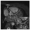
Fig. 2.
(A) Targeted images of the left anterior descending artery (LAD), (B) right coronary artery (RCA) and (C) whole-heart scan in healthy volunteers. With the targeted approach, high-resolution images of the coronary arteries and some branches can be obtained in a relatively short scan time. By using a whole-heart approach, all coronary arteries are imaged in one scan, and the data set allows for post-processing and display of anatomy similar to multi-detector computed tomography. LCX: left circumflex artery, Ao: aorta.
Fig. 3.
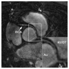
Fig. 3.
Three-dimensional balanced steady-state free precession (bSSFP) coronary MRI at 1.5 T in a 51-year-old female with aberrant right coronary artery (RCA). The RCA originates from the left coronary artery sinus and traverses between the aorta (Ao) and the right ventricular outflow tract (RVOT). LM indicates the left main stem.
Fig. 4.
Fig. 4.
(A) Targeted Cartesian balanced steady-state free precession (bSSFP) and (B) whole-heart source image in a patient with a coronary arteriovenous fistula. Note the enlarged LCX compared to the left anterior descending artery (LAD) and right coronary artery (RCA). There is a connection (arrowhead) between the left circumflex artery (LCX) and the coronary sinus. GCV: great cardiac vein, Ao: aorta.
Fig. 5.
Fig. 5.
Post-processed whole-heart scan of a 12-year-old boy diagnosed with Kawasaki disease. Dilatation of the left and right coronary artery can be seen. Note the kinking and aneurysmatic distal part (diameter 4.4 mm) of the left main stem (LM) in comparison with the diameter of the ostium (diameter 2.8 mm). Ao: aorta, LAD: left anterior descending artery, LCX: left circumflex artery, RCA: right coronary artery.
Fig. 6.
Fig. 6.
Correlation between coronary X-ray angiography and MRI. X-ray angiography (A) and a targeted Cartesian balanced steady-state free precession (bSSFP) MR sequence (B) in a 62-year-old patient with left coronary artery disease. X-ray angiography (C) and radial bSSFP images (D) in a different 53-year-old patient with stable angina. A diffuse, long and severe stenosis of the proximal right coronary artery (RCA) can be seen with both techniques. In both cases, there is a good correlation between angiography and MRI (arrows).
Fig. 7.
Fig. 7.
Coronary angiogram (A, B) and targeted Cartesian balanced steady-state free precession (bSSFP) MRI with mid-diastolic (C) and systolic imaging (D) in a 52-year-old man with acute anterior wall myocardial infarction. Coronary angiography revealed single-vessel disease of the left anterior descending artery (LAD). In A, distal occlusion of the LAD can be seen (arrow). B, systolic squeezing of the intramyocardial mid-LAD (arrowheads). C, the proximal and mid-segment of the LAD can be seen during diastole, however, this segment disappears during systole (D, arrowheads). Ao: aorta, LM: left main stem.
Fig. 8.
Fig. 8.
Radial balanced steady-state free precession (bSSFP) (A) and vessel wall scan (B) of the right coronary artery (RCA) in a 62-year-old healthy female. The proximal and middle parts of the RCA were free of atherosclerotic disease. The vessel wall (arrowheads) is well delineated, thin and has a uniform signal intensity. BSSFP (C) and vessel wall scan (D) in a 59-year-old female without history of coronary artery disease. In D, note the thickening and bright signal intensity of the posterior wall of the RCA (arrowhead) compared to the anterior wall (arrows).

References

    1. Thom T, Haase N, Rosamond W, Howard VJ, Rumsfeld J, Manolio T. et al. Heart disease and stroke statistics − 2006 update: a report from the American Heart Association Statistics Committee and Stroke Statistics Subcommittee. Circulation. 2006;113:e85–e151. - PubMed
    1. Mackay J, Mensah GA. The Atlas of Heart Disease and Stroke. 1st edn. Geneva: World Health Organization; 2004.
    1. Kim WY, Danias PG, Stuber M, Flamm SD, Plein S, Nagel E. et al. Coronary magnetic resonance angiography for the detection of coronary stenoses. N Engl J Med. 2001;345:1863–1869. - PubMed
    1. Budoff MJ, Georgiou D, Brody A, Agatston AS, Kennedy J, Wolfkiel C. et al. Ultrafast computed tomography as a diagnostic modality in the detection of coronary artery disease: a multicenter study. Circulation. 1996;93:898–904. - PubMed
    1. Runge VM. Safety of approved MR contrast media for intravenous injection. J Magn Reson Imag. 2000;12:205–213. - PubMed

Publication types

MeSH terms