

The effect of fibroblast growth factor-2 on autologous osteochondral transplantation
- PMID: 17940768
- PMCID: PMC2899235
- DOI: 10.1007/s00264-007-0459-x
The effect of fibroblast growth factor-2 on autologous osteochondral transplantation
Abstract
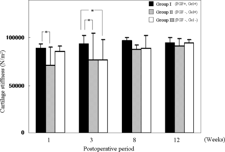
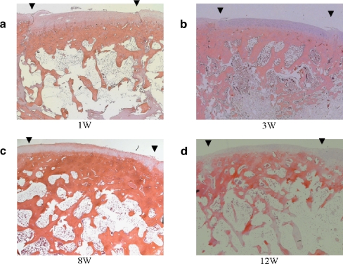
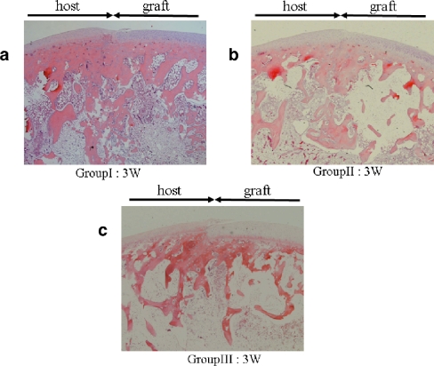
In this study, we performed a mechanical analysis of the effect of fibroblast growth factor-2 (FGF-2) on autologous osteochondral transplantation in a rabbit model. A full-thickness cartilage defect (diameter: 5 mm; depth: 5 mm) made in the right femoral condyle was treated with osteochondral transplantation using an osteochondral plug (diameter: 6 mm; depth: 5 mm) taken from the left femoral condyle. The animals were divided into three groups: Group I, the defect was filled with 0.1 ml of gelatin hydrogel containing 1 microg of FGF-2; Group II, the defect was filled with 0.1 ml of gelatin hydrogel only; Group III, the defect was left untreated. Thereafter, osteochondral plugs were transplanted and the transplanted osteochondral grafts were evaluated mechanically and histologically at postoperative weeks 1, 3, 8 and 12. The structural property of the osteochondral graft was significantly greater in Group I than in Groups II and III at postoperative week 3. Histological analysis at 3 weeks revealed a tendency towards increased subchondral bone trabeculae in Group I compared with the other groups. Autologous osteochondral grafts transplanted with gelatin hydrogel containing FGF-2 acquired adequate stiffness at an early postoperative phase.
Le but de ce travail est de mettre en évidence les effets du « fibroblast growth factor-2 » (FGF-2) sur les greffes autologues ostéo cartilagineuses chez le lapin. Une lésion cartilagineuse de 5 mm de diamètre et de 5 mm de profondeur a été réalisée sur l’extrémité inférieure du fémur droit du lapin. Le traitement a été réalisé par une greffe ostéo cartilagineuse, utilisant un implant ostéochondral de type bouchon de 6 mm de diamètre et de 5 mm d’épaisseur prélevé sur le condyle fémoral gauche. Les animaux ont été divisés en 3 groupes : groupe 1 le défect étant traité par mise en place d’un hydrogel de gélatine de 0.1ml et contenant 1 fEg FGF-2. Le groupe 2 est traité seulement avec un gel de gélatine hydrogel isolé et le groupe 3 n’a pas été traité. Par la suite, les transplants ont été prélevés, et évalués mécaniquement, histologiquement à 1, 3, 8 et 12 semaines post-opératoires. En trois semaines, l’aspect structurel de la greffe est bien meilleur dans le groupe 1 que dans les groupes 2 et 3. Néanmoins, en ce qui concerne l’analyse histologique, toujours à 3 semaines, il existe une tendance à l’amélioration de l’ossification sous chondrale avec ossification trobéculaire. En conclusion, la transplantation de greffes ostéo autologues, de greffes ostéo cartilagineuses en association avec un hydrogel de gélatine contenant du FGF-2, permet d’avoir une meilleure résistance du transplant dans la phase post-opératoire précoce.
Figures
Similar articles
-
The structural properties of an osteochondral cylinder graft-recipient construct on autologous osteochondral transplantation.Arthroscopy. 2006 Apr;22(4):422-7. doi: 10.1016/j.arthro.2005.09.025. Arthroscopy. 2006. PMID: 16581455
-
Histologic analysis of the implanted cartilage in an exact-fit osteochondral transplantation model.Arthroscopy. 2001 Sep;17(7):747-51. doi: 10.1053/jars.2001.24705. Arthroscopy. 2001. PMID: 11536095
-
The use of a single osteochondral autograft plug in the treatment of a large osteochondral lesion in the femoral condyle: an experimental study in sheep.Am J Sports Med. 2006 Feb;34(2):247-55. doi: 10.1177/0363546505279914. Epub 2005 Oct 11. Am J Sports Med. 2006. PMID: 16219943
-
The effect of platelet-rich plasma on autologous osteochondral transplantation: an in vivo rabbit model.J Bone Joint Surg Am. 2013 Dec 18;95(24):2185-93. doi: 10.2106/JBJS.L.01497. J Bone Joint Surg Am. 2013. PMID: 24352772
-
[Autologous osteochondral transplantation].Acta Orthop Traumatol Turc. 2007;41 Suppl 2:70-8. Acta Orthop Traumatol Turc. 2007. PMID: 18180587 Review. Turkish.
Cited by
-
Platelet-Rich Plasma and Hyaluronic Acid Are Not Synergistic When Used as Biological Adjuncts with Autologous Osteochondral Transplantation.Cartilage. 2018 Jul;9(3):321-328. doi: 10.1177/1947603517690022. Epub 2017 Feb 8. Cartilage. 2018. PMID: 29156980 Free PMC article.
-
Strategies for osteochondral repair: Focus on scaffolds.J Tissue Eng. 2014 Jul 8;5:2041731414541850. doi: 10.1177/2041731414541850. eCollection 2014. J Tissue Eng. 2014. PMID: 25343021 Free PMC article. Review.
-
Current and future options for dental pulp therapy.Jpn Dent Sci Rev. 2019 Nov;55(1):5-11. doi: 10.1016/j.jdsr.2018.09.001. Epub 2018 Sep 29. Jpn Dent Sci Rev. 2019. PMID: 30733839 Free PMC article. Review.
-
Ascorbic acid iontophoresis for chondral gain in rats with arthritis.Acta Ortop Bras. 2014;22(4):202-5. doi: 10.1590/1413-78522014220400769. Acta Ortop Bras. 2014. PMID: 25246850 Free PMC article.
-
Seamless and early gap healing of osteochondral defects by autologous mosaicplasty combined with bioactive supramolecular nanofiber-enabled gelatin methacryloyl (BSN-GelMA) hydrogel.Bioact Mater. 2022 Apr 5;19:88-102. doi: 10.1016/j.bioactmat.2022.03.038. eCollection 2023 Jan. Bioact Mater. 2022. PMID: 35441114 Free PMC article.
References
-
- Chow JCY, Hantes ME, Houle JB, Zalavras CG. Arthroscopic autogenous osteochondral transplantation for treating knee cartilage defects: a 2- to 5-year follow-up study. Arthroscopy. 2004;20:681–690. - PubMed
-
- Hangody L, Füles P. Autologous osteochondral mosaicplasty for the treatment of full-thickness defects of weight-bearing joints: ten years of experimental and clinical experience. J Bone Joint Surg Am. 2003;85:S25–S32. - PubMed
MeSH terms
Substances
LinkOut - more resources
Full Text Sources
Medical
