Requirement of TrkB for synapse elimination in developing cerebellar Purkinje cells
- PMID: 17940915
- PMCID: PMC3303929
- DOI: 10.1007/s11068-006-9002-z
Requirement of TrkB for synapse elimination in developing cerebellar Purkinje cells
Abstract
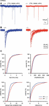
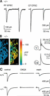
The receptor tyrosine kinase TrkB and its ligands, brain-derived neurotrophic factor (BDNF) and neurotrophin-4/5 (NT-4/5), are critically important for growth, survival and activity-dependent synaptic strengthening in the central nervous system. These TrkB-mediated actions occur in a highly cell-type specific manner. Here we report that cerebellar Purkinje cells, which are richly endowed with TrkB receptors, develop a normal morphology in trkB-deficient mice. Thus, in contrast to other types of neurons, Purkinje cells do not need TrkB for dendritic growth and spine formation. Instead, we find a moderate delay in the maturation of GABAergic synapses and, more importantly, an abnormal multiple climbing fiber innervation in Purkinje cells in trkB-deficient mice. Thus, our results demonstrate an involvement of TrkB receptors in synapse elimination and reveal a new role for receptor tyrosine kinases in the brain.
Figures


Similar articles
-
TrkB is necessary for pruning at the climbing fibre-Purkinje cell synapse in the developing murine cerebellum.J Physiol. 2007 Jul 15;582(Pt 2):629-46. doi: 10.1113/jphysiol.2007.133561. Epub 2007 Apr 26. J Physiol. 2007. PMID: 17463037 Free PMC article.
-
TrkB receptor signaling is required for establishment of GABAergic synapses in the cerebellum.Nat Neurosci. 2002 Mar;5(3):225-33. doi: 10.1038/nn808. Nat Neurosci. 2002. PMID: 11836532 Free PMC article.
-
Brain-derived neurotrophic factor modulates cerebellar plasticity and synaptic ultrastructure.J Neurosci. 2002 Feb 15;22(4):1316-27. doi: 10.1523/JNEUROSCI.22-04-01316.2002. J Neurosci. 2002. PMID: 11850459 Free PMC article.
-
Activity-dependent modulation of inhibition in Purkinje cells by TrkB ligands.Cerebellum. 2006;5(3):220-6. doi: 10.1080/14734220600621344. Cerebellum. 2006. PMID: 16997754 Review.
-
Climbing fiber synapse elimination in cerebellar Purkinje cells.Eur J Neurosci. 2011 Nov;34(10):1697-710. doi: 10.1111/j.1460-9568.2011.07894.x. Eur J Neurosci. 2011. PMID: 22103426 Review.
Cited by
-
Task Force Paper On Cerebellar Transplantation: Are We Ready to Treat Cerebellar Disorders with Cell Therapy?Cerebellum. 2019 Jun;18(3):575-592. doi: 10.1007/s12311-018-0999-1. Cerebellum. 2019. PMID: 30607797 Review.
-
Tissue plasminogen activator regulates Purkinje neuron development and survival.Proc Natl Acad Sci U S A. 2013 Jun 25;110(26):E2410-9. doi: 10.1073/pnas.1305010110. Epub 2013 May 14. Proc Natl Acad Sci U S A. 2013. PMID: 23674688 Free PMC article.
-
Brain-Derived Neurotrophic Factor Is Required for the Neuroprotective Effect of Mifepristone on Immature Purkinje Cells in Cerebellar Slice Culture.Int J Mol Sci. 2019 Jan 12;20(2):285. doi: 10.3390/ijms20020285. Int J Mol Sci. 2019. PMID: 30642045 Free PMC article.
-
Genetic perturbation of postsynaptic activity regulates synapse elimination in developing cerebellum.Proc Natl Acad Sci U S A. 2009 Sep 22;106(38):16475-80. doi: 10.1073/pnas.0907298106. Epub 2009 Sep 4. Proc Natl Acad Sci U S A. 2009. PMID: 19805323 Free PMC article.
-
Homosynaptic long-term synaptic potentiation of the "winner" climbing fiber synapse in developing Purkinje cells.J Neurosci. 2008 Jan 23;28(4):798-807. doi: 10.1523/JNEUROSCI.4074-07.2008. J Neurosci. 2008. PMID: 18216188 Free PMC article.
References
-
- Adcock KH, Metzger F, Kapfhammer JP. Purkinje cell dendritic tree development in the absence of excitatory neurotransmission and of brain-derived neurotrophic factor in organotypic slice cultures. Neuroscience. 2004;127:137–145. - PubMed
-
- Aiba A, Kano M, Chen C, Stanton ME, Fox GD, Herrup K, Zwingman TA, Tonegawa S. Deficient cerebellar long-term depression and impaired motor learning in mGluR1 mutant mice. Cell. 1994;79:377–388. - PubMed
-
- Altman J. Postnatal development of the cerebellar cortex in the rat. II. Phases in the maturation of Purkinje cells and of the molecular layer. J Comp Neurol. 1972;145:399–463. - PubMed
Publication types
MeSH terms
Substances
Grants and funding
LinkOut - more resources
Full Text Sources
Molecular Biology Databases
Research Materials

