

Mosquitoes inoculate high doses of West Nile virus as they probe and feed on live hosts
- PMID: 17941708
- PMCID: PMC1976553
- DOI: 10.1371/journal.ppat.0030132
Mosquitoes inoculate high doses of West Nile virus as they probe and feed on live hosts
Abstract
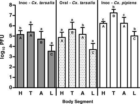
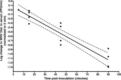
West Nile virus (WNV) is transmitted to vertebrate hosts by mosquitoes as they take a blood meal. The amount of WNV inoculated by mosquitoes as they feed on a live host is not known. Previous estimates of the amount of WNV inoculated by mosquitoes (10(1.2)-10(4.3) PFU) were based on in vitro assays that do not allow mosquitoes to probe or feed naturally. Here, we developed an in vivo assay to determine the amount of WNV inoculated by mosquitoes as they probe and feed on peripheral tissues of a mouse or chick. Using our assay, we recovered approximately one-third of a known amount of virus inoculated into mouse tissues. Accounting for unrecovered virus, mean and median doses of WNV inoculated by four mosquito species were 10(4.3) PFU and 10(5.0) PFU for Culex tarsalis, 10(5.9) PFU and 10(6.1) PFU for Cx. pipiens, 10(4.7) PFU and 10(4.7) PFU for Aedes japonicus, and 10(3.6) PFU and 10(3.4) PFU for Ae. triseriatus. In a direct comparison, in vivo estimates of the viral dose inoculated by Cx. tarsalis were approximately 600 times greater than estimates obtained by an in vitro capillary tube transmission assay. Virus did not disperse rapidly, as >99% of the virus was recovered from the section fed or probed upon by the mosquito. Furthermore, 76% (22/29) of mosquitoes inoculated a small amount of virus ( approximately 10(2) PFU) directly into the blood while feeding. Direct introduction of virus into the blood may alter viral tropism, lead to earlier development of viremia, and cause low rates of infection in co-feeding mosquitoes. Our data demonstrate that mosquitoes inoculate high doses of WNV extravascularly and low doses intravascularly while probing and feeding on a live host. Accurate estimates of the viral dose inoculated by mosquitoes are critical in order to administer appropriate inoculation doses to animals in vaccine, host competence, and pathogenesis studies.
Conflict of interest statement
Figures

Similar articles
-
Mosquito saliva causes enhancement of West Nile virus infection in mice.J Virol. 2011 Feb;85(4):1517-27. doi: 10.1128/JVI.01112-10. Epub 2010 Dec 8. J Virol. 2011. PMID: 21147918 Free PMC article.
-
The potential of Aedes triseriatus (Diptera: Culicidae) as an enzootic vector of West Nile virus.J Med Entomol. 2006 Sep;43(5):966-70. doi: 10.1603/0022-2585(2006)43[966:tpoatd]2.0.co;2. J Med Entomol. 2006. PMID: 17017235
-
Vector competence of Aedes vexans (Diptera: Culicidae) for West Nile virus and potential as an enzootic vector.J Med Entomol. 2008 May;45(3):452-7. doi: 10.1603/0022-2585(2008)45[452:VCOAVD]2.0.CO;2. J Med Entomol. 2008. PMID: 18533439
-
The contribution of Culex pipiens complex mosquitoes to transmission and persistence of West Nile virus in North America.J Am Mosq Control Assoc. 2012 Dec;28(4 Suppl):137-51. doi: 10.2987/8756-971X-28.4s.137. J Am Mosq Control Assoc. 2012. PMID: 23401954 Review.
-
On the Fly: Interactions Between Birds, Mosquitoes, and Environment That Have Molded West Nile Virus Genomic Structure Over Two Decades.J Med Entomol. 2019 Oct 28;56(6):1467-1474. doi: 10.1093/jme/tjz112. J Med Entomol. 2019. PMID: 31549720 Free PMC article. Review.
Cited by
-
ZIKA--How fast does this virus mutate?Dongwuxue Yanjiu. 2016 Mar 18;37(2):110-5. doi: 10.13918/j.issn.2095-8137.2016.2.110. Dongwuxue Yanjiu. 2016. PMID: 27029869 Free PMC article.
-
Activation of the innate immune response against DENV in normal non-transformed human fibroblasts.PLoS Negl Trop Dis. 2011 Dec;5(12):e1420. doi: 10.1371/journal.pntd.0001420. Epub 2011 Dec 20. PLoS Negl Trop Dis. 2011. PMID: 22206025 Free PMC article.
-
Fetal loss in pregnant rhesus macaques infected with high-dose African-lineage Zika virus.PLoS Negl Trop Dis. 2022 Aug 4;16(8):e0010623. doi: 10.1371/journal.pntd.0010623. eCollection 2022 Aug. PLoS Negl Trop Dis. 2022. PMID: 35926066 Free PMC article.
-
West Nile virus vector competency of Culex quinquefasciatus mosquitoes in the Galapagos Islands.Am J Trop Med Hyg. 2011 Sep;85(3):426-33. doi: 10.4269/ajtmh.2011.10-0739. Am J Trop Med Hyg. 2011. PMID: 21896799 Free PMC article.
-
Translational Model of Zika Virus Disease in Baboons.J Virol. 2018 Jul 31;92(16):e00186-18. doi: 10.1128/JVI.00186-18. Print 2018 Aug 15. J Virol. 2018. PMID: 29875247 Free PMC article.
References
-
- Centers for Disease Control and Prevention. CDC West Nile virus homepage. 2007. Available: http://www.cdc.gov/ncidod/dvbid/westnile/index.htm. Accessed 15 August 2007.
-
- Granwehr BP, Lillibridge KM, Higgs S, Mason PW, Aronson JF, et al. West Nile virus: Where are we now? Lancet Infect Dis. 2004;4:547–556. - PubMed
-
- Styer LM, Bernard KA, Kramer LD. Enhanced early West Nile virus infection in young chickens infected by mosquito bite: Effect of viral dose. Am J Trop Med Hyg. 2006;75:337–345. - PubMed
-
- Reisen WK, Fang Y, Martinez VM. Avian host and mosquito (Diptera: Culicidae) vector competence determine the efficiency of West Nile and St. Louis encephalitis virus transmission. J Med Entomol. 2005;42:367–375. - PubMed
-
- Colton L, Nasci RS. Quantification of West Nile virus in the saliva of Culex species collected from the southern United States. J Am Mosq Control Assoc. 2006;22:57–63. - PubMed
Publication types
MeSH terms
Grants and funding
LinkOut - more resources
Full Text Sources
Medical
