. 2008 Jan;27(1):9-17.
doi: 10.1089/dna.2007.0673.

PIN: a novel protein involved in IFN-gamma accumulation of NOS-1 in neurons

Affiliations

PIN: a novel protein involved in IFN-gamma accumulation of NOS-1 in neurons

Jingjun Yang et al. DNA Cell Biol. 2008 Jan.

Abstract

In this study we investigate the role of the protein inhibitor of NOS-1 (PIN) in the interferon-gamma (IFN-gamma)-mediated posttranscriptional accumulation of nitric oxide synthase-1 (NOS-1) and the anti-vesicular stomatitis virus response in neuronal cells. IFN-gamma-induced enhancement of NOS-1 activity is crucial for its antiviral activity in the central nervous system. IFN-gamma treatment of neuronal cells results in an increase of total NOS-1 and decrease of total PIN proteins without alteration in their respective mRNA levels. PIN/NOS-1 complexes decreased after IFN-gamma treatment. Transfection of cells with small interfering RNA (siRNA) for PIN results in a higher constitutive activity of NOS-1 and inhibition of viral replication. IFN-gamma treatment did not change the amount of NOS-1 detectable by Western blot, when PIN is diminished by RNAi treatment. Overexpression of PIN results in lower constitutive NOS-1 expression and activity, and diminishes activation of NOS-1 by IFN-gamma. Our findings indicate that in neurons, IFN-gamma upregulates NOS-1 through proteasomal degradation of PIN.


Figures

FIG. 1
FIG. 1
IFN-γ treatment in neuronal cells results in an increase followed by decrease of PIN/NOS-1 complexes. NB41A3 cells were treated with media or IFN-γ for 8, 16, or 24 h. Amounts of NOS-1, PIN, or NOS-1/PIN complex were determined by double-antibody sandwich ELISA. HRP-conjugated secondary antibody and TMB substrate were used in all reactions. (A) Expression of NOS-1 increases with IFN-γ treatment. Ninety-six–well plates were coated with anti-NOS-1 pAb (Capture Ab). Equal amounts of cell lysates from above treated samples were added in triplicate into precoated well and detected with anti-NOS-1 mAb. (B) Expression of PIN decreases with IFN-γ treatment. Anti-PIN antibody was used for both capturing and detecting. (C) An increase followed by decrease of NOS-1/PIN complexes was observed along with IFN-γ treatment in neuronal cells. Anti-NOS-1 antibody was used in capturing steps, and anti-PIN antibody was used in detecting steps. Same results were observed when using anti-PIN antibody for capturing and anti-NOS-1 antibody for detecting. Results are representative of three independent experiments (*p < 0.01; **p < 0.05).
FIG. 2
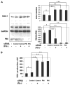
FIG. 2
IFN-γ treatment does not change the amount of NOS-1 when PIN is knocked down by RNAi. siRNA of PIN or negative control siRNA (Neg, random sequence that does not target any genes) was introduced to NB41A3 cells using siPORT Amine transfection reagent. siRNA-transfected cells and mock-transfected cells were treated with IFN-γ for 24 h. (A) Expressions of PIN and NOS-1 protein were determined by Western blot. (B) NO2 production was measured by Griess assay. Values are mean ± SE of three independent experiments (*p > 0.05; **p < 0.01; ***p < 0.05).
FIG. 3
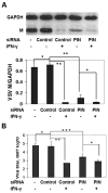
FIG. 3
RNAi of PIN in NB41A3 cells results in inhibition of viral replication. siRNA of PIN or Neg siRNA was introduced to NB41A3 cells using siPORT Amine transfection reagent. siRNA-transfected cells and mock-transfected cells were treated with IFN-γ for 24 h and infected with VSV at M.O.I = 3. Cell lysates were extracted at 5 h postinfection. Supernatants were collected at 7 h postinfection. (A) Expression of the VSV M protein relative to GAPDH was determined by Western blot. (B) Viral replication was measured by plaque assay. Values are mean ± SE of three samples. Results are representative of three independent experiments (*p > 0.05; **p < 0.01; ***p < 0.05).
FIG. 4
FIG. 4
Overexpression of PIN results in lower constitutive NOS-1 expression and activity, increased PIN/NOS-1 complexes, and diminished activation of NOS-1 by IFN-γ. NB41A3 cells were plated in six-well plates and cultured overnight prior to transfection with PcDNA3.1/His A/PIN or PcDNA3.1/is A/MUTA. The untransfected control NB41A3 cells were treated with the same amount of transfection reagent. Twenty-four hours posttransfection, cells were treated with media or IFN-γ for indicated time. (A) Overexpression of Xpress-PIN was confirmed by co-IP of Xpress and PIN. Anti-PIN antibody was used at IP step, and anti-Xpress antibody was used for Western blot (or vice verse, data not shown). Original lysates (no IP step involved) collected from NB41A3 cells transfected with pcDNA3.1/His/LacZ plasmid (Invitrogen) served as a positive control for anti-Xpress antibody in the Western blot. (B) Amount of NOS-1/PIN complex was measured by sandwich ELISA using original lysates. (C) Expression of NOS-1 protein was determined by Western blot. (D) In a parallel experiment, NB41A3 cells were transfected with PcDNA3.1/His A/PIN or PcDNA3.1/His A/MUTA, and then treated with media or IFN-γ for 48 h. Supernatant medium was collected, and production of NO2 was measured by Griess assay. Values are mean ± SEM of three independent experiments (*p > 0.05; **p < 0.01; ***p < 0.05).
FIG. 5
FIG. 5
PIN overexpression resulted in increased viral replication in NB41A3 cells. In PIN overexpressed cells, IFN-γ treatment did not inhibit viral replication as significantly as the control group. NB41A3 cells were plated in 12-well plates (2.5×10⁵ cells/well) and cultured overnight prior to transfection with PcDNA3.1/His A/PIN or PcDNA3.1/His A/MUTA. The untransfected control NB41A3 cells were treated with the same amount of transfection reagent. Twenty-four hours posttransfection, cells were treated with media or IFN-γ. Following 24 h IFN-γ treatment, cells were infected with VSV at M.O.I = 3. Supernatant and cell lysates were collected at 5 h postinfection. (A) VSV M protein was measured by Western blot with GAPDH as housekeeping control. (B) The viral titer was determined by plaque assay. This experiment is representative of three independent experiments (*p < 0.01; **p > 0.05).

Similar articles

Cited by

References

    1. Alderton WK, Cooper CE, Knowles RG. Nitric oxide synthases: structure, function and inhibition. Biochem J. 2001;357:593–615. - PMC - PubMed
    1. Bender AT, Demady DR, Osawa Y. Ubiquitination of neuronal nitric-oxide synthase in vitro and in vivo. J Biol Chem. 2000;275:17407–17411. - PubMed
    1. Bi Z, Reiss CS. Inhibition of vesicular stomatitis virus infection by nitric oxide. J Virol. 1995;69:2208–2213. - PMC - PubMed
    1. Chang TL, Reiss CS, Huang AS. Inhibition of vesicular stomatitis virus RNA synthesis by protein hyper-phosphorylation. J Virol. 1994;68:4980–4987. - PMC - PubMed
    1. Chen N, Reiss CS. Distinct roles of eicosanoids in the immune response to viral encephalitis: or why you should take NSAIDS. Viral Immunol. 2002;15:133–146. - PubMed

Publication types

MeSH terms

LinkOut - more resources