

Laser capture sampling and analytical issues in proteomics
- PMID: 17941818
- PMCID: PMC2639751
- DOI: 10.1586/14789450.4.5.627
Laser capture sampling and analytical issues in proteomics
Abstract
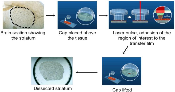
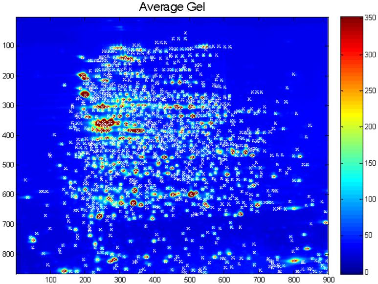
Proteomics holds the promise of evaluating global changes in protein expression and post-translational modification in response to environmental stimuli. However, difficulties in achieving cellular anatomic resolution and extracting specific types of proteins from cells have limited the efficacy of these techniques. Laser capture microdissection has provided a solution to the problem of anatomical resolution in tissues. New extraction methodologies have expanded the range of proteins identified in subsequent analyses. This review will examine the application of laser capture microdissection to proteomic tissue sampling, and subsequent extraction of these samples for differential expression analysis. Statistical and other quantitative issues important for the analysis of the highly complex datasets generated are also reviewed.
Figures
Similar articles
-
Microproteomics: analysis of protein diversity in small samples.Mass Spectrom Rev. 2008 Jul-Aug;27(4):316-30. doi: 10.1002/mas.20161. Mass Spectrom Rev. 2008. PMID: 18271009 Free PMC article. Review.
-
Combining laser capture microdissection and proteomics: methodologies and clinical applications.Proteomics Clin Appl. 2010 Feb;4(2):116-23. doi: 10.1002/prca.200900138. Epub 2009 Nov 19. Proteomics Clin Appl. 2010. PMID: 21137037 Review.
-
Laser capture microdissection and colorectal cancer proteomics.Methods Mol Biol. 2005;293:245-53. doi: 10.1385/1-59259-853-6:245. Methods Mol Biol. 2005. PMID: 16028424 Review.
-
[Laser microdissection for biology and medicine].Tsitologiia. 2012;54(5):381-9. Tsitologiia. 2012. PMID: 22827035 Review. Russian.
-
Combining laser capture microdissection and proteomics techniques.Methods Mol Biol. 2008;428:159-78. doi: 10.1007/978-1-59745-117-8_9. Methods Mol Biol. 2008. PMID: 18287773 Review.
Cited by
-
Comparisons of protein profiles of beech bark disease resistant and susceptible American beech (Fagus grandifolia).Proteome Sci. 2013 Jan 14;11(1):2. doi: 10.1186/1477-5956-11-2. Proteome Sci. 2013. PMID: 23317283 Free PMC article.
-
Enrichment of single neurons and defined brain regions from human brain tissue samples for subsequent proteome analysis.J Neural Transm (Vienna). 2015 Jul;122(7):993-1005. doi: 10.1007/s00702-015-1414-4. Epub 2015 Jun 30. J Neural Transm (Vienna). 2015. PMID: 26123835
-
Biomarkers of lung-related diseases: current knowledge by proteomic approaches.J Cell Physiol. 2009 Dec;221(3):535-43. doi: 10.1002/jcp.21893. J Cell Physiol. 2009. PMID: 19681054 Free PMC article. Review.
-
Evaluating the performance of new approaches to spot quantification and differential expression in 2-dimensional gel electrophoresis studies.J Proteome Res. 2010 Jan;9(1):595-604. doi: 10.1021/pr9005603. J Proteome Res. 2010. PMID: 19919108 Free PMC article.
-
Microproteomics: quantitative proteomic profiling of small numbers of laser-captured cells.Cold Spring Harb Protoc. 2011 Feb 1;2011(2):pdb.prot5573. doi: 10.1101/pdb.prot5573. Cold Spring Harb Protoc. 2011. PMID: 21285273 Free PMC article. No abstract available.
References
-
- Palkovits M, Eskay RL. Distribution and possible origin of β-endorphin and ACTH in discrete brainstem nuclei of rats. Neuropeptides. 1987:123–137. - PubMed
-
- Cuello AC, Carson S. Microdissection of fresh rat brain tissue slices. In: Cuello AC, editor. Brain Microdissection Techniques. John Wiley and Sons; Chichester: 1983. pp. 37–126.
-
- Christensen M, Estevez A, Yin X, et al. A primary culture system for functional analysis of C. elegans neurons and muscle cells. Neuron. 2002:33–4. 503–514. - PubMed
-
- Safarik I, Safarikova M. Use of magnetic techniques for the isolation of cells. Journal of Chromatography B, Biomedical Sciences & Applications. 1999;722(12):33–53. - PubMed
Publication types
MeSH terms
Substances
Grants and funding
LinkOut - more resources
Full Text Sources
Other Literature Sources