. 2007 Nov;88(2):182-90.
doi: 10.1016/j.cmpb.2007.08.006. Epub 2007 Oct 17.

Topology correction of segmented medical images using a fast marching algorithm

Affiliations

Topology correction of segmented medical images using a fast marching algorithm

Pierre-Louis Bazin et al. Comput Methods Programs Biomed. 2007 Nov.

Abstract

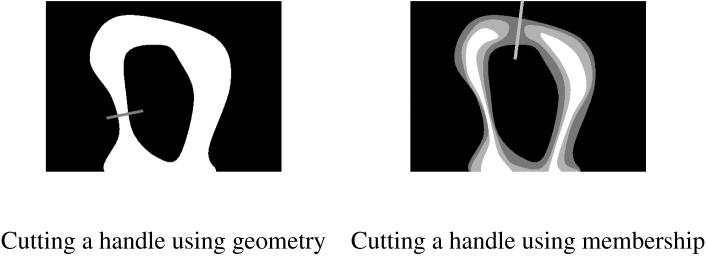
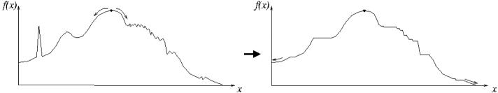
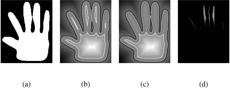
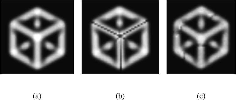
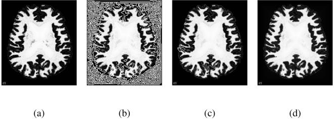
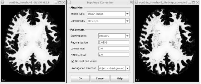
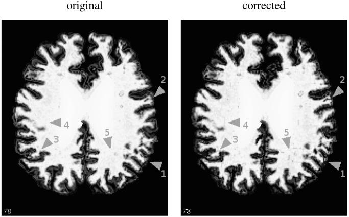
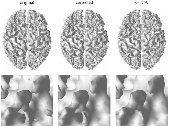
We present here a new method for correcting the topology of objects segmented from medical images. Whereas previous techniques alter a surface obtained from a binary segmentation of the object, our technique can be applied directly to the image intensities of a probabilistic or fuzzy segmentation, thereby propagating the topology for all isosurfaces of the object. From an analysis of topological changes and critical points in implicit surfaces, we derive a topology propagation algorithm that enforces any desired topology using a fast marching technique. The method has been applied successfully to the correction of the cortical gray matter/white matter interface in segmented brain images and is publicly released as a software plug-in for the MIPAV package.


Figures

Fig. 1
Fig. 1
An illustration of the interest of membership functions: cutting a handle at the thinnest point (left) can sever thin structures, but a membership function (right) can provide the additional information needed to preserve them.
Fig. 2
Fig. 2
A 1D example of topology-preserving approximation: starting from the highest point of f (left), the propagation algorithm follows its slope downward and thresholds the function when f goes upward. The resulting function g (right) has one global maximum, its minima are located at the boundary of the domain and there is no local minima or maxima.
Fig. 3
Fig. 3
A 2D example: from the outline of a hand (a), we build a signed distance function (b). Notice from the outlined isocontours how the topology of the object changes as the fingers get separated from the palm or merge. The topology-preserving approximation (c) is very similar to the original distance function, but maintains the same topology for all isovalues. The difference f - g (d) is zero almost everywhere, and small otherwise (the amplitude of the difference image is about 5% of the amplitude of the original distance function).
Fig. 4
Fig. 4
A 3D example: (a) a solid shape made of the edges of a cube and blurred into a volumetric scalar image, (b) the same object corrected to have spherical topology, (c) the corrected object after adding noise to (a). The propagation is started from a single point of the hidden corner on the back. After the propagation, the corrected object has cuts that preserve the spherical topology of the original point. The cuts respect the symmetry of the original object because the propagation is identical in all directions. If we add some noise to the image, the symmetry is broken and the cuts adopt a more conventional shape.
Fig. 5
Fig. 5
Influence of noise and flat regions: (a) a membership function from our experimental study, (b) the approximated function with spherical topology computed from the lowest to highest membership values, with δ = 0, (c) the same approximation with δ = 1E - 12, (d) the approximation with δ = 1E - 3. The propagation is started from the boundary of the image, and evolves at random through the background region if δ = 0. The smallest increase to will remove most of the critical points, and the remaining artificial structures due to background noise will also disappear with a higher δ.
Fig. 6
Fig. 6
The topology correction software: original image, user interface and result.
Fig. 7
Fig. 7
An example of topology correction (image number 18 of the experiments, which has the highest numbers of changed voxels): a slice of the filled white matter membership function before and after the topology correction, Note the few visible changes in the membership, pointed by gray arrows: parts with high membership but disconnected from the white matter are removed (1), loops are cut (2, 3) and some patterns appear near the filled region (4, 5).
Fig. 8
Fig. 8
An example of topology correction (image number 18 of the experiments): top) the 3D surface extracted from the original and corrected memberships, bottom) a close-up of the surface in an area with three interconnected handles, before and after correction. The corrected surface obtained with GTCA is also presented for comparison. The corrected surface is very close to the original and to the surface produced by GTCA.

References

    1. van Essen DC, Maunsell HR. Two dimensional maps of the cerebral cortex. Journal of Comparative Neurology. 1980;191:255–281. - PubMed
    1. Mangin J-F, Frouin V, Bloch I, Regis J, Lopez-Krahe J. From 3D magnetic resonance images to structural representations of the cortex topography using topology preserving deformations. Journal of Mathematical Imaging and Vision. 1995;5:297–318.
    1. Han Xiao, Xu Chenyang, Prince Jerry L. A topology preserving level set method for geometric deformable models. IEEE Transactions on Pattern Analysis and Machine Intelligence. 2003;25(6):755–768.
    1. Bischoff Stefan, Kobbelt Leif. Sub-voxel topology control for level-set surfaces. Computer Graphics Forum. 2003;22(3):273–280.
    1. Bazin P-L, Pham DL. Topology-preserving tissue classification of magnetic resonance brain images. IEEE Transactions on Medical Imaging. 2007;26(4):487–496. Special Issue on Computational Neuroanatomy. - PubMed