. 2008 Jan;82(1):196-206.
doi: 10.1128/JVI.01640-07. Epub 2007 Oct 17.

Lymphocytoid choriomeningitis virus activates plasmacytoid dendritic cells and induces a cytotoxic T-cell response via MyD88

Affiliations

Lymphocytoid choriomeningitis virus activates plasmacytoid dendritic cells and induces a cytotoxic T-cell response via MyD88

Andreas Jung et al. J Virol. 2008 Jan.

Abstract

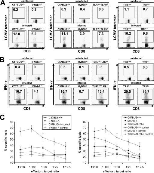
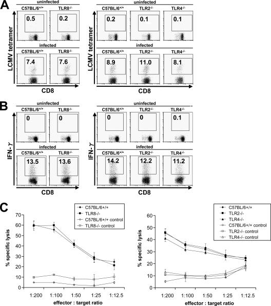
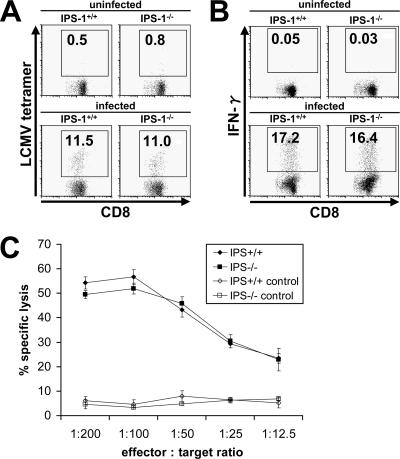
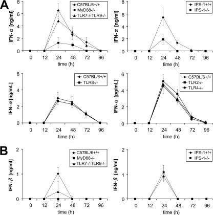
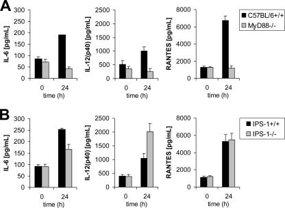
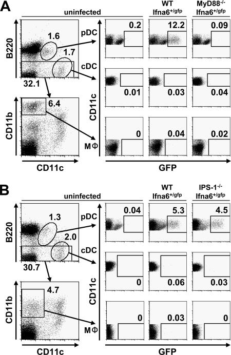
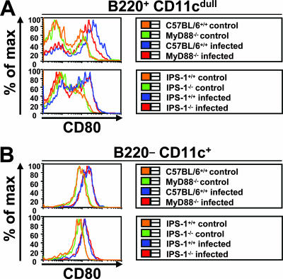
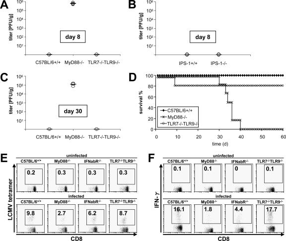
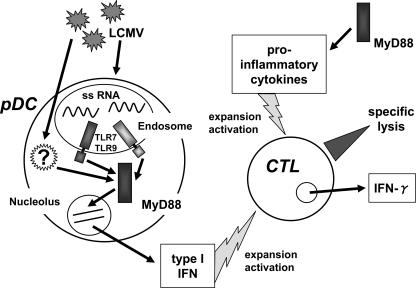
Toll-like receptors (TLRs) and retinoic acid-inducible gene I-like helicases (RLHs) are two major machineries recognizing RNA virus infection of innate immune cells. Intracellular signaling for TLRs and RLHs is mediated by their cytoplasmic adaptors, i.e., MyD88 or TRIF and IPS-1, respectively. In the present study, we investigated the contributions of TLRs and RLHs to the cytotoxic T-lymphocyte (CTL) response by using lymphocytoid choriomeningitis virus (LCMV) as a model virus. The generation of virus-specific cytotoxic T lymphocytes was critically dependent on MyD88 but not on IPS-1. Type I interferons (IFNs) are known to be important for the development of the CTL response to LCMV infection. Serum levels of type I IFNs and proinflammatory cytokines were mainly dependent on the presence of MyD88, although IPS-1(-/-) mice showed a decrease in IFN-alpha levels but not in IFN-beta and proinflammatory cytokine levels. Analysis of Ifna6(+/GFP) reporter mice revealed that plasmacytoid dendritic cells (DCs) are the major source of IFN-alpha in LCMV infection. MyD88(-/-) mice were highly susceptible to LCMV infection in vivo. These results suggest that recognition of LCMV by plasmacytoid DCs via TLRs is responsible for the production of type I IFNs in vivo. Furthermore, the activation of a MyD88-dependent innate mechanism induces a CTL response, which eventually leads to virus elimination.


Figures

FIG. 1.
FIG. 1.
Induction of LCMV-specific T-cell response via the TLR system. Wild-type, MyD88−/−, IFN-α/β receptor−/−, TLR7−/− TLR9−/−, and TRIF−/− mice were infected intravenously with 3 × 10⁵ PFU LCMV WE, and splenocytes were harvested on day 8 after infection. Cells were stained with LCMV major histocompatibility complex class I tetramer, CD4, and CD8α antibodies (A) or stimulated with gp33-41 prior to CD8α antibody staining (B). Induction of LCMV-specific T lymphocytes and IFN-γ production was analyzed by flow cytometry. (C) Ex vivo CTL activity in splenocytes was determined by a 5-h 51Cr release assay, using gp33-41-loaded EL4 cells as targets. Values are means ± standard deviations (SD) for three mice. Data are representative of three independent experiments.
FIG. 2.
FIG. 2.
Involvement of other TLRs in T-cell response. Wild-type, TLR2−/−, TLR4−/−, and TLR8−/− mice were infected intravenously with 3 × 10⁵ PFU LCMV WE, and splenocytes were harvested on day 8 after infection. Induction of LCMV-specific T lymphocytes (A), IFN-γ production (B), and CTL activity (C) were analyzed. Values are means ± SD for three mice.
FIG. 3.
FIG. 3.
T-cell response via the RLH system. Wild-type and IPS-1−/− mice were infected intravenously with 3 × 10⁵ PFU LCMV WE, and splenocytes were harvested on day 8 after infection. Induction of LCMV-specific T lymphocytes (A), IFN-γ production (B), and CTL activity (C) were analyzed as described in the legend to Fig. 1. Values are means ± SD for three mice. Data are representative of three independent experiments.
FIG. 4.
FIG. 4.
Type I IFN response to LCMV infection. The indicated mice were infected intravenously with 3 × 10⁵ PFU LCMV WE, and IFN-α (A) and IFN-β (B) levels in sera were measured by enzyme-linked immunosorbent assay between 12 and 96 h after infection. Data show means ± SD for three mice and are representative of three independent experiments.
FIG. 5.
FIG. 5.
Cytokine production. IL-6, IL-12(p40), and RANTES levels in sera of MyD88−/− (A) and IPS-1−/− (B) mice after intravenous infection with 3 × 10⁵ PFU LCMV WE were assessed after 24 h by a multiplex bead-based assay. Data show means ± SD for three mice.
FIG. 6.
FIG. 6.
Expression of GFP in splenic DCs and macrophages. Ifna6+/GFP mice lacking MyD88 (A) or IPS-1 (B) as well as wild-type mice were infected intravenously with 3 × 10⁵ PFU LCMV WE. Splenocytes were harvested 24 and 48 h after infection, and the expression of GFP in pDCs, cDCs, and macrophages was analyzed by fluorescence-activated cell sorter analysis. Data are representative of two experiments.
FIG. 7.
FIG. 7.
Activation of splenic DCs. Mice were infected intravenously with 3 × 10⁵ PFU LCMV WE. Splenocytes of MyD88- and IPS-1-deficient mice were investigated for the activation of CD80 on pDCs (A) and cDCs (B) by flow cytometry 48 h after infection. Data shown are representative of three mice.
FIG. 8.
FIG. 8.
Long-term sequelae after LCMV infection. Wild-type, MyD88−/−, and TLR7−/− TLR9−/− mice or IPS-1−/− mice were infected intravenously with 3 × 10⁵ PFU LCMV WE, and virus titers were measured in spleens on day 8 (A and B) and day 30 (C) by a methylcellulose titration assay. Results for three mice are displayed, with means. Data are representative of three independent experiments. (D) Survival rates were assessed after infecting six mice per indicated genotype with 2 × 10⁶ PFU LCMV WE in two independent experiments. (E and F) Generation of LCMV-specific memory CD8+ T lymphocytes in MyD88−/−, TLR7−/− TLR9−/−, and IFN-α/β receptor−/− mice following infection with 1 × 10⁵ PFU LCMV WE was analyzed as described above. Indicated values are means ± SD for three mice and are representative of two independent experiments.
FIG. 9.
FIG. 9.
Innate immune modulation of CTL response to LCMV through MyD88 signaling. MyD88 mediates recognition of LCMV by TLR7/TLR9 and possibly other, unknown receptors via pDCs. Type I IFNs produced by infected pDCs result in activation and clonal expansion of virus-specific CTLs. In an alternative pathway, CTLs are activated by proinflammatory cytokines in an MyD88-dependent manner. Activated CTLs mount effector functions that finally contribute to virus elimination.

References

    1. Aichele, P., H. Unsoeld, M. Koschella, O. Schweier, U. Kalinke, and S. Vucikuja. 2006. Cutting edge: CD8 T cells specific for lymphocytic choriomeningitis virus require type I IFN receptor for clonal expansion. J. Immunol. 1764525-4529. - PubMed
    1. Akira, S., S. Uematsu, and O. Takeuchi. 2006. Pathogen recognition and innate immunity. Cell 124783-801. - PubMed
    1. Belz, G. T., K. Shortman, M. J. Bevan, and W. R. Heath. 2005. CD8α dendritic cells selectively present MHC class I-restricted noncytological viral and intracellular bacterial antigens in vivo. J. Immunol. 175196-200. - PMC - PubMed
    1. Beutler, B. 2004. Interferences, questions and possibilities in Toll-like receptor signaling. Nature 230257-263. - PubMed
    1. Binder, D., M. F. van den Broek, D. Kaegi, H. Bluethmann, J. Fehr, H. Hnegartner, and R. M. Zinkernagel. 1989. Aplastic anemia rescued by exhaustion of cytokine-secreting CD8+ T cells in persistent infection with lymphocytic choriomeningitis virus. J. Exp. Med. 1871902-1920. - PMC - PubMed

Publication types

MeSH terms

LinkOut - more resources