. 2008 Jan;82(1):394-407.
doi: 10.1128/JVI.01681-07. Epub 2007 Oct 17.

Inhibition of the cyclin-dependent kinases at the beginning of human cytomegalovirus infection specifically alters the levels and localization of the RNA polymerase II carboxyl-terminal domain kinases cdk9 and cdk7 at the viral transcriptosome

Affiliations

Inhibition of the cyclin-dependent kinases at the beginning of human cytomegalovirus infection specifically alters the levels and localization of the RNA polymerase II carboxyl-terminal domain kinases cdk9 and cdk7 at the viral transcriptosome

Anokhi J Kapasi et al. J Virol. 2008 Jan.

Abstract

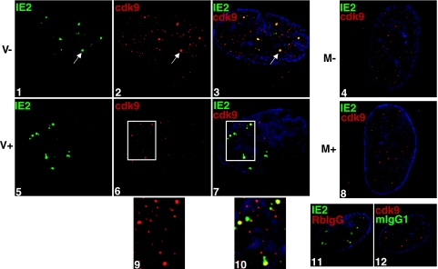
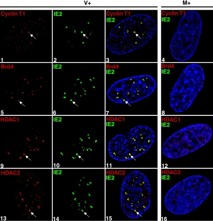
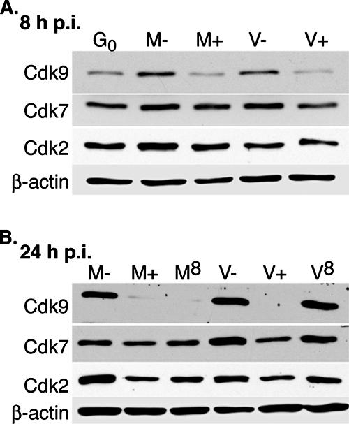
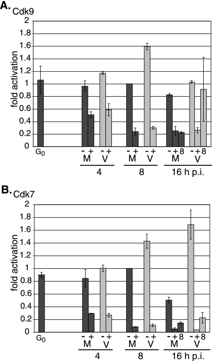
We previously reported that defined components of the host transcription machinery are recruited to human cytomegalovirus immediate-early (IE) transcription sites, including cdk9 and cdk7 (S. Tamrakar, A. J. Kapasi, and D. H. Spector, J. Virol. 79:15477-15493, 2005). In this report, we further document the complexity of this site, referred to as the transcriptosome, through identification of additional resident proteins, including viral UL69 and cellular cyclin T1, Brd4, histone deacetylase 1 (HDAC1), and HDAC2. To examine the role of cyclin-dependent kinases (cdks) in the establishment of this site, we used roscovitine, a specific inhibitor of cdk1, cdk2, cdk7, and cdk9, that alters processing of viral IE transcripts and inhibits expression of viral early genes. In the presence of roscovitine, IE2, cyclin T1, Brd4, HDAC1, and HDAC2 accumulate at the transcriptosome. However, accumulation of cdk9 and cdk7 was specifically inhibited. Roscovitine treatment also resulted in decreased levels of cdk9 and cdk7 RNA. There was a corresponding reduction in cdk9 protein but only a modest decrease in cdk7 protein. However, overexpression of cdk9 does not compensate for the effects of roscovitine on cdk9 localization or viral gene expression. Delaying the addition of roscovitine until 8 h postinfection prevented all of the observed effects of the cdk inhibitor. These data suggest that IE2 and multiple cellular factors needed for viral RNA synthesis accumulate within the first 8 h at the viral transcriptosome and that functional cdk activity is required for the specific recruitment of cdk7 and cdk9 during this time interval.


Figures

FIG. 1.
FIG. 1.
Localization of several proteins at the sites of viral IE transcription in HCMV-infected cells. G0-synchronized cells were released into G1, infected with HCMV Towne at an MOI of 5 or with mock supernatant, and seeded onto glass coverslips. At 8 h p.i., cells were washed with PBS, fixed in formaldehyde, permeabilized, and immunostained with antibodies specific for the proteins indicated in each panel. Specific antibody staining was detected with fluorescein isothiocyanate- or Cy3-conjugated isotype-specific secondary antibodies. For controls, one of the specific antibodies of the pair was replaced with an isotype-specific normal IgG. Nuclei were stained with Hoechst dye. The white arrow in each panel indicates a region of colocalization, although more are present. All of the images are confocal optical sections of 0.2 μm at a magnification of ×1,000 under oil immersion.
FIG. 2.
FIG. 2.
Treatment with roscovitine impairs accumulation of cdk9 at the sites of viral IE transcription. G0-synchronized cells were released into G1, infected with HCMV Towne at an MOI of 5 or with mock supernatant, and seeded onto glass coverslips. Cells were treated with 16 μM roscovitine or DMSO at the time of infection. At 8 h p.i., cells were washed with PBS, fixed in formaldehyde, permeabilized, and immunostained with IE2 (IgG1) and cdk9. Specific antibody staining was detected with fluorescein isothiocyanate- or Cy3-conjugated isotype-specific secondary antibodies. For controls, one of the specific antibodies of the pair was replaced with an isotype-specific normal IgG. Nuclei were stained with Hoechst dye. The white arrow in each panel indicates a region of colocalization, although more are present. The white boxes in panels 6 and 7 are enlarged in panels 9 and 10, respectively, with the modification of a 200% magnification and a 200% Cy3 intensity using Adobe Photoshop v. 7.0. All of the images are confocal optical sections of 0.2 μm at a magnification of ×1,000 under oil immersion. M, mock treated; V, HCMV infected; −, DMSO treated; +, roscovitine treated at the time of infection.
FIG. 3.
FIG. 3.
Treatment with roscovitine impairs cdk9 aggregation in infected cells. G0-synchronized cells were released into G1, infected with HCMV Towne at an MOI of 5 or with mock supernatant, and seeded onto glass coverslips. Cells were treated with 16 μM roscovitine or DMSO at the time of infection or with roscovitine at 8 h p.i. At 14 h p.i., cells were washed with PBS, fixed in formaldehyde, permeabilized, and immunostained with anti-cdk9 and anti-IE2 antibodies followed by Cy3- and fluorescein isothiocyanate-conjugated secondary antibodies. For controls, isotype-specific normal IgG was used in place of the specific antibody (not shown). The white arrow in each panel indicates a cdk9 aggregate, although more are present. All of the images are confocal optical sections of 0.2 μm at a magnification of ×1,000 under oil immersion. M, mock treated; V, HCMV infected; −R, DMSO treated; +R0, roscovitine treated at the time of infection; +R8, roscovitine treated at 8 h p.i.
FIG. 4.
FIG. 4.
Treatment with roscovitine inhibits recruitment of cdk7 at the sites of viral IE transcription. G0-synchronized cells were released into G1, infected with HCMV Towne at an MOI of 5 or with mock supernatant, and seeded onto glass coverslips. Cells were treated with DMSO (panels 1 to 4) or 16 μM roscovitine (panels 5 to 8) at the time of infection. At 8 h p.i., cells were washed with PBS, fixed in formaldehyde, permeabilized, and immunostained with cdk7 (IgG2B) and IE2 (IgG1). Specific antibody staining was detected with fluorescein isothiocyanate- or Cy3-conjugated isotype-specific secondary antibodies. For controls, one of the specific antibodies of the pair was replaced with an isotype-specific normal IgG (panels 9 and 10). Nuclei were stained with Hoechst dye. The white arrow in each panel indicates a region of colocalization, although more are present. All of the images are confocal optical sections of 0.2 μm at a magnification of ×1,000 under oil immersion. M, mock treated; V, HCMV infected; −, DMSO treated; +, roscovitine treated at the time of infection.
FIG. 5.
FIG. 5.
Cyclin T1, Brd4, HDAC1, and HDAC2 are able to localize to the sites of viral IE transcription during infection in the presence of roscovitine. G0-synchronized cells were released into G1, infected with HCMV Towne at an MOI of 5 (panels 1 to 3, 5 to 7, and 13 to 15) or with mock supernatant (panels 4, 8, 12, and 16), and seeded onto glass coverslips. Cells were treated with 16 μM roscovitine or DMSO (see Fig. 1) at the time of infection. Only the roscovitine-treated samples are shown in this figure. At 8 h p.i., cells were washed with PBS, fixed in formaldehyde, permeabilized, and immunostained with an antibody specific for the protein indicated in each panel. Specific antibody staining was detected with fluorescein isothiocyanate- or Cy3-conjugated isotype-specific secondary antibodies. For controls, one of the specific antibodies of the pair was replaced with an isotype-specific normal IgG (not shown). Nuclei were stained with Hoechst dye. The white arrow in each panel indicates a region of colocalization, although more are present. All of the images are confocal optical sections of 0.2 μm at a magnification of ×1,000 magnification under oil immersion. M, mock treated; V, HCMV infected; +, roscovitine treated at the time of infection.
FIG. 6.
FIG. 6.
Effect of roscovitine on the steady-state levels of cdk9 and cdk7 protein. G0-synchronized cells were released into G1 and infected with HCMV Towne at an MOI of 5 or with mock supernatant. Cells were treated with 16 μM roscovitine or DMSO at the time of infection or at 8 h p.i. and were harvested at 8 h p.i. (A) or 24 h p.i. (B). For mock- and virus-infected cells that were harvested at 24 h p.i. and treated with roscovitine or DMSO at the beginning of the infection, the medium was removed at 8 h p.i., and fresh medium containing roscovitine or DMSO, respectively, was added. Cell lysates from equal numbers of cells were run on an 8% SDS-polyacrylamide gel and subjected to Western blotting using antibodies for cdk9, cdk7, cdk2, and β-actin (loading control). G0, cells released from G0; M, mock treated; V, HCMV infected; −, DMSO treated; +, roscovitine treated at the time of infection; 8, roscovitine treated at 8 h p.i.
FIG. 7.
FIG. 7.
Effect of roscovitine on cdk9 and cdk7 RNA levels. G0-synchronized cells were released into G1 and infected with HCMV Towne at an MOI of 5 or with mock supernatant. Cells were treated with 16 μM roscovitine or DMSO at the time of infection or at 8 h p.i. and were harvested at 4, 8, and 16 h p.i. Total RNA was isolated and analyzed by quantitative real-time RT-PCR using primers and probes listed in Table 1 for cdk9 (A) and cdk7 (B) RNAs. The values for cdk9 and cdk7 were normalized to that of G6PD RNA for each sample. For each time point, two separate experiments were performed and duplicate reactions were analyzed. A standard curve for each gene was used to calculate the relative amount of specific RNA present in a sample, which was then converted to fold activation relative to DMSO-treated, mock-infected 8 h p.i. samples. The graphs shows the averages from the two experiments, and the range bars indicate the highest and lowest values. G0, cells released from G0; M, mock treated; V, HCMV infected; −, DMSO treated; +, roscovitine treated at the time of infection; 8, roscovitine treated at 8 h p.i.
FIG. 8.
FIG. 8.
Roscovitine treatment affects steady-state levels of endogenous cdk9 and not those of overexpressed cdk9HA. (A) The G0-synchronized cdk9HA-overexpressing HFF were released into G1, HCMV infected at an MOI of 5, and seeded onto coverslips. At 12 h p.i., the cells were washed with PBS, fixed in formaldehyde, permeabilized, and immunostained with IE2 (IgG1) and HA (IgG2a). As a control, as second anti-HA antibody (rabbit polyclonal) was used to confirm specific staining (not shown). Specific antibody staining was detected with fluorescein isothiocyanate- or tetramethyl rhodamine isocyanate-conjugated isotype-specific secondary antibodies. Nuclei were stained with Hoechst dye. The white arrow in each panel indicates a region of colocalization, although more are present. All of the images are confocal optical sections of 0.2 μm at a magnification of ×1,000 under oil immersion. (B and C) The immunocomplexes from mock cells containing the exogenously expressed cdk9 were assayed for kinase activity towards purified GST-CTD (B), and the corresponding immunoprecipitates were run on an 8% SDS-polyacrylamide gel and subjected to Western blot with an antibody specific for cdk9 (C). Pre, preimmunoprecipitate; IP, immunoprecipitate; Post, postimmunoprecipitate. (D) The G0-synchronized cdk9HA-overexpressing HFF and untransduced HFF were HCMV infected at an MOI of 5 or mock infected in the presence of 16 μM roscovitine or DMSO. Samples were collected at 8 and 24 h p.i. Lysates consisting of an equal number of cells were loaded on a 10% SDS-polyacrylamide gel. Western blot analysis was performed as described above for cdk9. M, mock treated; V, HCMV infected; −, DMSO treated; +, roscovitine treated at the time of infection; F, untransduced HFF cells; 9, cdk9HA-overexpressing cells; *, endogenous cdk9; **, cdk9HA.
FIG. 9.
FIG. 9.
Overexpression of cdk9 does not compensate for alterations in the expression of viral IE proteins and RNAP II during infection in the presence of roscovitine. The G0-synchronized cdk9HA-overexpressing HFF and untransduced HFF were HCMV infected at an MOI of 5 or mock treated in the presence of 16 μM roscovitine or DMSO. Samples were collected at 8 and 24 h p.i. and analyzed by Western blotting using antibodies against the viral proteins IE1/2 (CH16.0) and β-actin (loading control) (A) or antibodies against total RNAP II (ARNA3) and serine 2-phosphorylated RNAP II CTD (H5) (B). β-Catenin was used as a loading control. M, mock treated; V, HCMV infected; −, DMSO treated; +, roscovitine treated at the time of infection; F, untransduced HFF cells; 9, cdk9HA-overexpressing cells.
FIG. 10.
FIG. 10.
Impaired cdk9 localization in the presence of cdk inhibitor occurs in infected cells overexpressing cdk9HA. The G0-synchronized cdk9HA-overexpressing HFF and untransduced HFF were HCMV-infected at an MOI of 5 or mock treated in the presence of 16 μM roscovitine or DMSO. At 8 h p.i., cells were washed with PBS, fixed in formaldehyde, permeabilized, and immunostained with cdk9 antibody followed by fluorescein isothiocyanate- or Cy3-conjugated isotype-specific secondary antibodies. For controls, one of the specific antibodies of the pair was replaced with isotype-specific normal IgG (not shown). Nuclei were stained with Hoechst dye. The white arrow in each panel indicates a region of colocalization, but more are present. All of the images are confocal optical sections of 0.2 μm at a magnification of ×1,000 under oil immersion. M, mock treated; V, HCMV infected; −R, DMSO treated; +R, roscovitine treated at the time of infection.

Similar articles

Cited by

References

    1. Ahn, J.-H., W.-J. Jang, and G. S. Hayward. 1999. The human cytomegalovirus IE2 and UL112-113 proteins accumulate in viral DNA replication compartments that initiate from the periphery of promyelocytic leukemia protein-associated nuclear bodies (PODs or ND10). J. Virol. 7310458-10471. - PMC - PubMed
    1. Ahn, J. H., E. R. Brignole, and G. S. Hayward. 1998. Disruption of PML subnuclear domains by the acidic IE1 protein of human cytomegalovirus is mediated through interaction with PML and may modulate a RING finger-dependent cryptic transactivator function of PML. Mol. Cell. Biol. 184899-4913. - PMC - PubMed
    1. Ahn, J. H., and G. S. Hayward. 1997. The major immediate-early proteins IE1 and IE2 of human cytomegalovirus colocalize with and disrupt PML-associated nuclear bodies at very early times in infected permissive cells. J. Virol. 714599-4613. - PMC - PubMed
    1. Baek, M.-C., P. M. Krosky, A. Pearson, and D. M. Coen. 2004. Phosphorylation of the RNA polymerase II carboxyl-terminal domain in human cytomegalovirus-infected cells and in vitro by the viral UL97 protein kinase. Virol. 324184-193. - PubMed
    1. Bain, J., H. McLauchlan, M. Elliott, and P. Cohen. 2003. The specificities of protein kinase inhibitors: an update. Biochem. J. 371199-204. - PMC - PubMed

Publication types

MeSH terms

Substances

LinkOut - more resources