. 2007 Dec;81(24):13681-93.
doi: 10.1128/JVI.01702-07. Epub 2007 Oct 17.

Altered pathogenesis of porcine respiratory coronavirus in pigs due to immunosuppressive effects of dexamethasone: implications for corticosteroid use in treatment of severe acute respiratory syndrome coronavirus

Affiliations

Altered pathogenesis of porcine respiratory coronavirus in pigs due to immunosuppressive effects of dexamethasone: implications for corticosteroid use in treatment of severe acute respiratory syndrome coronavirus

Kwonil Jung et al. J Virol. 2007 Dec.

Abstract

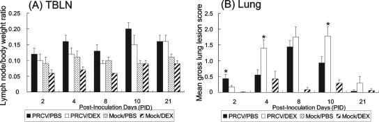
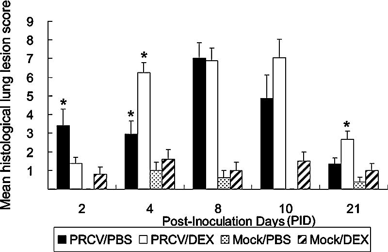
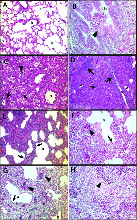
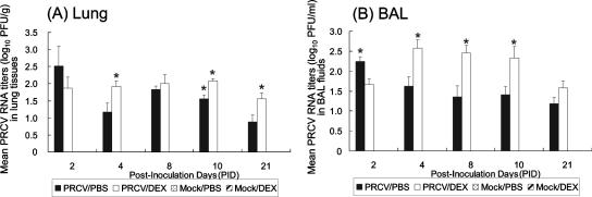
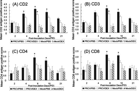
The pathogenesis and optimal treatments for severe acute respiratory syndrome (SARS) are unclear, although corticosteroids were used to reduce lung and systemic inflammation. Because the pulmonary pathology of porcine respiratory coronavirus (PRCV) in pigs resembles SARS, we used PRCV as a model to clarify the effects of the corticosteroid dexamethasone (DEX) on coronavirus (CoV)-induced pneumonia. Conventional weaned pigs (n = 130) in one of four groups (PRCV/phosphate-buffered saline [PBS] [n = 41], PRCV/DEX [n = 41], mock/PBS [n = 23], and mock/DEX [n = 25]) were inoculated intranasally and intratracheally with the ISU-1 strain of PRCV (1 x 10(7) PFU) or cell culture medium. DEX was administered (once daily, 2 mg/kg of body weight/day, intramuscularly) from postinoculation day (PID) 1 to 6. In PRCV/DEX pigs, significantly milder pneumonia, fewer PRCV-positive cells, and lower viral RNA titers were present in lungs early at PID 2; however, at PID 4, 10, and 21, severe bronchointerstitial pneumonia, significantly higher numbers of PRCV-positive cells, and higher viral RNA titers were observed compared to results for PRCV/PBS pigs. Significantly lower numbers of CD2(+), CD3(+), CD4(+), and CD8(+) T cells were also observed in lungs of PRCV/DEX pigs than in those of PRCV/PBS pigs at PID 8 and 10, coincident with fewer gamma interferon (IFN-gamma)-secreting cells in the tracheobronchial lymph nodes as determined by enzyme-linked immunospot assay. Our results confirm that DEX treatment alleviates PRCV pneumonia early (PID 2) in the infection but continued use through PID 6 exacerbates later stages of infection (PID 4, 10, and 21), possibly by decreasing cellular immune responses in the lungs (IFN-gamma-secreting T cells), thereby creating an environment for more-extensive viral replication. These data have potential implications for corticosteroid use with SARS-CoV patients and suggest a precaution against prolonged use based on their unproven efficacy in humans, including possible detrimental secondary effects.


Figures

FIG. 1.
FIG. 1.
Mean LN/BW ratios (A) and gross lung lesion scores (B) for the four experimental pig groups at different PID: n = 4 to 5 at PID 2 and 10, n = 5 at PID 4 and 8, and n = 5 to 6 at PID 21 for mock/PBS and mock/DEX groups; n = 8 at PID 2 to 10 and n = 9 at PID 21 for PRCV/PBS and PRCV/DEX groups. (A) The left TBLNs from each pig were weighed, and the LN/BW ratios of the lymph nodes were calculated. The left TBLNs of mock/DEX and PRCV/DEX pigs were atrophic by 60 to 80% at PID 2, 4, 8, and 21 and 69 to 83% at PID 2, 4, 8, and 10 compared to those of mock/PBS and PRCV/PBS pigs, respectively. However, the differences in LN/BW ratios were not statistically significant (Kruskal-Wallis test) due to animal variability. (B) Gross lung lesions were evaluated based on the percentage of virus-induced lesions (consolidation) of all six lobes. PRCV/DEX pigs had significantly milder lesions than PRCV/PBS pigs at PID 2. However, at PID 4 and 10, the percentages of overall lung tissue consolidation were significantly higher for PRCV/DEX pigs than for PRCV/PBS pigs. Each bar represents the mean ± SEM. An asterisk above a bar represents significant differences (P < 0.05) between results for PRCV/PBS and PRCV/DEX pigs by the Kruskal-Wallis test.
FIG. 2.
FIG. 2.
Mean histological lung lesion scores for the four experimental pig groups at different PID with numbers of pigs at each time point as indicated in the legend to Fig. 1. Histological lung lesion scores ranged from 0 (no abnormalities) to 12 (most severe) based on the combined score of three parameters: (i) thickening of the alveolar septa (0 to 6); (ii) accumulation of necrotic cells and inflammatory leucocytes in alveolar and bronchiolar lumina (0 to 3); and (iii) peribronchiolar or perivascular lymphohistiocytic inflammation (0 to 3). PRCV/PBS pigs had significantly higher histological lesion scores than PRCV/DEX pigs at PID 2 (*), with the opposite effect at PID 4 (**) and 21 (*). A higher lesion score was also observed at PID 10 for PRCV/DEX pigs. Each bar represents means ± SEM. *, P < 0.05; **, P < 0.01 (asterisk[s] above bar represents significant differences between results for PRCV/PBS and PRCV/DEX pigs by the Kruskal-Wallis test).
FIG. 3.
FIG. 3.
Lung histopathology. Histopathological lesions in the lungs of mock-infected pigs (A) or PRCV-infected pigs with (C, D, G, and H) or without (B, E, and F) DEX treatment. Note that the lungs of PRCV/DEX-treated pigs had more-severe bronchopneumonia than those of PRCV/PBS-treated pigs at PID 4. (A) Mock/PBS-treated control at PID 4. No abnormalities; original magnification, ×100. (B) PRCV/PBS pig at PID 2 showing moderate to severe diffuse necrosis of the bronchiolar epithelium, with the presence of necrotic cells with pycnotic and karyorrhectic nuclei and exfoliated epithelial cells (arrowhead) in the lumen; original magnification, ×300. (C) PRCV/DEX pig at PID 4 showing moderate diffuse bronchointerstitial pneumonia characterized by the thickening of alveolar septa with type 2 pneumocyte hyperplasia and inflammatory leucocytes such as alveolar macrophages and lesser lymphocytes (solid-headed arrow), accumulation of inflammatory cells (alveolar macrophages and lesser lymphocytes), desquamated epithelial necrotic cells (arrowhead), and perivascular lymphohistiocytic inflammation (open-headed arrow); original magnification, ×100. (D) PRCV/DEX pig at PID 10 showing severe diffuse bronchointerstitial pneumonia with severe peribronchiolar and perivascular lymphohistiocytic inflammation. Note that normal alveolar spaces were rarely observed throughout the pulmonary parenchyma; original magnification, ×50. (E) PRCV/PBS pig at PID 4 showing mild to moderate thickening of the alveolar septa (solid-headed arrow) with mild to moderate type 2 pneumocyte hyperplasia and macrophage infiltration; original magnification, ×150. (F) PRCV/PBS pig at PID 4 (higher magnification of panel E) showing mild to moderate thickening of alveolar septa (solid-headed arrow) and alveolar spaces containing small amounts of cellular debris and mononuclear cells (arrowhead); original magnification, ×300. (G) PRCV/DEX pig at PID 4 showing alveolar septa markedly thickened by type 2 pneumocyte hyperplasia and hypertrophy and by infiltrates of alveolar macrophages (solid-headed arrow). Alveolar spaces contain larger amounts of cellular debris and mononuclear cells (arrowhead) than observed for the PRCV/PBS pigs; original magnification, ×200. (H) PRCV/DEX pig at PID 4 (higher magnification of panel G) showing alveolar spaces containing larger amounts of cellular debris and mononuclear cells (arrowhead) than was the case for the PRCV/PBS pigs; original magnification, ×300. Hematoxylin and eosin stain was used. a, alveolar space; b, bronchiolar lumen; v, pulmonary blood vessel.
FIG. 4.
FIG. 4.
Localization of PRCV antigens (A to F) and IHC staining scores (G) in the lungs of pigs in the four experimental groups. (A) Mock/PBS-treated control pig at PID 4. No positive cells evident; original magnification, ×100. (B) Pig in PRCV/PBS group at PID 4. The PRCV antigen (brown stain; arrowhead) was observed in a few scattered bronchiolar epithelial cells with markedly thickened alveolar septa; original magnification, ×200. (C) Pig in PRCV/DEX group at PID 4. Note high numbers of PRCV antigen-positive (arrowheads) bronchiolar epithelial cells; original magnification, ×200. (D) Pig in PRCV/PBS group at PID 10. The PRCV antigen (open-headed arrow) was occasionally observed in interstitial macrophages within the thickened alveolar septa; original magnification, ×400. (E) Pig in PRCV/PBS group at PID 10. PRCV antigen-positive cells were scattered within the mild thickened alveolar septa; original magnification, ×400. (F) Pig in PRCV/DEX group at PID 10. PRCV antigens were found in hypertrophied type 2 pneumocytes in a single line pattern (open-headed arrow) and in alveolar macrophage-like mononuclear cells (solid-headed arrows) within the thickened alveolar septa. For PRCV/DEX pigs, numbers of PRCV antigen-positive cells were higher than those for PRCV/PBS pigs (E) at the same time; original magnification, ×400. IHC, 3,3′-diaminobenzidine, and Mayer's hematoxylin counterstain were used (G) IHC staining scores at each PID with pig numbers at each time indicated as described in the legend to Fig. 1. PRCV antigen-positive scores were computed by estimating the number of PRCV-positive cells in the lung by the following criteria: 0, no positive cells; 1, few (less than 10 positive cells); 2, moderate (from 11 to 25 positive cells); and 3, high (more than 26 positive cells). Note that PRCV/DEX pigs had significantly lower numbers of PRCV antigen-positive cells than PRCV/PBS pigs at PID 2 (*). In contrast, at PID 4 (**) and PID 10 and 21 (*), numbers of PRCV antigen-positive cells were significantly higher for PRCV/DEX pigs. Each bar represents means ± SEM. *, P < 0.05; **, P < 0.01 (asterisk[s] above bar represents significant differences between PRCV/PBS and PRCV/DEX pigs by the Kruskal-Wallis test).
FIG. 5.
FIG. 5.
Mean PRCV RNA titers as determined by real-time RT-PCR in lung tissues (A) and BAL fluids (B). The four experimental pig groups were examined at the indicated PID, with pig numbers at each time as indicated in the legend to Fig. 1. Mean viral RNA titers were expressed as log10 PFU per gram of lung tissue or log10 PFU per ml of BAL fluid. (A) Lungs of PRCV/PBS pigs had higher (1.35-fold) viral RNA levels than those of PRCV/DEX pigs at PID 2 but with the opposite effect at PID 4 (*), 10 (*), and 21 (*), with significantly higher viral RNA levels for PRCV/DEX pigs. (B) BAL fluids of PRCV/PBS pigs had significantly higher viral RNA levels than PRCV/DEX pigs at PID 2 (*) but with the opposite effect at PID 4, 8, and 10 (*), with significantly higher viral RNA levels for the PRCV/DEX pigs and 1.33-fold higher levels also at PID 21. Each bar represents the mean ± SEM. *, P < 0.05; **, P < 0.01; an asterisk(s) above a bar represents significant differences between PRCV/PBS and PRCV/DEX pigs as determined by the Kruskal-Wallis test.
FIG. 6.
FIG. 6.
IHC staining scores of CD2+ (A), CD3+ (B), CD4+ (C), or CD8+ (D) T cells in the lungs of the four experimental pig groups at various PID with pig numbers at each time point as indicated in the legend to Fig. 1. (A and B) Lungs of PRCV/DEX pigs had fewer CD2+ T cells than those of PRCV/PBS pigs at PID 4, 8, and 10 and fewer CD3+ T cells at PID 8 and 10. (C and D) Lungs of PRCV/DEX pigs had fewer CD4+ and CD8+ T cells than those of PRCV/PBS pigs at PID 8 and 10. Each bar represents the mean ± SEM. *, P < 0.05; **, P < 0.01; asterisk(s) above bar represents significant differences between results for PRCV/PBS and PRCV/DEX pigs as determined by the Kruskal-Wallis test.
FIG. 7.
FIG. 7.
Distribution and localization of T cells in lungs of PRCV-infected pigs with or without DEX treatment at PID 8. (A) Pig in PRCV/DEX group, stained with anti-CD3 serum. A number of CD3+ T-cells (red stain; arrowhead) were observed around the bronchioles and were scattered within the bronchiolar epithelium; original magnification, ×200. (B) Pig in PRCV/PBS group, stained with anti-CD3 serum. Higher numbers of CD3+ T cells (arrowhead) were observed around the bronchioles and were scattered within the bronchiolar epithelium than was the case with the PRCV/DEX pigs at the same time point; original magnification, ×200. (C) Pig in PRCV/DEX group stained with anti-CD8 serum. A few CD8+ T cells (solid-headed arrows) were scattered within the moderately thickened alveolar septa; original magnification, ×200. (D) Pig in PRCV/PBS group stained with anti-CD8 serum. Higher numbers of CD8+ T cells (open-headed arrows) were observed within the mildly to moderately thickened alveolar septa than was the case with the PRCV/DEX pigs at the same time point; original magnification, ×200. IHC, alkaline phosphatase, red substrate, and Mayer's hematoxylin counterstain were used.

Similar articles

Cited by

References

    1. Auyeung, T. W., J. S. Lee, W. K. Lai, C. H. Choi, H. K. Lee, J. S. Lee, P. C. Li, K. H. Lok, Y. Y. Ng, W. M. Wong, and Y. M. Yeung. 2005. The use of cortiocosteroid as treatment in SARS was associated with adverse outcomes: a retrospective cohort study. J. Infect. 51:98-102. - PMC - PubMed
    1. Bastien, N., K. Anderson, L. Hart, P. Van Caeseele, K. Brandt, D. Milley, T. Hatchette, E. C. Weiss, and Y. Li. 2005. Human coronavirus NL63 infection in Canada. J. Infect. Dis. 191:503-506. - PMC - PubMed
    1. Beasley, M. B., T. J. Franks, J. R. Galvin, B. Gochuico, and W. D. Travis. 2002. Acute fibrinous and organizing pneumonia: a histological pattern of lung injury and possible variant of diffuse alveolar damage. Arch. Pathol. Lab. Med. 126:1064-1070. - PubMed
    1. Bohl, E. H., R. K. Gupta, M. V. Olquin, and L. J. Saif. 1972. Antibody responses in serum, colostrum, and milk of swine after infection or vaccination with transmissible gastroenteritis virus. Infect. Immun. 6:289-301. - PMC - PubMed
    1. Cameron, M. J., J. F. Bermejo-Martin, A. Danesh, M. P. Muller, and D. J. Kelvin. 2007. Human immunopathogenesis of severe acute respiratory syndrome (SARS). Virus Res. Doi:10.1016/j.virusres.2007.02.014. - DOI - PMC - PubMed

Publication types

MeSH terms