. 2007 Oct 23;104(43):16828-33.
doi: 10.1073/pnas.0708109104. Epub 2007 Oct 17.

Potent D-peptide inhibitors of HIV-1 entry

Affiliations

Potent D-peptide inhibitors of HIV-1 entry

Brett D Welch et al. Proc Natl Acad Sci U S A. .

Abstract

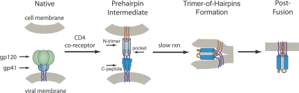
During HIV-1 entry, the highly conserved gp41 N-trimer pocket region becomes transiently exposed and vulnerable to inhibition. Using mirror-image phage display and structure-assisted design, we have discovered protease-resistant D-amino acid peptides (D-peptides) that bind the N-trimer pocket with high affinity and potently inhibit viral entry. We also report high-resolution crystal structures of two of these D-peptides in complex with a pocket mimic that suggest sources of their high potency. A trimeric version of one of these peptides is the most potent pocket-specific entry inhibitor yet reported by three orders of magnitude (IC(50) = 250 pM). These results are the first demonstration that D-peptides can form specific and high-affinity interactions with natural protein targets and strengthen their promise as therapeutic agents. The D-peptides described here address limitations associated with current L-peptide entry inhibitors and are promising leads for the prevention and treatment of HIV/AIDS.


Conflict of interest statement

The authors declare no conflict of interest.

Figures

Fig. 1.
Fig. 1.
HIV entry pathway. Upon cellular receptor recognition, gp120 and gp41 undergo conformational changes resulting in exposure of the N-trimer and its hydrophobic pocket in the prehairpin intermediate. Formation of the trimer-of-hairpins structure juxtaposes cellular and viral membranes and causes fusion. The gp41 fusion peptide (red) and transmembrane domain (purple) are also shown. For clarity, gp120 is omitted from the prehairpin intermediate. Adapted from ref. .
Fig. 2.
Fig. 2.
Structural analysis of the IQN17:2K-PIE1 inhibitor complex. (A) IQN17, consisting of IQ (orange) and gp41 (N17, gray) segments, with inhibitors (green, yellow, and purple) located in the canonical gp41 binding pockets. The purple inhibitor is mostly occluded in this view. (B) Omit map for 2K-PIE1 contoured at 3.0 × rmsd. Five of the eight pocket residues (gray, HXB2 numbering) that make hydrophobic contacts with 2K-PIE1 (green) are shown. Two hydrogen bonds (black) at the binding interface are also shown. (C) Overlay of D10-p1 (slate) and 2K-PIE1 (green) superposed by alignment of the IQN17 trimers. Intramolecular disulfide bonds (solid yellow) are also shown. (D) A slab view through the center of 2K-PIE1 (green) reveals an intact hydrophobic core (black) that excludes water. (E) A similar view of D10-p1 (slate) reveals the presence of several water molecules (red) in its core that nearly form a water channel. (F) End-on view of the complex (same color scheme as A) in which the surface from the last three residues of IQN17 have been removed. This view illustrates the packing of the inhibitor into the deep hydrophobic pocket. dK2 (blue), equivalent to the N-terminal Lys in PIE7 used for cross-linking, is highlighted.
Fig. 3.
Fig. 3.
Representative viral entry inhibition data. Each point represents the average of quadruplicate measurements normalized to uninhibited control. Error bars represent the SEM. (A) IC50 curves for various inhibitors against HXB2. (B) IC50 curves for PIE7 and (PIE7)3 against JRFL, BaL, and HXB2.
Fig. 4.
Fig. 4.
Structural analysis of the IQN17:2K-PIE1 and IQN17:PIE7 inhibitor complexes. Shown is a comparison of unique polar contacts observed in the 2K-PIE1 (A) and PIE7 (B) costructures (described in the text).

References

    1. Chan DC, Fass D, Berger JM, Kim PS. Cell. 1997;89:263–273. - PubMed
    1. Weissenhorn W, Dessen A, Harrison SC, Skehel JJ, Wiley DC. Nature. 1997;387:426–430. - PubMed
    1. Tan K, Liu J, Wang J, Shen S, Lu M. Proc Natl Acad Sci USA. 1997;94:12303–12308. - PMC - PubMed
    1. Eckert DM, Kim PS. Annu Rev Biochem. 2001;70:777–810. - PubMed
    1. Chan DC, Kim PS. Cell. 1998;93:681–684. - PubMed

Publication types