

HOXB13 promotes ovarian cancer progression
- PMID: 17942676
- PMCID: PMC2040435
- DOI: 10.1073/pnas.0707938104
HOXB13 promotes ovarian cancer progression
Abstract
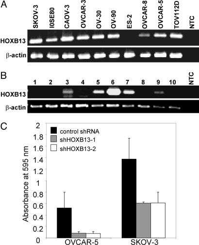
Deregulated expression of HOXB13 in a subset of estrogen receptor-positive breast cancer patients treated with tamoxifen monotherapy is associated with an aggressive clinical course and poor outcome. Because the ovary is another hormone-responsive organ, we investigated whether HOXB13 plays a role in ovarian cancer progression. We show that HOXB13 is expressed in multiple human ovarian cancer cell lines and tumors and that knockdown of endogenous HOXB13 by RNA interference in human ovarian cancer cell lines is associated with reduced cell proliferation. Ectopic expression of HOXB13 is capable of transforming p53(-/-) mouse embryonic fibroblasts and promotes cell proliferation and anchorage-independent growth in mouse ovarian cancer cell lines that contain genetic alterations in p53, myc, and ras. In this genetically defined cell line model of ovarian cancer, we demonstrate that HOXB13 collaborates with activated ras to markedly promote tumor growth in vivo and that HOXB13 confers resistance to tamoxifen-mediated apoptosis. Taken together, our results support a pro-proliferative and pro-survival role for HOXB13 in ovarian cancer.
Conflict of interest statement
The authors declare no conflict of interest.
Figures


Similar articles
-
HOXB13 and ALX4 induce SLUG expression for the promotion of EMT and cell invasion in ovarian cancer cells.Oncotarget. 2015 May 30;6(15):13359-70. doi: 10.18632/oncotarget.3673. Oncotarget. 2015. PMID: 25944620 Free PMC article.
-
Targeting the BRD4-HOXB13 Coregulated Transcriptional Networks with Bromodomain-Kinase Inhibitors to Suppress Metastatic Castration-Resistant Prostate Cancer.Mol Cancer Ther. 2018 Dec;17(12):2796-2810. doi: 10.1158/1535-7163.MCT-18-0602. Epub 2018 Sep 21. Mol Cancer Ther. 2018. PMID: 30242092 Free PMC article.
-
HOXB13-to-IL17BR expression ratio is related with tumor aggressiveness and response to tamoxifen of recurrent breast cancer: a retrospective study.J Clin Oncol. 2007 Feb 20;25(6):662-8. doi: 10.1200/JCO.2006.07.3676. J Clin Oncol. 2007. PMID: 17308270
-
Epigenetic repression of the estrogen-regulated Homeobox B13 gene in breast cancer.Carcinogenesis. 2008 Jul;29(7):1459-65. doi: 10.1093/carcin/bgn115. Epub 2008 May 21. Carcinogenesis. 2008. PMID: 18499701 Free PMC article.
-
HOXB13 in cancer development: molecular mechanisms and clinical implications.Front Med. 2025 Jun;19(3):439-455. doi: 10.1007/s11684-024-1119-x. Epub 2025 Mar 11. Front Med. 2025. PMID: 40067581 Review.
Cited by
-
Downstream of the HOX genes: Explaining conflicting tumour suppressor and oncogenic functions in cancer.Int J Cancer. 2022 Jun 15;150(12):1919-1932. doi: 10.1002/ijc.33949. Epub 2022 Feb 15. Int J Cancer. 2022. PMID: 35080776 Free PMC article. Review.
-
HOXB13 mutations and binding partners in prostate development and cancer: Function, clinical significance, and future directions.Genes Dis. 2017 Jun;4(2):75-87. doi: 10.1016/j.gendis.2017.01.003. Epub 2017 Feb 16. Genes Dis. 2017. PMID: 28798948 Free PMC article.
-
Anterior Hox genes interact with components of the neural crest specification network to induce neural crest fates.Stem Cells. 2011 May;29(5):858-70. doi: 10.1002/stem.630. Stem Cells. 2011. PMID: 21433221 Free PMC article.
-
Underexpression of HOXA11 Is Associated with Treatment Resistance and Poor Prognosis in Glioblastoma.Cancer Res Treat. 2017 Apr;49(2):387-398. doi: 10.4143/crt.2016.106. Epub 2016 Jul 19. Cancer Res Treat. 2017. PMID: 27456940 Free PMC article.
-
HOXB13 and ALX4 induce SLUG expression for the promotion of EMT and cell invasion in ovarian cancer cells.Oncotarget. 2015 May 30;6(15):13359-70. doi: 10.18632/oncotarget.3673. Oncotarget. 2015. PMID: 25944620 Free PMC article.
References
-
- Abate-Shen C. Nat Rev Cancer. 2002;2:777–785. - PubMed
-
- Miller GJ, Miller HL, van Bokhoven A, Lambert JR, Werahera PN, Schirripa O, Lucia MS, Nordeen SK. Cancer Res. 2003;63:5879–5888. - PubMed
-
- Raman V, Martensen SA, Reisman D, Evron E, Odenwald WF, Jaffee E, Marks J, Sukumar S. Nature. 2000;405:974–978. - PubMed
Publication types
MeSH terms
Substances
Grants and funding
LinkOut - more resources
Full Text Sources
Medical
Research Materials
Miscellaneous
