

A unique Extradenticle recruitment mode in the Drosophila Hox protein Ultrabithorax
- PMID: 17942685
- PMCID: PMC2040397
- DOI: 10.1073/pnas.0705832104
A unique Extradenticle recruitment mode in the Drosophila Hox protein Ultrabithorax
Abstract
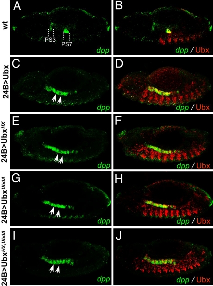
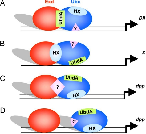
Hox transcription factors are essential for shaping body morphology in development and evolution. The control of Hox protein activity in part arises from interaction with the PBC class of partners, pre-B cell transcription factor (Pbx) proteins in vertebrates and Extradenticle (Exd) in Drosophila. Characterized interactions occur through a single mode, involving a short hexapeptide motif in the Hox protein. This apparent uniqueness in Hox-PBC interaction provides little mechanistic insight in how the same cofactors endow Hox proteins with specific and diverse activities. Here, we identify in the Drosophila Ultrabithorax (Ubx) protein a short motif responsible for an alternative mode of Exd recruitment. Together with previous reports, this finding highlights that the Hox protein Ubx has multiple ways to interact with the Exd cofactor and suggests that flexibility in Hox-PBC contacts contributes to specify and diversify Hox protein function.
Conflict of interest statement
The authors declare no conflict of interest.
Figures
Similar articles
-
Hox repression of a target gene: extradenticle-independent, additive action through multiple monomer binding sites.Development. 2002 Jul;129(13):3115-26. doi: 10.1242/dev.129.13.3115. Development. 2002. PMID: 12070087
-
Selection of distinct Hox-Extradenticle interaction modes fine-tunes Hox protein activity.Proc Natl Acad Sci U S A. 2011 Feb 8;108(6):2276-81. doi: 10.1073/pnas.1006964108. Epub 2011 Jan 24. Proc Natl Acad Sci U S A. 2011. PMID: 21262810 Free PMC article.
-
Hox proteins display a common and ancestral ability to diversify their interaction mode with the PBC class cofactors.PLoS Biol. 2012;10(6):e1001351. doi: 10.1371/journal.pbio.1001351. Epub 2012 Jun 26. PLoS Biol. 2012. PMID: 22745600 Free PMC article.
-
Roles for intrinsic disorder and fuzziness in generating context-specific function in Ultrabithorax, a Hox transcription factor.Adv Exp Med Biol. 2012;725:86-105. doi: 10.1007/978-1-4614-0659-4_6. Adv Exp Med Biol. 2012. PMID: 22399320 Review.
-
Hox cofactors in vertebrate development.Dev Biol. 2006 Mar 15;291(2):193-206. doi: 10.1016/j.ydbio.2005.10.032. Epub 2006 Mar 3. Dev Biol. 2006. PMID: 16515781 Review.
Cited by
-
A Hox Transcription Factor Collective Binds a Highly Conserved Distal-less cis-Regulatory Module to Generate Robust Transcriptional Outcomes.PLoS Genet. 2016 Apr 8;12(4):e1005981. doi: 10.1371/journal.pgen.1005981. eCollection 2016 Apr. PLoS Genet. 2016. PMID: 27058369 Free PMC article.
-
An efficient algorithm for improving structure-based prediction of transcription factor binding sites.BMC Bioinformatics. 2017 Jul 17;18(1):342. doi: 10.1186/s12859-017-1755-0. BMC Bioinformatics. 2017. PMID: 28715997 Free PMC article.
-
Molecular insights into the origin of the Hox-TALE patterning system.Elife. 2014 Mar 18;3:e01939. doi: 10.7554/eLife.01939. Elife. 2014. PMID: 24642410 Free PMC article.
-
Distinct molecular strategies for Hox-mediated limb suppression in Drosophila: from cooperativity to dispensability/antagonism in TALE partnership.PLoS Genet. 2013;9(3):e1003307. doi: 10.1371/journal.pgen.1003307. Epub 2013 Mar 7. PLoS Genet. 2013. PMID: 23505377 Free PMC article.
-
Hox genes collaborate with helix-loop-helix factor Grainyhead to promote neuroblast apoptosis along the anterior-posterior axis of the Drosophila larval central nervous system.Genetics. 2022 Aug 30;222(1):iyac101. doi: 10.1093/genetics/iyac101. Genetics. 2022. PMID: 35792854 Free PMC article.
References
-
- Pearson JC, Lemons D, McGinnis W. Nat Rev Genet. 2005;6:893–904. - PubMed
-
- Gebelein B, McKay DJ, Mann RS. Nature. 2004;431:653–659. - PubMed
-
- Mann RS, Chan S-K. Trends Genet. 1996;12:258–262. - PubMed
-
- Chan SK, Ryoo HD, Gould A, Krumlauf R, Mann RS. Development (Cambridge, UK) 1997;124:2007–2014. - PubMed
Publication types
MeSH terms
Substances
LinkOut - more resources
Full Text Sources
Molecular Biology Databases
