. 2007 Oct 23;104(43):17087-92.
doi: 10.1073/pnas.0704144104. Epub 2007 Oct 17.

Inhibition of 5-HT neuron activity and induction of depressive-like behavior by high-frequency stimulation of the subthalamic nucleus

Affiliations

Inhibition of 5-HT neuron activity and induction of depressive-like behavior by high-frequency stimulation of the subthalamic nucleus

Yasin Temel et al. Proc Natl Acad Sci U S A. .

Abstract

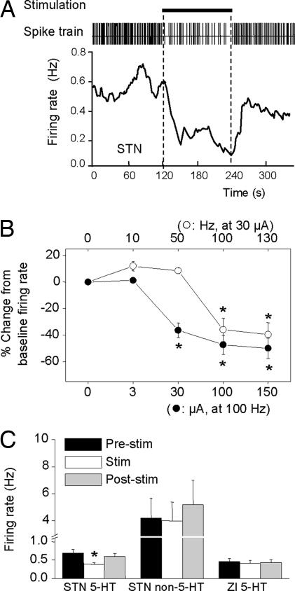
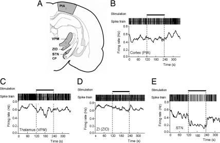
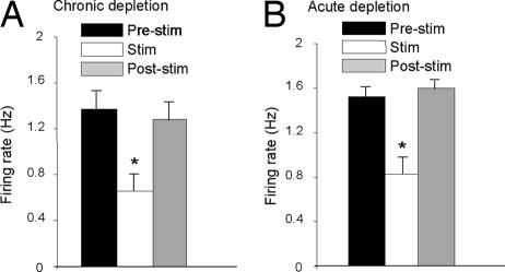
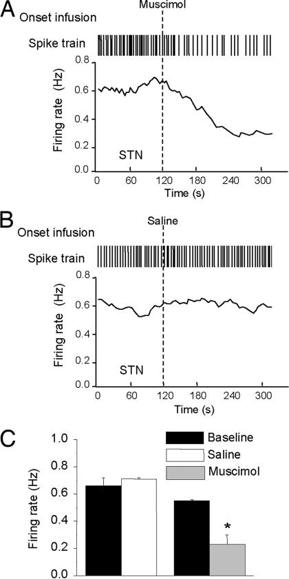
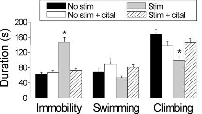
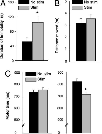
Bilateral, high-frequency stimulation (HFS) of the subthalamic nucleus (STN) is the surgical therapy of choice for movement disability in advanced Parkinson's disease (PD), but this procedure evokes debilitating psychiatric effects, including depressed mood, of unknown neural origin. Here, we report the unexpected finding that HFS of the STN inhibits midbrain 5-hydroxytryptamine (5-HT) neurons to evoke depression-related behavioral changes. We found that bilateral HFS of the STN consistently inhibited (40-50%) the firing rate of 5-HT neurons in the dorsal raphe nucleus of the rat, but not neighboring non-5-HT neurons. This effect was apparent at clinically relevant stimulation parameters (> or =100 Hz, > or =30 microA), was not elicited by HFS of either neighboring or remote structures to the STN, and was still present in rat models of PD. We also found that bilateral HFS of the STN evoked clear-cut, depressive-like behavior in a widely used experimental paradigm of depression (forced swim test), and this effect was also observed in a PD model. Importantly, the depressive-like behavior elicited by HFS of the STN was reversed by a selective 5-HT-enhancing antidepressant, thereby linking the behavioral change to decreased 5-HT neuronal activity. Overall, these findings link reduced 5-HT function to the psychiatric effects of HFS of the STN observed in PD patients and provide a rational basis for their clinical management. More generally, the powerful interaction between the STN and 5-HT system uncovered here offers insights into the high level of comorbidity of basal ganglia disease and mood disorder.


Conflict of interest statement

The authors declare no conflict of interest.

Figures

Fig. 1.
Fig. 1.
Effect of HFS of the STN on 5-HT neuronal firing. (A) Spike train and instantaneous firing rate of a 5-HT neuron in response to STN HFS. Horizontal bar indicates stimulation period (100 Hz, 100 μA, 2 min). Note inhibition of single-unit activity during stimulation. (B) Effect of stimulation frequency and current on 5-HT neuron firing rate. Each point is a mean ± SEM value (n = 6). (C) Firing rate of 5-HT neurons (Left) and non-5-HT neurons (Center) before, during, and after STN stimulation (100 Hz, 100 μA). Also, firing rate of 5-HT neurons (Right) before, during, and after stimulation of the zona incerta (ZI). *, P < 0.05 versus relevant prestimulus baseline control values.
Fig. 2.
Fig. 2.
Histological evaluation of electrode localizations. (A) Illustrative coronal section showing the histological verification of the electrode location in the STN. (Scale bar, 200 μm.) (B) Locations of stimulation sites in or close to the STN, as verified by post hoc histology. Filled circles, stimulation sites that produced consistent inhibition of 5-HT cell firing in the DRN; open circles, stimulation sites that produced no significant change in 5-HT cell firing.
Fig. 3.
Fig. 3.
Effect of HFS at various sites during descent to the STN while recording the same 5-HT neuron in the DRN. (A) Illustration of stimulation sites during electrode descent to the STN. PtA, parietal association cortex; VPM, ventroposteromedial thalamus; ZID, dorsal zona incerta. (B–E) Typical effects of electrical stimulation at the level of the cortex (B), thalamus (C), zona incerta (D), and STN (E) on the firing rate of the same 5-HT neuron in the DRN. Horizontal bars indicate stimulation periods (100 Hz, 100 μA, 2 min).
Fig. 4.
Fig. 4.
Effect of HFS of the STN on 5-HT neuronal firing after chronic and acute dopamine depletions. (A) Firing rate of 5-HT neurons before, during, and after STN stimulation (100 Hz, 100 μA) in animals chronically depleted of dopamine by i.c.v. injection of 6-hydroxy-dopamine. (B) Firing rate of 5-HT neurons before, during, and after STN stimulation (100 Hz, 100 μA) in animals acutely depleted of dopamine after coadministration of reserpine and α-methyl-p-tyrosine. *, P < 0.05 versus relevant prestimulus baseline control values.
Fig. 5.
Fig. 5.
Effect of muscimol and saline vehicle infusion into STN on 5-HT neuronal firing. (A) Spike train and instantaneous firing rate of a 5-HT neuron in response to bilateral muscimol (0.1 μg per side) infusion into STN. (B). Spike train and instantaneous firing rate of a 5-HT neuron in response to saline infusion into STN. (C) Grouped data for all 5-HT neurons tested with bilateral intra-STN infusion of muscimol or saline. Data are presented as mean ± SEM values. *, P < 0.05 versus relevant prestimulus or predrug baseline control values.
Fig. 6.
Fig. 6.
Effect of HFS of the STN on different behavioral measures in the FST. Four groups of rats (six per group) were implanted with STN electrodes and tested as follows: (i) no STN stimulation during test (No stim), (ii) pretreatment with citalopram, followed by no STN stimulation during test (No stim + cital), (iii) STN stimulation during test (Stim), and (iv) pretreatment with citalopram, followed by STN stimulation during test (Stim + cital). Stimulation parameters were 130 Hz and 150 μA for the duration of the test. Citalopram pretreatment comprised 10 mg/kg s.c. for 14 days. Bars represent mean ± SEM values (n = 6). The effect of STN stimulation was significantly different from other groups with respect to immobility and climbing: F values >11.4, *, P < 0.05 (two-way ANOVA with repeated measures, post hoc Duncan's test).
Fig. 7.
Fig. 7.
Effect of STN HFS on behaviors in the rat. (A) Immobility time of 6-hydroxydopamine-lesioned rats in the FST. (B) Locomotor activity in the open field of nonlesioned rats. Note that stimulation had no effect. (C) Motor time in nonlesioned (Left) and 6-hydroxydopamine-lesioned (Right) rats in a serial choice reaction time task (data redrawn from previous studies; refs. and 32). Note that effects of stimulation were present only in lesioned animals. STN stimulation parameters were 130 Hz and 30–150 μA for the duration of the tasks. Bars represent mean ± SEM values (n = 6). *, P < 0.05, post hoc analyses revealed that STN stimulation induced a significant effect.

Similar articles

Cited by

References

    1. Wichmann T, Delong MR. Neuron. 2006;52:197–204. - PubMed
    1. Deuschl G, Schade-Brittinger C, Krack P, Volkmann J, Schafer H, Botzel K, Daniels C, Deutschlander A, Dillmann U, Eisner W, et al. N Engl J Med. 2006;355:896–908. - PubMed
    1. Benabid AL, Chabardes S, Seigneuret E. Curr Opin Neurol. 2005;18:623–630. - PubMed
    1. Bevan MD, Magill PJ, Terman D, Bolam JP, Wilson CJ. Trends Neurosci. 2002;25:525–531. - PubMed
    1. Lozano AM, Dostrovsky J, Chen R, Ashby P. Lancet Neurol. 2002;1:225–231. - PubMed

Publication types

MeSH terms