Comparative Study
. 2007 Oct 17;27(42):11214-27.
doi: 10.1523/JNEUROSCI.3588-07.2007.

The heat shock response and chaperones/heat shock proteins in brain tumors: surface expression, release, and possible immune consequences

Affiliations
Comparative Study

The heat shock response and chaperones/heat shock proteins in brain tumors: surface expression, release, and possible immune consequences

Michael W Graner et al. J Neurosci. .

Abstract

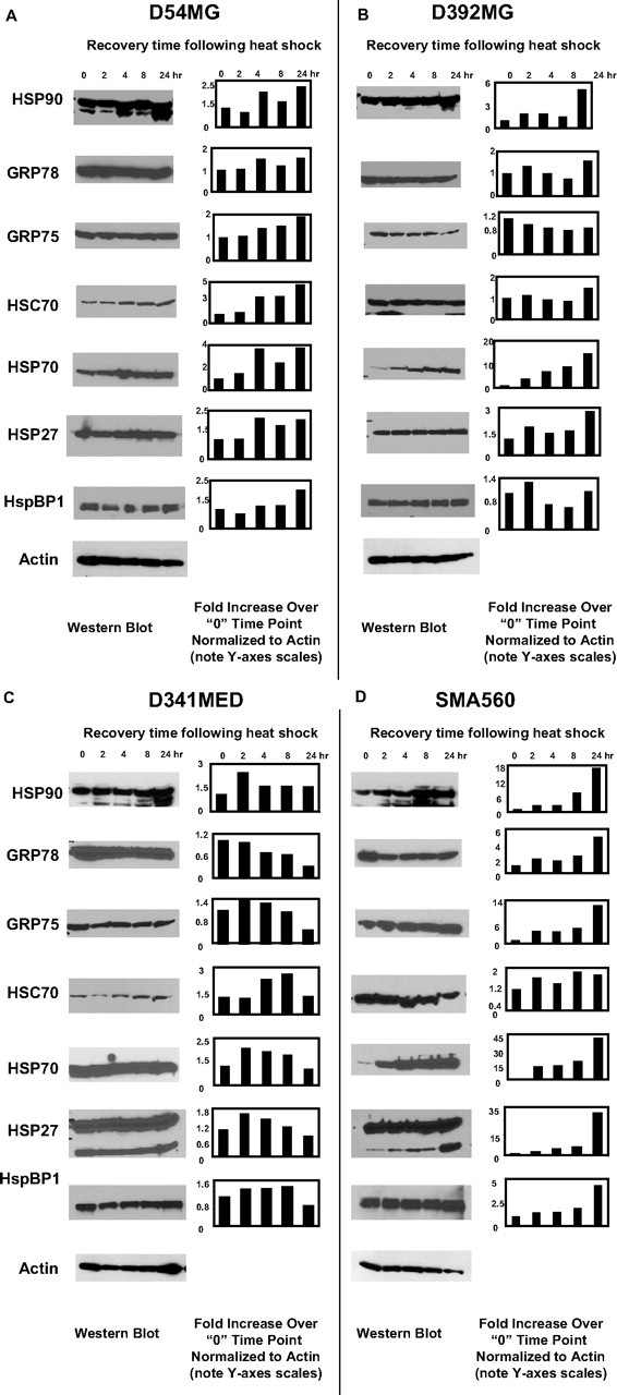
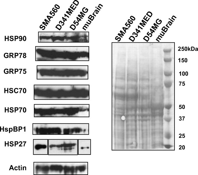
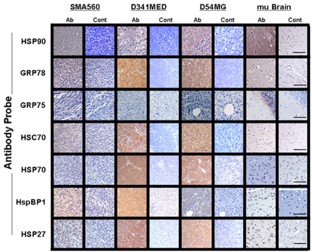
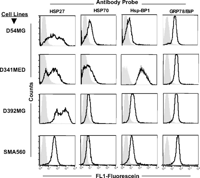
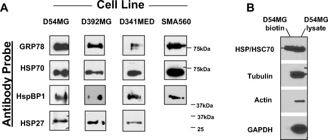
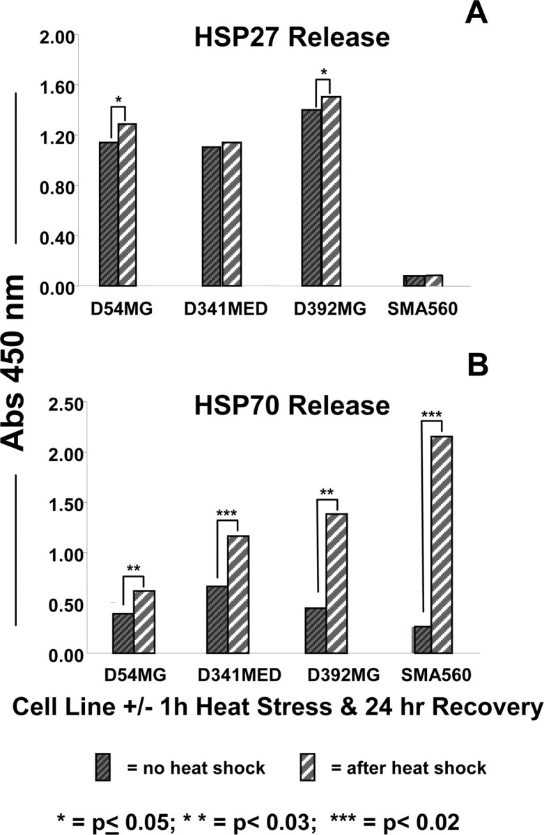
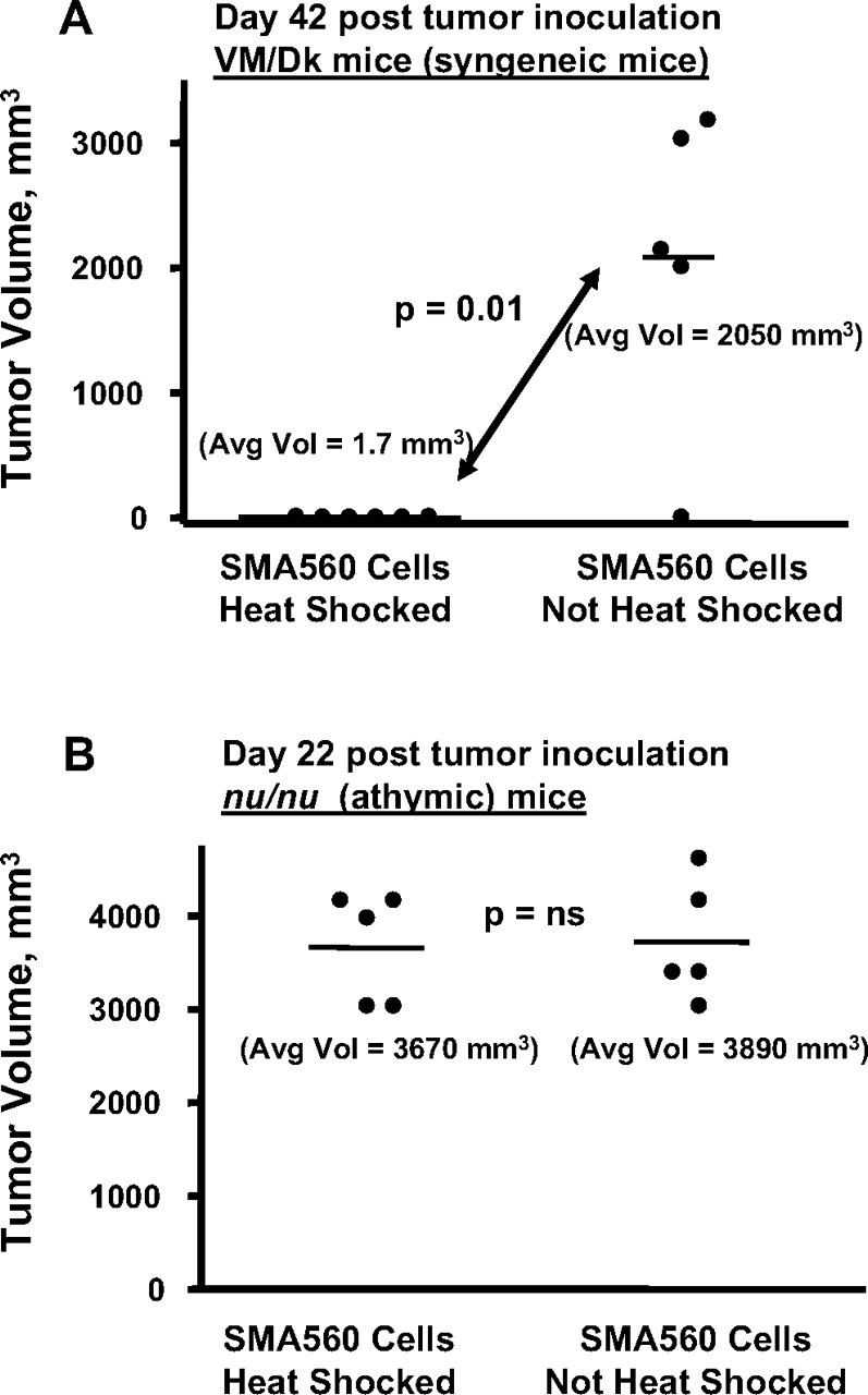
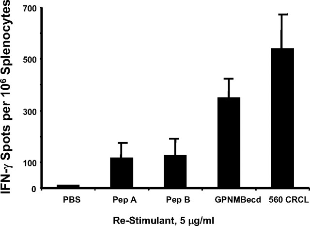
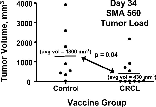
The heat shock response is a highly conserved "stress response" mechanism used by cells to protect themselves from potentially damaging insults. It often involves the upregulated expression of chaperone and heat shock proteins (HSPs) to prevent damage and aggregation at the proteome level. Like most cancers, brain tumor cells often overexpress chaperones/HSPs, probably because of the stressful atmosphere in which tumors reside, but also because of the benefits of HSP cytoprotection. However, the cellular dynamics and localization of HSPs in either stressed or unstressed conditions has not been studied extensively in brain tumor cells. We have examined the changes in HSP expression and in cell surface/extracellular localization of selected brain tumor cell lines under heat shock or normal environments. We herein report that brain tumor cell lines have considerable heat shock responses or already high constitutive HSP levels; that those cells express various HSPs, chaperones, and at least one cochaperone on their cell surfaces; and that HSPs may be released into the extracellular environment, possibly as exosome vesicular content. In studies with a murine astrocytoma cell line, heat shock dramatically reduces tumorigenicity, possibly by an immune mechanism. Additional evidence indicative of an HSP-driven immune response comes from immunization studies using tumor-derived chaperone protein vaccines, which lead to antigen-specific immune responses and reduced tumor burden in treated mice. The heat shock response and HSPs in brain tumor cells may represent an area of vulnerability in our attempts to treat these recalcitrant and deadly tumors.


Figures

Figure 1.
Figure 1.
Heat shock generally increases chaperone/heat shock protein expression in brain tumor cell lines. A–D, Cell lines derived from patient GBMs (D54MG, A; D392MG, B), from a medulloblastoma (D341MED, C), or from a murine astrocytoma (SMA560, D) were subjected to sublethal heat shock (42°C, 1 h) and allowed to recover for the time periods indicated after heat shock (2–24 h). Lysates were prepared from cells at the time points indicated (“0” refers to before heat shock); proteins were separated by LDS-PAGE and Western blotted. Antibody probes are listed on the left. Quantitative comparisons (bar graphs) were made by densitometry relative to actin staining (average of two independent experiments). Error bars indicate SD. All other SDs were <10%. Cells were >95% viable whether heat shocked or not.
Figure 2.
Figure 2.
Heat shock and chaperone/cochaperone proteins are highly expressed by brain tumor xenografts or syngeneic tumors grown in vivo compared with normal brain. Western blots of tumor or brain lysates were probed with the antibodies listed on the left; actin probe is shown as a loading control, as is the Ponceau Red-stained blot shown on the right. The panel for HSP27 is broken into three parts to indicate that the murine tissues (SMA560 and brain) were probed with different HSP27 antibodies than were the human xenograft tumors (D54MG and D341MED).
Figure 3.
Figure 3.
Brain tumors express high chaperone levels as seen in IHC. IHC of in vivo-grown brain tumors and normal mouse brain was performed on formalin-fixed, paraffin-embedded sections with the antibodies listed on the left (staining shown in Ab columns) or with control (Cont) antibodies. “mu Brain” indicates sections from nu/nu mouse brain cortex. Scale bars, 100 μm.
Figure 4.
Figure 4.
Flow cytometry of brain tumor cell surface HSPs 27, 70, GRP78, and cochaperone HspBP1. Formalin-fixed cells were incubated with the antibodies listed (directly fluoresceinated) and were analyzed by FACS (dark tracing). The isotype control is indicated by gray fill.
Figure 5.
Figure 5.
Cell surface biotinylation reveals that GRP78, HSPs 27 and 70, and HspBP1 are on the surfaces of brain tumor cell lines. A, Using membrane-impermeant biotin, the cell lines shown were surface biotinylated, and surface proteins were enriched on streptavidin matrices. Biotinylated proteins were separated on gels and Western blotted. Antibody probes are shown at left. B, To verify that only surface proteins were labeled, the fractions unbound on the streptavidin beads (D54MG lysate) and the bound/eluted fractions (D54MG biotin) were probed with antibodies against cytosolic proteins tubulin, actin, and glyceraldehyde-3 phosphate dehydrogenase (GAPDH). HSP/HSC70 was found in both the nonbiotinylated cytosolic fraction and the labeled membrane fraction (as expected, from the results in A).
Figure 6.
Figure 6.
HSPs 27 and 70 are released into culture medium by brain tumor cell lines under normal and heat shock conditions. The cell lines listed were left untreated (dark hatched bars) or were heat stressed (42°C, 1 h; light striped bars). Media were exchanged after heat shock, and cells recovered for 24 h. Media were then harvested for ELISA analyses for HSP27 (A) and HSP70 (B) using commercial kits. Statistical differences in HSP output were determined using t tests, with the p values indicated by asterisks. Those unmarked were not significant. SDs of replicate measurements were always <10% of the average and would be essentially unreadable as error bars; thus, they are not shown.
Figure 7.
Figure 7.
Brain tumor cell lines release exosomes into the media. Exosomes were isolated from the spent media from cell lines D54MG and SMA560 by differential centrifugation. A, Exosomes recovered in the final pellets were observed by electron microscopy after uranyl acetate negative staining (arrows). B, Western blots of exosome material show that HSPs 90, 70, 60, and 27 are present. For the SMA560 line, the asterisk indicates a rodent mitochondrial small HSP of ∼37 kDa rather than the expected 20–30 kDa.
Figure 8.
Figure 8.
Heat-shocked SMA560 cells show reduced tumorigenicity in immune-competent syngeneic mice. SMA560 cells were heat shocked (42°C, 1 h) or were left untreated. A, After a 24 h recovery, groups of 5 VM/Dk mice were injected with 100,000 cells, either heat shocked or untreated, subcutaneously into the right flanks, and tumor volumes were measured 42 d later. Cells in both groups were of equal viability. p value (t test) between heat-shocked cell tumor growth versus control is shown. In this experiment, the solitary mouse in the control group without tumor later grew one and had to be killed 90 d after tumor inoculation. B, The same heat shock experiment was conducted using immunoincompetent athymic (nu/nu) mice as tumor recipients. Both heat-shocked and non-heat-shocked tumor cells grew in nude mice at statistically indistinguishable rates (p value, not significant), and mice were killed on day 22.
Figure 9.
Figure 9.
Mice receiving brain tumor-derived CRCL vaccines develop specific splenocyte reactivity against brain tumor antigens as well as against undefined antigens within the whole vaccine. VM/Dk mice were vaccinated on day −14 and day −7 with 20 μg of SMA560-derived CRCL each time. On day 0, splenocytes were harvested, plated, and restimulated with the antigens shown (GPNMBecd; Pep A and Pep B are GPNMB 18-mer and 22-mer peptides, respectively; see Materials and Methods for sequences). Restimulation was with antigen concentrations of 5 μg/ml. Interferon-γ output was quantified by ELISPOT assay; shown are averages and SD of triplicate wells. Error bars represent SEM.
Figure 10.
Figure 10.
Vaccination with SMA560 CRCL reduces tumor burden in syngeneic, tumor-bearing mice. Groups of VM/Dk mice (10 per group, initially) were inoculated subcutaneously with 100,000 SMA60 cells. Five days later, one group of mice received subcutaneous injections of CRCL vaccines (50 μg) derived from SMA560 tumor. Control mice received equal volumes of saline. Injections were given on the flank opposite the tumor, and tumor growth was monitored thereafter. Before day 34, two mice in the control group had died from their tumors, and their tumor volumes were censored from the averages and statistics shown here. avg vol, Average volume.

References

    1. Arap MA, Lahdenranta J, Mintz PJ, Hajitou A, Sarkis AS, Arap W, Pasqualini R. Cell surface expression of the stress response chaperone GRP78 enables tumor targeting by circulating ligands. Cancer Cell. 2004;6:275–284. - PubMed
    1. Asea A. Stress proteins and initiation of immune response: chaperokine activity of hsp72. Exerc Immunol Rev. 2005;11:34–45. - PMC - PubMed
    1. Bausero MA, Page DT, Osinaga E, Asea A. Surface expression of Hsp25 and Hsp72 differentially regulates tumor growth and metastasis. Tumour Biol. 2004;25:243–251. - PMC - PubMed
    1. Binder RJ, Vatner R, Srivastava P. The heat-shock protein receptors: some answers and more questions. Tissue Antigens. 2004;64:442–451. - PubMed
    1. Bullard DE, Schold SC, Jr, Bigner SH, Bigner DD. Growth and chemotherapeutic response in athymic mice of tumors arising from human glioma-derived cell lines. J Neuropathol Exp Neurol. 1981;40:410–427. - PubMed

Publication types

MeSH terms