

Orbital prefrontal cortex is required for object-in-place scene memory but not performance of a strategy implementation task
- PMID: 17942727
- PMCID: PMC2092501
- DOI: 10.1523/JNEUROSCI.3369-07.2007
Orbital prefrontal cortex is required for object-in-place scene memory but not performance of a strategy implementation task
Abstract
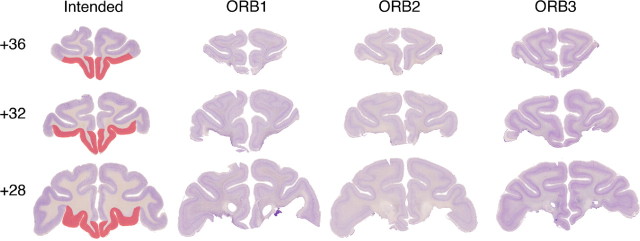
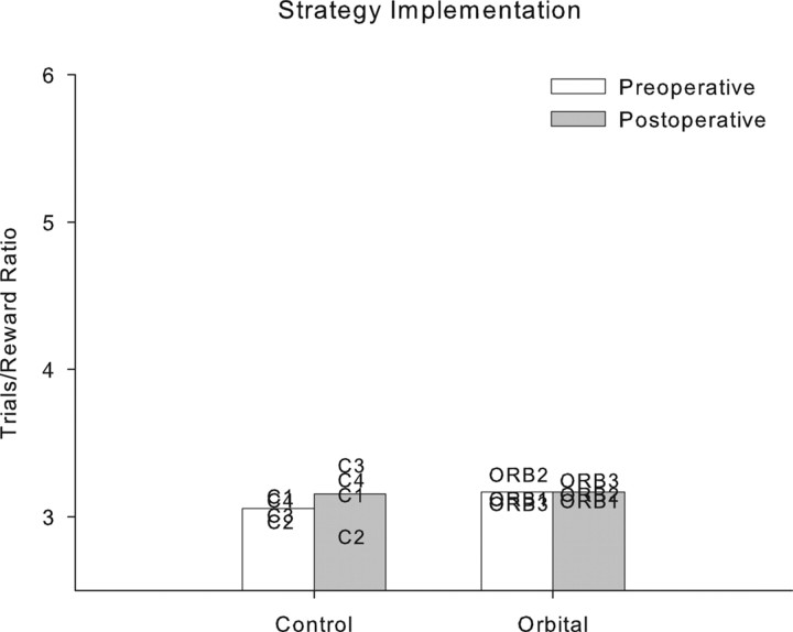
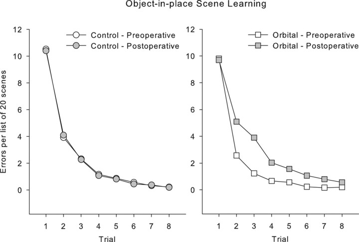
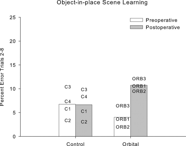
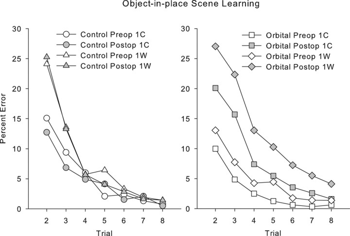
The orbital prefrontal cortex is thought to be involved in behavioral flexibility in primates, and human neuroimaging studies have identified orbital prefrontal activation during episodic memory encoding. The goal of the present study was to ascertain whether deficits in strategy implementation and episodic memory that occur after ablation of the entire prefrontal cortex can be ascribed to damage to the orbital prefrontal cortex. Rhesus monkeys were preoperatively trained on two behavioral tasks, the performance of both of which is severely impaired by the disconnection of frontal cortex from inferotemporal cortex. In the strategy implementation task, monkeys were required to learn about two categories of objects, each associated with a different strategy that had to be performed to obtain food reward. The different strategies had to be applied flexibly to optimize the rate of reward delivery. In the scene memory task, monkeys learned 20 new object-in-place discrimination problems in each session. Monkeys were tested on both tasks before and after bilateral ablation of orbital prefrontal cortex. These lesions impaired new scene learning but had no effect on strategy implementation. This finding supports a role for the orbital prefrontal cortex in memory but places limits on the involvement of orbital prefrontal cortex in the representation and implementation of behavioral goals and strategies.
Figures
References
-
- Aggleton JP, McMackin D, Carpenter K, Hornak J, Kapur N, Halpin S, Wiles CM, Kamel H, Brennan P, Carton S, Gaffan D. Differential cognitive effects of colloid cysts in the third ventricle that spare or compromise the fornix. Brain. 2000;123:800–815. - PubMed
-
- Browning PG, Easton A, Buckley MJ, Gaffan D. The role of prefrontal cortex in object-in-place learning in monkeys. Eur J Neurosci. 2005;22:3281–3291. - PubMed
-
- Carmichael ST, Price JL. Limbic connections of the orbital and medial prefrontal cortex in macaque monkeys. J Comp Neurol. 1995;363:615–641. - PubMed
-
- Chiavaras MM, Petrides M. Orbitofrontal sulci of the human and macaque monkey brain. J Comp Neurol. 2000;422:35–54. - PubMed
Publication types
MeSH terms
Grants and funding
LinkOut - more resources
Full Text Sources
Medical