Comparative Study
. 2007 Oct 17;27(42):11354-65.
doi: 10.1523/JNEUROSCI.0723-07.2007.

Conditional knock-out of Kir4.1 leads to glial membrane depolarization, inhibition of potassium and glutamate uptake, and enhanced short-term synaptic potentiation

Affiliations
Comparative Study

Conditional knock-out of Kir4.1 leads to glial membrane depolarization, inhibition of potassium and glutamate uptake, and enhanced short-term synaptic potentiation

Biljana Djukic et al. J Neurosci. .

Abstract

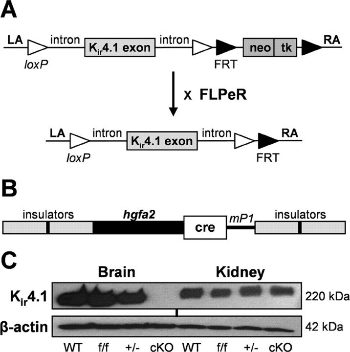
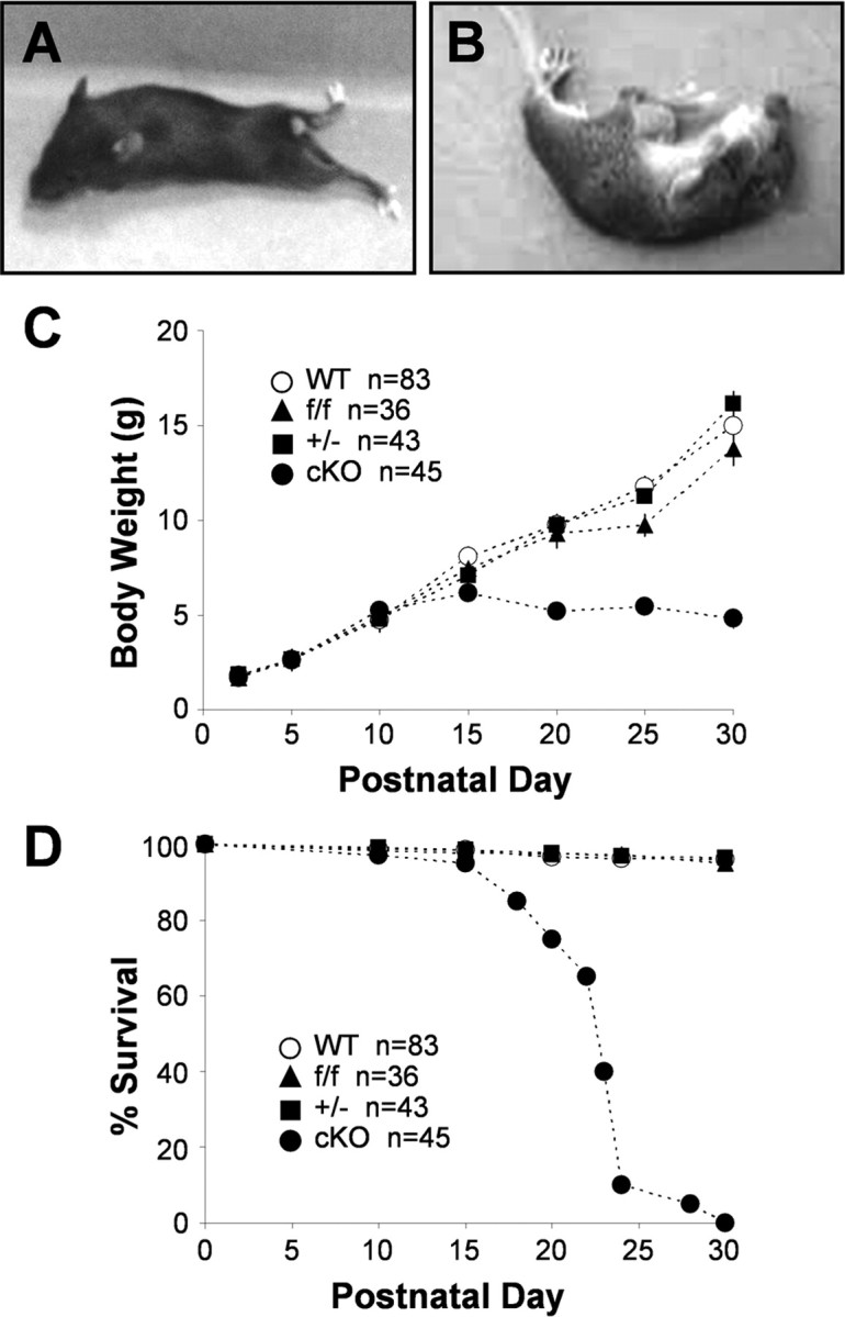
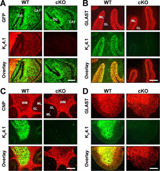
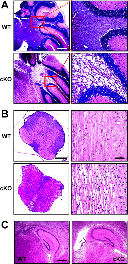
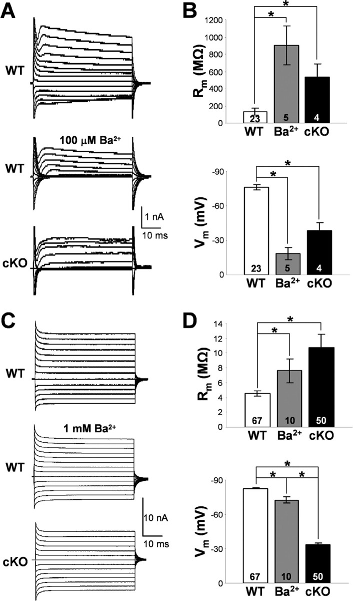
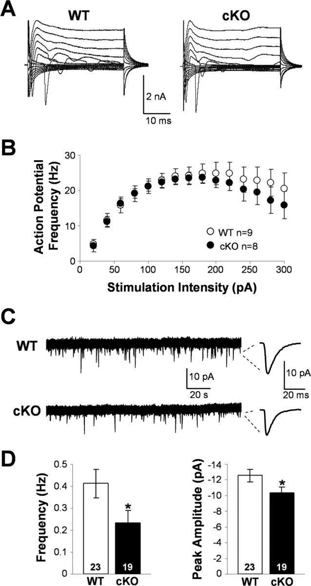
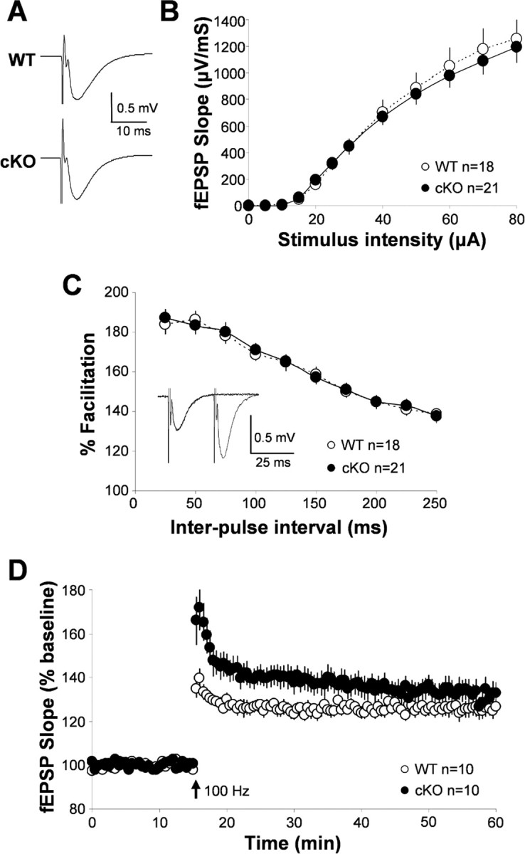
During neuronal activity, extracellular potassium concentration ([K+]out) becomes elevated and, if uncorrected, causes neuronal depolarization, hyperexcitability, and seizures. Clearance of K+ from the extracellular space, termed K+ spatial buffering, is considered to be an important function of astrocytes. Results from a number of studies suggest that maintenance of [K+]out by astrocytes is mediated by K+ uptake through the inward-rectifying Kir4.1 channels. To study the role of this channel in astrocyte physiology and neuronal excitability, we generated a conditional knock-out (cKO) of Kir4.1 directed to astrocytes via the human glial fibrillary acidic protein promoter gfa2. Kir4.1 cKO mice die prematurely and display severe ataxia and stress-induced seizures. Electrophysiological recordings revealed severe depolarization of both passive astrocytes and complex glia in Kir4.1 cKO hippocampal slices. Complex cell depolarization appears to be a direct consequence of Kir4.1 removal, whereas passive astrocyte depolarization seems to arise from an indirect developmental process. Furthermore, we observed a significant loss of complex glia, suggestive of a role for Kir4.1 in astrocyte development. Kir4.1 cKO passive astrocytes displayed a marked impairment of both K+ and glutamate uptake. Surprisingly, membrane and action potential properties of CA1 pyramidal neurons, as well as basal synaptic transmission in the CA1 stratum radiatum appeared unaffected, whereas spontaneous neuronal activity was reduced in the Kir4.1 cKO. However, high-frequency stimulation revealed greatly elevated posttetanic potentiation and short-term potentiation in Kir4.1 cKO hippocampus. Our findings implicate a role for glial Kir4.1 channel subunit in the modulation of synaptic strength.


Figures

Figure 1.
Figure 1.
Generation of Kir4.1 cKO mice. A, Targeting construct for the recombinant Kir4.1f/f mouse line. neo/tk selection cassette was removed by crossing Kir4.1f/f and FLPeR mice. LA, Left arm homology; RA, right arm homology. B, Construct for the transgenic hGFAP–Cre line. mP1, Poly(A) signal from mouse protamine1 gene. C, Kir4.1 Western blot from P20 littermate WT, Kir4.1f/f, Kir4.1+/−, and Kir4.1 cKO brain and kidney tissue. Blotting for β-actin was used as a loading control.
Figure 2.
Figure 2.
Kir4.1 cKO phenotype. Kir4.1 cKO mice exhibit ataxia and hindleg paralysis (A), stress-induced seizures (B), growth retardation (C), and premature lethality (D).
Figure 3.
Figure 3.
Loss of Kir4.1 from the CNS gray and white matter. A, Fluorescent GFP and Kir4.1 immunostaining in WT/S100β–eGFP and Kir4.1 cKO/S100β–eGFP hippocampus. DG, Dentate gyrus; CA1, CA1 pyramidal cell layer. Scale bar, 50 μm. B, C, Fluorescent immunostaining for Kir4.1, astrocyte marker GLAST (B) and oligodendrocyte marker CNP (C) in WT and Kir4.1 cKO cerebellum. GL, Granule cell layer; ML, molecular layer; WM, white matter. Scale bar, 500 μm. D, Fluorescent immunostaining for Kir4.1 and astrocyte marker GLAST in WT and Kir4.1 cKO spinal cord. Scale bar, 100 μm.
Figure 4.
Figure 4.
Morphological changes and white-matter vacuolization in the Kir4.1 cKO CNS. A, Paraffin-embedded sagittal sections of the WT and Kir4.1 cKO cerebellum stained with solochrome and eosin. Scale bar, 1 mm. B, Left, Transverse thoracic spinal cord sections stained with solochrome and eosin. Scale bar, 200 μm. Right, Longitudinal thoracic spinal cord sections stained with hematoxylin and eosin. Scale bar, 50 μm. C, Sagittal sections of the hippocampus stained with hematoxylin and eosin. Scale bar, 500 μm.
Figure 5.
Figure 5.
Membrane properties of the hippocampal passive and complex glia. A, Representative whole-cell currents of WT and Kir4.1 cKO complex glia in CA1 stratum radiatum. Middle trace depicts Ba2+ block of whole-cell currents in a WT complex cell. B, Rm and Vm of complex glia. Mean ± SEM; *p < 0.05. C, Representative whole-cell currents of WT and Kir4.1 cKO passive glial cells in CA1 stratum radiatum. Middle trace depicts Ba2+ block of whole-cell currents in a passive WT cell. D, Rm and Vm of passive glia. Mean ± SEM; *p < 0.05.
Figure 6.
Figure 6.
Potassium and glutamate uptake by astrocytes. A, Whole-cell current traces of WT and Kir4.1 cKO passive astrocytes during Schaffer collateral stimulation in control condition (black trace), after Ba2+ block (dark gray trace), and after Ba2+/TBOA block (light gray trace). Peak amplitude of K+ uptake (Ba2+-sensitive) current (B) and peak amplitude of GluT (TBOA-sensitive) current (C) in WT and cKO cells. Mean ± SEM; *p < 0.05.
Figure 7.
Figure 7.
Spontaneous activity, membrane, and action potential properties of wild-type and Kir4.1 cKO pyramidal neurons. A, Representative whole-cell currents of WT and Kir4.1 cKO CA1 pyramidal neurons. B, Influence of the stimulation intensity on the action potential frequency in WT and Kir4.1 cKO neurons. Mean ± SEM. C, Representative sEPSC recordings. D, sEPSC frequency and peak amplitude in WT and Kir4.1 cKO CA1 pyramidal neurons. Mean ± SEM; *p < 0.05.
Figure 8.
Figure 8.
Synaptic transmission and plasticity in the wild-type and Kir4.1 cKO hippocampus. A, Representative WT and Kir4.1 cKO fEPSPs. B, Input/output curve: influence of stimulation intensity on fEPSP slope. C, Paired-pulse facilitation (representative trace from WT shown as inset). D, Long-term potentiation in WT and Kir4.1 cKO hippocampus.

References

    1. Akopian G, Kuprijanova E, Kressin K, Steinhuser C. Analysis of ion channel expression by astrocytes in red nucleus brain stem slices of the rat. Glia. 1997;19:234–246. - PubMed
    1. Amzica F, Massimini M, Manfridi A. Spatial buffering during slow and paroxysmal sleep oscillations in cortical networks of glial cells in vivo. J Neurosci. 2002;22:1042–1053. - PMC - PubMed
    1. Balestrino M, Aitken PG, Somjen GG. The effects of moderate changes of extracellular K+ and Ca2+ on synaptic and neural function in the CA1 region of the hippocampal slice. Brain Res. 1986;377:229–239. - PubMed
    1. Ballanyi K, Grafe P, ten Bruggencate G. Ion activities and potassium uptake mechanisms of glial cells in guinea-pig olfactory cortex slices. J Physiol (Lond) 1987;382:159–174. - PMC - PubMed
    1. Barbour B, Keller BU, Llano I, Marty A. Prolonged presence of glutamate during excitatory synaptic transmission to cerebellar Purkinje cells. Neuron. 1994;12:1331–1343. - PubMed

Publication types

MeSH terms