Comparative Study
. 2007 Oct 17;27(42):11389-400.
doi: 10.1523/JNEUROSCI.3473-07.2007.

Role of kinase suppressor of Ras-1 in neuronal survival signaling by extracellular signal-regulated kinase 1/2

Affiliations
Comparative Study

Role of kinase suppressor of Ras-1 in neuronal survival signaling by extracellular signal-regulated kinase 1/2

Erzsebet Szatmari et al. J Neurosci. .

Abstract

Scaffolding proteins including kinase suppressor of Ras-1 (KSR1) determine specificity of signaling by extracellular signal-regulated kinase 1/2 (ERK1/2), enabling it to couple diverse extracellular stimuli to various cellular responses. The scaffolding protein(s) that contributes to ERK1/2-mediated neuronal survival has not yet been identified. In cultured rat cortical neurons, BDNF activates ERK1/2 to enhance neuronal survival by suppressing DNA damage- or trophic deprivation-induced apoptosis. Here we report that in this system, BDNF increased KSR1 association with activated ERK1/2, whereas KSR1 knockdown with a short hairpin (sh) RNA reduced BDNF-mediated activation of ERK1/2 and protection against a DNA-damaging drug, camptothecin (CPT). In contrast, BDNF suppression of trophic deprivation-induced apoptosis was unaffected by shKSR1 although blocked by shERK1/2. Also, overexpression of KSR1 enhanced BDNF protection against CPT. Therefore, KSR1 is specifically involved in antigenotoxic activation of ERK1/2 by BDNF. To test whether KSR1 contributes to ERK1/2 activation by other neuroprotective stimuli, we used a cAMP-elevating drug, forskolin. In cortical neurons, ERK1/2 activation by forskolin was protein kinase A (PKA) dependent but TrkB (receptor tyrosine kinase B) independent and was accompanied by the increased association between KSR1 and active ERK1/2. Forskolin suppressed CPT-induced apoptosis in a KSR1 and ERK1/2-dependent manner. Inhibition of PKA abolished forskolin protection, whereas selective PKA activation resulted in an ERK1/2- and KSR1-mediated decrease in apoptosis. Hence, KSR1 is critical for the antiapoptotic activation of ERK1/2 by BDNF or cAMP/PKA signaling. In addition, these novel data indicate that stimulation of cAMP signaling is a candidate neuroprotective strategy to intervene against neurotoxicity of DNA-damaging agents.


Figures
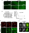
Figure 1.
Figure 1.
KSR1 associates with active ERK1/2 in BDNF-stimulated rat cortical neurons. A, KSR1 expression in the adult rat brain. KSR1 immunoreactivity in coronal rat brain sections was visualized by immunofluorescence with a rabbit anti-KSR1 antibody (b′, c′, e′, f′); IgG from nonimmunized rabbits was used to control the specificity of staining (a′, d′). Low-magnification views of the neocortex (a′, b′) and the hippocampus (d′, e′) are shown. In c′ and f′, higher-magnification views of the neocortical layers 4/5 and the CA3 subfield of the hippocampus are presented as indicated. Single confocal sections are shown; a similar pattern of KSR1 staining was observed in sections from four different animals. B, Coimmunoprecipitation of KSR1 and active ERK1/2 from BDNF-stimulated neurons. Cortical neurons were treated as indicated. Proteins immunoprecipitated with a KSR1 antibody were analyzed by Western blotting for KSR1, active ERK1/2 (phosphorylated at Thr183 and Tyr185, pERK1/2), total ERK1/2 (ERK1/2) or MKK1/2. BDNF induced the association between KSR1 and pERK1/2. Note the presence of total ERK1/2 associated with KSR1 in unstimulated neurons. No pERK1/2 or ERK1/2 was found if the immunoprecipitation (IP) was performed using nonimmune IgG (data not shown). The interactions between KSR1 and MKK1/2 were unaffected by BDNF. Similar levels of KSR1 indicated consistent immunoprecipitation. The graph presents quantitative analysis of pERK1/2-KSR1 association from three independent experiments. Error bars are SEM. p < 0.001 (ANOVA). C, D, KSR1 colocalizes with pERK1/2 in BDNF-stimulated cultured rat cortical neurons. C, Immunofluorescence analysis of KSR1 and pERK1/2 at the indicated times after addition of 10 ng/ml BDNF. At 0, 2, or 5 min after BDNF addition, perikarial KSR1 immunoreactivity was concentrated in membrane-containing Golgi-like structures (arrows). BDNF increased pERK1/2 immunoreactivity at all time points. Z-stack confocal images are presented. D, Z-stack confocal images of representative neurons coimmunostained for KSR1 and pERK at 5 min after BDNF stimulation. Note the colocalization of pERK1/2 and KSR1 in membrane-containing structures with concentrated KSR1 immunostaining.
Figure 2.
Figure 2.
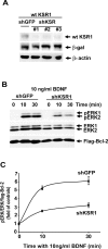
KSR1 contributes to BDNF-mediated activation of ERK1/2. A, Validation of shRNA constructs targeting KSR1. Freshly isolated neurons were coelectroporated with WT-KSR1 and β-gal expression constructs (1 μg of wtKSR1 and 0.2 μg of pON260 plasmid DNAs/2 × 106 cells, respectively) together with 1.5 μg of plasmid DNA/2 × 106 cells of each of the following plasmids: control shRNA (shGFP) or one of three KSR1-specific shRNAs cloned in pSUPER as indicated (shKSR1 #1, #2, or #3; see Materials and Methods for details). Forty-eight hours after electroporation, KSR1 levels were determined by Western blotting followed by reprobing the blot with an anti-β-gal or anti-β-actin antibodies to monitor transfection efficiency or protein loading, respectively. Each of the shKSR1 constructs inhibited WT-KSR1 expression. Similar results were obtained in another independent experiment. B, Freshly isolated cortical neurons were coelectroporated with control shRNA (shGFP) or shKSR (a 1:1:1 mix of shKSR1 #1, #2, and #3) together with a Flag-BclII expression vector (4 μg of shRNA and 0.5 μg of pcDNA3-Flag-BclII/5 × 106 neurons, respectively). Two days later, neurons were stimulated with BDNF as indicated. The BDNF-mediated ERK1/2 activation was analyzed by Western blotting for pERK1/2 followed by reprobing for ERK1/2. In addition, reprobing for flag-Bcl-2 was performed to monitor transfection efficiency. C, Analysis of pERK/ERK/Flag-Bcl-2 indicated that shRNA-mediated knockdown of KSR1 reduced ERK1/2 activation in BDNF-stimulated neurons. Data from two independent experiments are presented. Error bars indicate SEM.
Figure 3.
Figure 3.
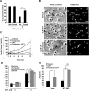
KSR1 is required for the BDNF-mediated protection against DNA damage. Cortical neurons were cotransfected with expression plasmids for β-gal (pON260) and shKSR1 or shERK1/2 as indicated (0.2 and 1 μg of plasmid DNAs/500,000 neurons, respectively); shGFP was used as a control. One day (A, B) or 3 d (D, E) after transfection, neurons were exposed to 5 μm CPT or TD for 24 h. In the TD experiment, control neurons were subjected to sham washes (Control; see Materials and Methods for details). The treatments were performed in the presence or absence of 10 ng/ml BDNF. Transfected cells were identified by β-gal immunostaining. The morphology of the nuclei was visualized by counterstaining with Hoechst 33258 (HOECHST). A, B, Effects of shERK1/2 on BDNF neuroprotection against CPT- or TD-induced apoptosis. BDNF-mediated protection against CPT and TD was antagonized by shERK1/2. C, Representative photomicrographs depicting neurons transfected with shKSR1. Arrows indicate viable neurons; arrowheads indicate neurons with nuclear alterations indicating apoptosis (nuclear shrinkage and condensation with or without fragmentation). D, E, Effects of shKSR1 on BDNF neuroprotection against CPT- or TD-induced apoptosis. BDNF-mediated protection against CPT but not TD was reduced by shKSR1. F, KSR1 appears to be specific for the antiapoptotic activation of ERK1/2 that protects against CPT but not TD. In A, B, and D, data represent averages of duplicate determinations from three independent experiments. In E, averages of duplicate determinations from two independent experiments are shown. Error bars indicate SEM. *p < 0.05; **p < 0.01; ***p < 0.001. ns, Nonsignificant.
Figure 4.
Figure 4.
Overexpression of wtKSR1 increases BDNF protection against CPT-induced apoptosis. Cortical neurons were cotransfected with expression plasmids for β-gal and WT-KSR1 or an empty cloning vector, pcDNA3.1 (0.2 and 0.03 μg of plasmid DNAs/5 × 105 neurons, respectively). Two days after transfection, neurons were exposed to 5 μm CPT in the presence of 0 or 1 or 10 ng/ml BDNF. After 24 h, WT-KSR1 increased antiapoptotic protection by low but not high concentration of BDNF. Data are averages of duplicate determinations ± SEM from two independent experiments.
Figure 5.
Figure 5.
Stimulation of cAMP signaling activates ERK1/2 in a TrkB-independent and PKA-dependent manner. Cortical neurons were treated with the adenylyl cyclase activator forskolin (10 μm) or BDNF as indicated. The levels of active (phosphorylated) forms of ERK1/2 (pERK1/2; A, C, D) or MKK1/2 (pMKK1/2; A) were analyzed by Western blotting. Levels of total ERK1/2 or MKK1/2 were determined by reprobing blots with appropriate antibodies. In B, the level of activated TrkB that was phosphorylated at the phospholipase C-γ binding site (Y816, pTrkB) was determined by Western blotting after immunoprecipitation of total TrkB with a pan-Trk antibody. Total levels of Trk were revealed by reprobing with the pan-Trk antibody. All results were replicated in two to three independent experiments. A, Forskolin activated MKK1/2 and ERK1/2. MKK1/2 inhibition with U0126 abolished ERK1/2 activation by forskolin, indicating a critical role of MKK1/2. B, ERK1/2 activation by forskolin was not accompanied by TrkB activation, suggesting that, in cortical neurons, TrkB is not involved in cAMP signaling to ERK1/2. In contrast, TrkB was strongly activated by 10 ng/ml BDNF. C, Forskolin failed to activate ERK1/2 in the presence of the PKA inhibitor H89 or myrPKI (PKI). D, BDNF-induced activation of ERK1/2 was not affected by PKA inhibition using H89. Effects of H89 were tested using both low and high concentrations of BDNF that were added for 30 min as indicated. E, ERK1/2 activation in response to cAMP and BDNF is mediated by different pathways.
Figure 6.
Figure 6.
KSR1 associates with active ERK1/2 after stimulation of cAMP signaling. Neurons were treated as indicated. The association between phosphorylated ERK1/2 (pERK1/2) and KSR1 was studied by coimmunoprecipitation (IP) as described for Figure 1. A, Activation of cAMP signaling by forskolin increased the association between pERK1/2 and KSR1 without affecting MKK1/2 binding to KSR1. B, Increased ratios between coimmunoprecipitated pERK1/2 and KSR1 from forskolin-stimulated neurons. Data in the graph represent three independent experiments. Error bars indicate SEM. p < 0.001 (ANOVA). C, Inhibition of PKA with H89 (10 μm) disrupted the forskolin (10 μm)-induced increase in KSR1–pERK1/2 association. H89 did not affect BDNF (10 ng/ml)-induced interactions between KSR1 and pERK1/2. These results suggest a role for PKA in cAMP-mediated activation of KSR1/ERK signaling. Similar results were obtained in two independent experiments.
Figure 7.
Figure 7.
Stimulation of cAMP signaling inhibits DNA damage-induced apoptosis in an ERK1/2-dependent manner. A, Neurons were treated with 5 μm CPT for 48 h in the presence or absence of 10 μm forskolin (Forsk) or 0.2% DMSO (Veh). Neuronal survival was determined by an MTT assay. Data represent averages of triplicate determinations from four independent experiments ± SEM. Forskolin reduced CPT-induced neurotoxicity (**p < 0.01). B, Morphology of neurons protected with forskolin against toxicity by a 24 h CPT exposure. Hoechst staining and corresponding phase-contrast images are presented. Arrows indicate viable neurons; arrowheads indicate neurons with signs of apoptosis. The CPT-induced increase in the number of apoptotic cells was attenuated by forskolin. C, Forskolin reduced CPT-induced caspase-3 activity. Data are averages of triplicate determinations from two independent experiments. D, Effects of an ERK1/2 pathway inhibitor, U0126, on forskolin neuroprotection. Cortical neurons were treated for 24 h with CPT in the presence or absence of 10 μm forskolin and/or 50 μm U0126. U0126 abolished the forskolin mediated-protection against CPT, indicating a critical role for the ERK1/2 pathway in the antiapoptotic effects of cAMP signaling in CPT-treated neurons. Data represent averages of duplicate determinations from four independent experiments ± SEM. **p < 0.01. E, Cortical neurons were cotransfected with expression plasmids for β-gal (pON260) and an ERK1/2-specific phosphatase, MKP3 (WT-MKP3; 0.2 and 0.3 μg of plasmid DNA/500,000 cells, respectively). The empty cloning vector pcDNA3.1 (Vector) was used as a control. After 48 h, neurons were treated for 24 h with vehicle (0.2% DMSO) or 5 μm CPT with or without 10 μm forskolin. Quantification of apoptosis in transfected neurons indicated removal of forskolin-mediated protection by WT-MKP3. These data also support the critical role for ERK1/2 in cAMP neuroprotection. Data represent averages of duplicate determinations from three independent experiments ± SEM. **p < 0.01. ns, Not significant.
Figure 8.
Figure 8.
Role of KSR1 in cAMP neuroprotection against DNA damage. Neurons were transfected as in Figure 2 B–D. Seventy-two hours after transfection, neurons were treated for 24 h as indicated. Quantification of apoptosis in transfected neurons revealed that shKSR1 reduced forskolin-mediated protection against CPT. Data represent averages of duplicate determinations from four independent experiments ± SEM. **p < 0.01; ***p < 0.001.
Figure 9.
Figure 9.
PKA is required for cAMP-mediated neuroprotection against CPT-induced apoptosis. A, A 24 h CPT treatment of cortical neurons was performed in the presence or absence of 10 μm forskolin and/or 10 μm PKA inhibitor H89 as indicated. H89 abolished antiapoptotic effects of forskolin. B, C, Cortical neurons were cotransfected with expression constructs for β-gal (pON260) and an endogenous protein inhibitor of PKA, PKIα (0.2 and 0.3 μg of plasmid DNAs/ 500,000 neurons, respectively). Empty cloning vector (Vector, pcDNA3.1) was used as a control for PKIα construct. Twenty-four hours after transfection, neurons were treated with 5 μm CPT in the presence or absence of 10 μm forskolin or 10 ng/ml BDNF as indicated. After 24 h, apoptosis was analyzed in transfected neurons. Forskolin but not BDNF neuroprotection against CPT was abolished by the overexpressed PKIα. Data in all panels represent averages of duplicate determinations from three independent experiments ± SEM. ***p < 0.001. ns, Not significant.
Figure 10.
Figure 10.
PKA is sufficient to activate protective signaling by KSR1–ERK1/2. A, Cortical neurons were treated with CPT for 48 h in the presence or absence of a selective PKA activator, Sp (100 μm). Neuronal survival was determined by the MTT assay. PKA activation reduced CPT toxicity. Data are averages of triplicate determinations from two independent experiments ± SEM. Veh, Vehicle. B, Cortical neurons were treated with 100 μm Sp in the presence or absence of H89 or U0126 as indicated. PKA activity was determined by Western blotting with an antibody specific for PKA substrate sites after PKA-mediated phosphorylation [p(PKA subtrates)]. To control for equal protein content, β-actin levels were determined by reprobing the blots. In addition, ERK1/2 activation was monitored by pERK1/2 levels. PKA activation was inhibited by H89 but not U0126. In contrast, Sp-induced ERK1/2 activation was blocked by either of these drugs. Hence, PKA is sufficient to activate ERK1/2. C, Cortical neurons were treated with Sp with or without H89 as in B. The association between KSR1 and pERK1/2 or MKK1/2 was studied by coimmunoprecipitation as described in Figure 1. Sp induced KSR1–pERK1/2 association, which was abolished by H89. Therefore, PKA is sufficient to activate ERK1/2 through KSR1. Results presented in B and C were replicated in independent experiments. D, Effects of an ERK1/2 pathway inhibitor, U0126, on Sp neuroprotection. Cortical neurons were treated for 24 h with CPT in the presence or absence of 100 μm Sp and/or 50 μm U0126. U0126 reduced the Sp mediated-protection against CPT-induced apoptosis, indicating that the ERK1/2 pathway plays a role in the antiapoptotic effects of PKA activation. E, Neurons were transfected as in Figure 3. Three days after transfection, neurons were treated for 24 h as indicated. shKSR1 reduced Sp-mediated protection against CPT-induced apoptosis. In D and E, data represent averages of duplicate determinations from three independent experiments ± SEM. *p < 0.05; ***p < 0.001. ns, Nonsignificant. F, Our data support a model in which the antiapoptotic activity of KSR1–ERK1/2 is as a convergence point for protective signaling by BDNF/TrkB and cAMP/PKA.

References

    1. Adams JP, Sweatt JD. Molecular psychology: roles for the ERK MAP kinase cascade in memory. Annu Rev Pharmacol Toxicol. 2002;42:135–163. - PubMed
    1. Anderson CN, Tolkovsky AM. A role for MAPK/ERK in sympathetic neuron survival: protection against a p53-dependent, JNK-independent induction of apoptosis by cytosine arabinoside. J Neurosci. 1999;19:664–673. - PMC - PubMed
    1. Bonni A, Brunet A, West AE, Datta SR, Takasu MA, Greenberg ME. Cell survival promoted by the Ras-MAPK signaling pathway by transcription-dependent and -independent mechanisms [see comments] Science. 1999;286:1358–1362. - PubMed
    1. Brummelkamp TR, Bernards R, Agami R. A system for stable expression of short interfering RNAs in mammalian cells. Science. 2002;296:550–553. - PubMed
    1. Cacace AM, Michaud NR, Therrien M, Mathes K, Copeland T, Rubin GM, Morrison DK. Identification of constitutive and ras-inducible phosphorylation sites of KSR: implications for 14-3-3 binding, mitogen-activated protein kinase binding, and KSR overexpression. Mol Cell Biol. 1999;19:229–240. - PMC - PubMed

Publication types

Substances

LinkOut - more resources