. 2007 Dec;13(12):2238-51.
doi: 10.1261/rna.610607. Epub 2007 Oct 17.

MBNL binds similar RNA structures in the CUG repeats of myotonic dystrophy and its pre-mRNA substrate cardiac troponin T

Affiliations

MBNL binds similar RNA structures in the CUG repeats of myotonic dystrophy and its pre-mRNA substrate cardiac troponin T

M Bryan Warf et al. RNA. 2007 Dec.

Abstract

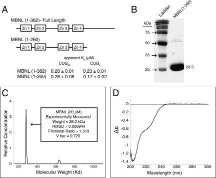
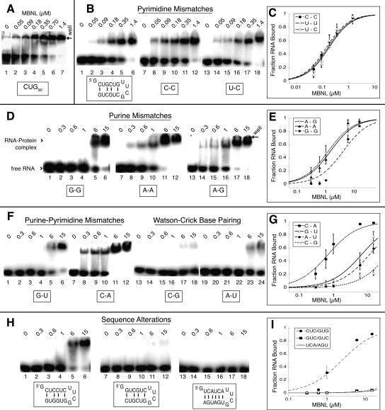
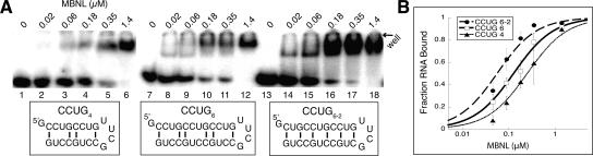
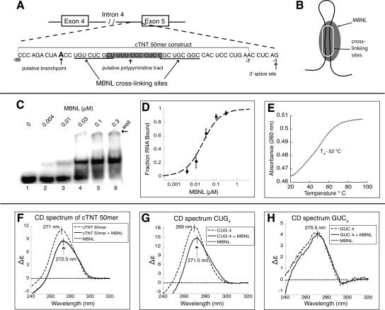
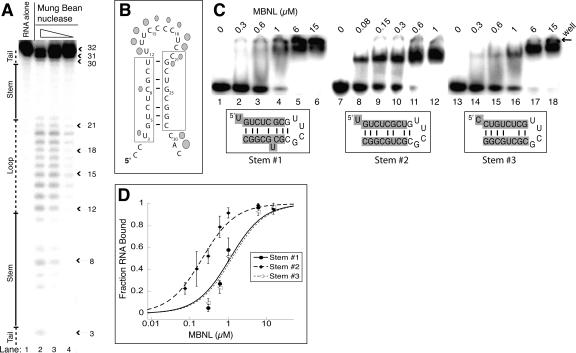
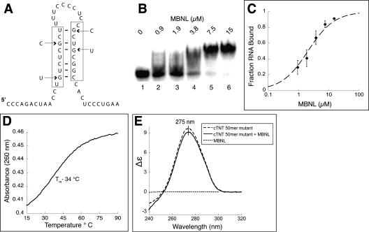
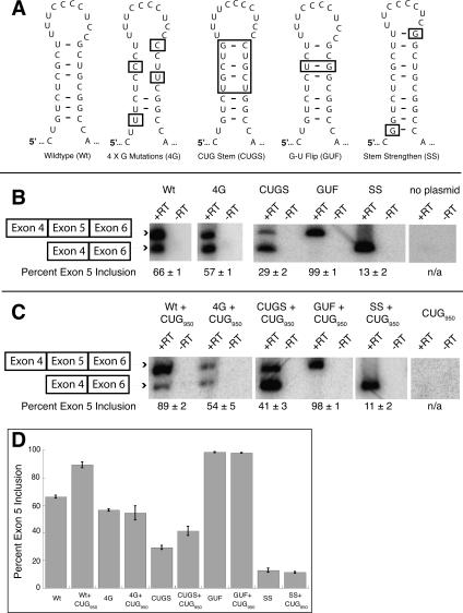
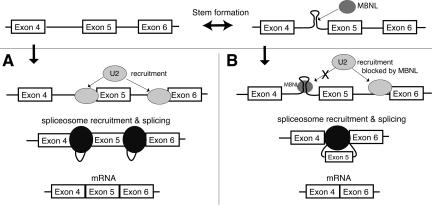
Myotonic dystrophy (DM) is a genetic disorder with multisystemic symptoms that is caused by expression (as RNA) of expanded repeats of CTG or CCTG in the genome. It is hypothesized that the RNA splicing factor muscleblind-like (MBNL) is sequestered to the expanded CUG or CCUG RNAs. Mislocalization of MBNL results in missplicing of a subset of pre-mRNAs that are linked to the symptoms found in DM patients. We demonstrate that MBNL can bind short structured CUG and CCUG repeats with high affinity and specificity. Only 6 base pairs are necessary for MBNL binding: two pyrimidine mismatches and four guanosine-cytosine base pairs in a stem. MBNL also has a preference for pyrimidine mismatches, but many other mismatches are tolerated with decreased affinity. We also demonstrate that MBNL binds the helical region of a stem-loop in the endogenous pre-mRNA target, the cardiac troponin T (cTNT) pre-mRNA. The stem-loop contains two mismatches and resembles both CUG and CCUG repeats. In vivo splicing results indicate that MBNL-regulated splicing is dependent upon the formation of stem-loops recognized by MBNL. These results suggest that MBNL may bind all of its RNA substrates, both normal and pathogenic, as structured stem-loops containing pyrimidine mismatches.


Figures

FIGURE 1.
FIGURE 1.
Structural characterization of MBNL. (A) A schematic of MBNL showing the truncated form MBNL(1–260) used in these studies. (B) Recombinant expressed MBNL has a molecular weight of 28.5 kDa on a 10% SDS-PAGE gel. Note there is a small amount of co-purifying contaminant with a molecular weight of ∼55–60 kDa. (C) Analytical ultracentrifugation of MBNL shows that it sediments with the weight of a monomer. MBNL has a slightly elongated shape, as its frictional ratio is 1.519. (D) Circular dichroism spectra of MBNL, showing a major peak at 203 and a minor peak at 220 nm, indicating that a portion of MBNL is disordered and MBNL lacks significant α-helical content in its structure (units Δε are molar circular-dichroic absorption).
FIGURE 2.
FIGURE 2.
MBNL binds short CUG repeats with affinity similar to longer repeats, and the presence of mismatches is necessary for binding. The concentration of MBNL is shown above each lane in micromolar in the gel shift assay. (A) MBNL binding CUG90 repeats (lanes 1–6) with an apparent Kd of 230 nm. (B) MBNL binding to pyrimidine–pyrimidine mismatches. The sequence of the (CUG)4 RNA is shown below lanes 1–6. The boxed C-C (lanes 7–12) and U-C (lanes 13–18) represent the replacement of the U-U mismatches with these mismatches. (D) MBNL binding to purine–purine mismatches in place of the U-U mismatch; boxed sequence indicates mismatch replacement base pairs. (F) MBNL binding to G-U (lanes 1–6) and C-A (lanes 7–12) mismatches and Watson–Crick base pairs in place of the U-U mismatches. (H) Binding of MBNL to RNAs containing sequence alterations to the cytosine and guanine positions in CUG4 and a control RNA with no sequence similarity to CUG repeats. (C,E,G,I) Binding curves of the different mismatch and sequence alteration RNAs.
FIGURE 3.
FIGURE 3.
MBNL binds CCUG expansions with high affinity, in two possible structures. (A) MBNL binding to CCUG4 (lanes 1–6), CCUG6 (lanes 7–12), and CCUG repeats in an alternate register (labeled CCUG6–2) in lanes 13–18. Concentration of MBNL is in micromolar labeled above each lane. (B) Binding curve for CCUG constructs.
FIGURE 4.
FIGURE 4.
MBNL binds a structured region in the 3′ end of intron 4 in the human cardiac troponin T (cTNT) pre-mRNA. (A) A schematic of intron 4 of the cTNT pre-mRNA. The cTNT 50mer used for binding studies is indicated. (B) A schematic model of MBNL binding this RNA as stem–loop. (C) Binding of MBNL to cTNT 50mer with the concentration of protein labeled above each lane. (D) Binding curve of cTNT 50mer. (E) Thermal melt of the cTNT 50mer, with a Tm of 52°C. (F) Circular dichroism (CD) spectra of the cTNT 50mer with and without MBNL present (units Δε are molar circular-dicroic absorption). (G) CD spectra of CUG4 with and without MBNL present. (H) CD spectra of GUC4 with and without MBNL present.
FIGURE 5.
FIGURE 5.
MBNL binds the cTNT 32mer as a stem–loop. (A) Mung bean nuclease cleavage pattern of cTNT 32mer. The concentration of mung bean nuclease is 100, 10, and 1 units per microliter, in lanes 2–4, respectively. (B) Schematic of the likely stem–loop within cTNT intron 4, showing cleavage locations of mung bean nuclease. Larger shapes indicate strong cleavage events while smaller shapes represent weak cleavage events. The cross-linking sites determined previously are boxed (Ho et al. 2004). (C) Binding curve of the three potential stem structures of the cTNT intron 4, showing MBNL prefers structure #2. (D) Gel shifts showing MBNL binding to the three potential stems of cTNT intron 4. Concentration of MBNL is labeled above each lane; note the lower concentrations in lanes 8–12 compared to lanes 2–6 and lanes 14–18. Sequences highlighted in gray are sequences from the cTNT intron in the three different potential base pair and mismatch configurations. The nongray sequence is the tetraloop cap and an additional base pair to stabilize the structure if necessary.
FIGURE 6.
FIGURE 6.
Four point mutations destabilize the stem in the cTNT 50mer and reduce MBNL binding. (A) Schematic of the stem–loop with the four point mutations indicated by arrows. (B) Gel shift assay shows MBNL binding to the mutated 50mer. (C) Binding curve of MBNL to the mutant cTNT 50mer. (D) UV melt of the cTNT 50mer mutant, with a Tm of 34°C. (E) Circular dichroism spectra of the cTNT 50mer mutant alone (1 μM) and with MBNL (1 μM).
FIGURE 7.
FIGURE 7.
The sequence and structure of the cTNT stem–loop upstream of exon 5 is important for regulated splicing by MBNL. (A) Schematic of mutations made to the stem–loop in intron 4 of the cTNT minigene. (B) RT-PCR results of the wild-type and mutant cTNT minigenes. (C) Cotransfection with a second minigene that contains 950 CUG repeats to test the role of MBNL sequestration on the splicing of the cTNT minigenes. (D) Graphical representation of exon 5 inclusion for the different constructs with and without the over expression of the 950 CUG repeats.
FIGURE 8.
FIGURE 8.
Model of MBNL's regulation of the cTNT pre-mRNA splicing. (A) If the stem–loop does not form, the splicing machinery represented by U2 (U2 snRNP) recognizes this 3′ splice site and exon 5 is included. (B) When the stem–loop forms and MBNL binds, this 3′ splice site is not recognized by the splicing machinery and exon 5 is skipped.

References

    1. Adereth, Y., Dammai, V., Kose, N., Li, R., Hsu, T. RNA-dependent integrin α3 protein localization regulated by the muscleblind-like protein MLP1. Nat. Cell Biol. 2005;7:1240–1247. - PMC - PubMed
    1. Auweter, S.D., Oberstrass, F.C., Allain, F.H. Sequence-specific binding of single-stranded RNA: Is there a code for recognition? Nucleic Acids Res. 2006;34:4943–4959. doi: 10.1093/nar/gkl620. - DOI - PMC - PubMed
    1. Begemann, G., Paricio, N., Artero, R., Kiss, I., Perez-Alonso, M., Mlodzik, M. Muscleblind, a gene required for photoreceptor differentiation in Drosophila, encodes novel nuclear Cys3His-type zinc-finger-containing proteins. Development. 1997;124:4321–4331. - PubMed
    1. Blencowe, B.J., Bowman, J.A., McCracken, S., Rosonina, E. SR-related proteins and the processing of messenger RNA precursors. Biochem. Cell Biol. 1999;77:277–291. - PubMed
    1. Brown, R.S. Zinc finger proteins: Getting a grip on RNA. Curr. Opin. Struct. Biol. 2005;15:94–98. - PubMed

Publication types