. 2008 Jan 15;111(2):750-60.
doi: 10.1182/blood-2007-03-077222. Epub 2007 Oct 17.

Multiple signaling pathways promote B lymphocyte stimulator dependent B-cell growth and survival

Affiliations

Multiple signaling pathways promote B lymphocyte stimulator dependent B-cell growth and survival

Robert T Woodland et al. Blood. .

Abstract

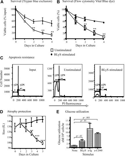
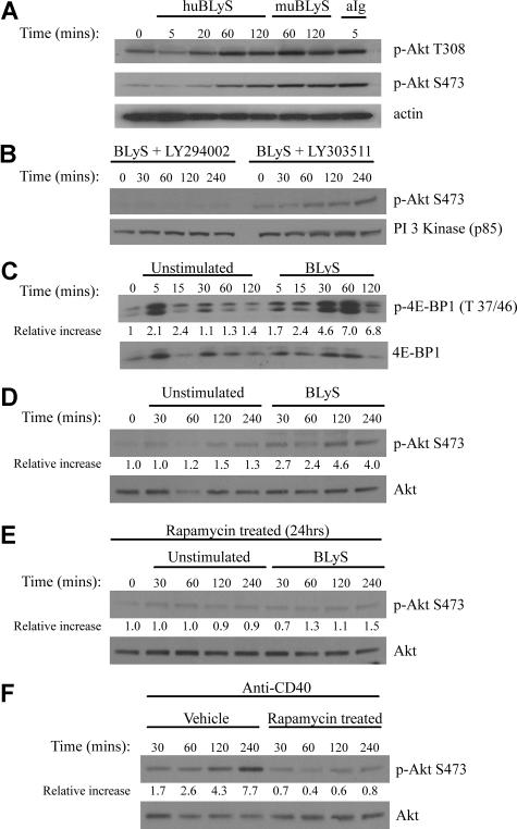
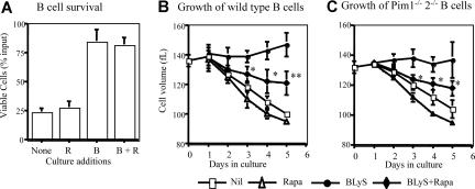
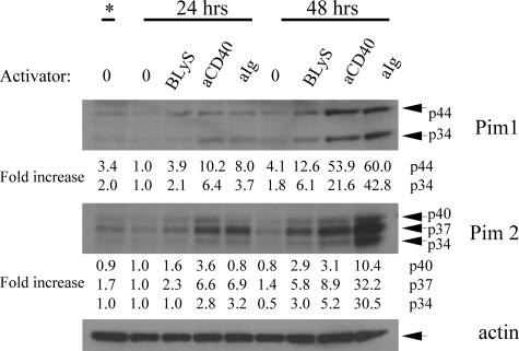
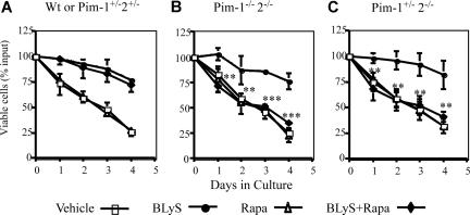
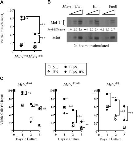
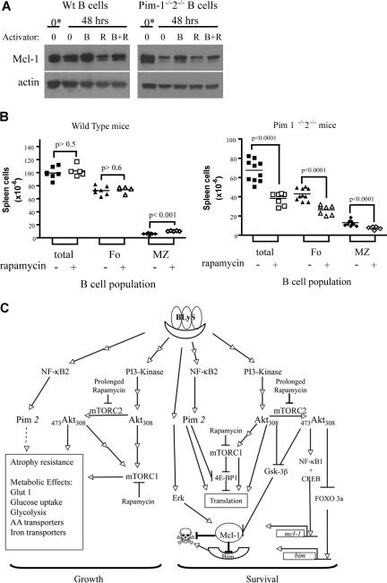
We investigated the mechanism by which B lymphocyte stimulator (BLyS)/BAFF, a tumor necrosis factor superfamily ligand, promotes B-cell survival and resistance to atrophy. BLyS stimulation activates 2 independent signaling pathways, Akt/mTOR and Pim 2, associated with cell growth and survival. BLyS blocks the cell volume loss (atrophy) that freshly isolated B cells normally undergo when maintained in vitro while concurrently increasing glycolytic activity and overall metabolism. This atrophy resistance requires Akt/mTOR. We used a genetic approach to resolve the contributions of Akt/mTOR and Pim kinase pathways to BLyS-mediated survival. Pim 2-deficient B cells are readily protected from death by BLyS stimulation, but this protection is completely abrogated by treatment with the mTOR inhibitor rapamycin. Furthermore, rapamycin treatment in vivo significantly reduces both follicular and marginal zone B cells in Pim-deficient but not healthy hosts. BLyS-dependent survival requires the antiapoptotic protein Mcl-1. Mcl-1 protein levels rise and fall in response to BLyS addition and withdrawal, respectively, and conditional deletion of the Mcl-1 gene renders B cells refractory to BLyS-mediated protection. Because BlyS is required for the normal homeostasis of all B cells, these data suggest a therapeutic strategy simultaneously inhibiting mTOR and Pim 2 could target pathogenic B cells.


Figures

Figure 1
Figure 1
BLyS stimulation promotes B-cell survival and growth. (A,B) BLyS sustains B cells ex vivo. Mature small resting murine splenic B cells positively selected with anti-CD23 and streptavidin magnetic beads were cultured for 4 days with or without 100 ng/mL huBLyS. The number of viable cells was determined daily by trypan blue exclusion (A) or by flow cytometry using blue fluorescent vital dye (B); data are the arithmetic mean (± SD) of 2 separate experiments. (C) B cells cultured with BLyS resist apoptosis. Apoptosis was determined by flow cytometry using PI on CD23+ B cells freshly prepared or cultured for 3 days with and without 100 ng/mL of huBLyS. The results are representative of 3 independent experiments. PI indicates propidium iodide. (D) B cells cultured with BLyS maintain cell size ex vivo. Percoll-purified B cells were cultured for 5 days in CM with and without 50 ng/mL of rhuBLyS and sized daily using a Coulter Z2 particle analyzer. Dead cells were excluded by sedimentation over Percoll. Data shown are the arithmetic means (±SD) for 3 independent experiments with statistical comparisons between unstimulated and BlyS-stimulated B cells indicated (**P < .01; ***P < .001). (E) BLyS promotes increased glucose metabolism. Glucose use by cultured CD23+ B cells left unstimulated or stimulated with 100 ng/mL rhuBLyS, 5 μg/mL anti-Ig or 0.5 μg/mL of anti-CD40 for 2 days. Results are the arithmetic means (± SD) for 2 independent experiments.
Figure 2
Figure 2
BLyS stimulates Akt and mTOR activation. (A) Phosphorylation of Akt. Purified B cells were stimulated for the indicated times with 100 ng/mL recombinant human or murine rBLyS or 5 μg/mL of anti-Ig at 37°C in pregassed medium. Lysates were prepared from iced samples and analyzed by Western Blot, using antibodies specific for the phosphoserine (S473) and phosphothreonine (T308) residues associated with Akt activation. (B) Inhibition of BLyS induced Akt phosphorylation by the PI 3-kinase inhibitor LY294002. Akt phosphorylation on Ser 473 was followed in purified B cells treated with LY294002 or the inactive analog LY303511 for 1 hour before BLyS stimulation. (C) BLyS induced phosphorylation of the mTOR substrate 4E-BP1. Phosphorylation of E4-BP1 on Thr 37/46 was followed by unstimulated or BLyS-stimulated purified B cells. Quantitation of Western signals represent fold increases of phosphorylated 4E-BP1 relative to that found in unstimulated B cells at time zero with a correction for sample loading performed on the same Western blot stripped and reprobed for total 4E-BP1 protein. Quantitation was performed with a Molecular Dynamics Densitometer as described in “Antibodies and Western blotting.” (D) BLyS induces Akt phosphorylation, whereas incubation of B cells in media only does not. Purified B cells were analyzed over 4 hours for Akt phosphorylation on S473 in the absence and presence of 100 ng/mL of rhuBLyS. Quantitation was as described in panel C. (E) Akt phosphorylation on S473 is inhibited in purified B cells precultured with rapamycin. B cells were cultured for 24 hours with 50 nM of rapamycin and then cultured for the indicated times with media only or media with 100 ng/mL of rhuBLyS. Panel D shows the response to BLyS of B cells incubated without rapamycin. (F) Akt phosphorylation on S473 is impaired in B cells precultured in rapamycin and activated with anti-CD40 antibody. Purified B cells were cultured for 24 hours in vehicle or 50 nM of rapamycin and then stimulated with 0.5 μg/mL of anti-CD40 antibody.
Figure 3
Figure 3
Rapamycin inhibits B-cell growth but not survival. (A) BLyS-dependent ex vivo B-cell survival is resistant to rapamycin. Small resting B cells from normal donors were cultured for 4 days with and without 100 ng/mL rhuBLyS, vehicle, or 50 nM rapamycin, which was used to pretreat B cells before culture, added directly to cultures on initiation and readded every 2 days. Viable cells were determined at day 4. Data are the arithmetic mean (±SD) and the results of 3 independent experiments. (B,C) Rapamycin inhibits BLyS-dependent maintenance of B cell size. CD23+ B cells from (B) normal or (C) Pim 1−/− 2−/− double-deficient donors were cultured with and without BLyS and with vehicle or rapamycin for 5 days. Cell size was determined for viable cells daily by a Coulter Z2 size analyzer. Results are the arithmetic mean (±SD) from 3 independent experiments. Statistical comparisons are between BLyS and BLyS plus rapamycin cell populations (* P < .05; ** P < .01).
Figure 4
Figure 4
BLyS induces both Pim 1 and Pim 2. Purified small resting B cells were cultured for 2 days unstimulated or with 100 ng/mL rhuBLyS, 5 μg/mL anti-Ig or 0.5 μg/mL anti-CD40. Lysates from the freshly isolated input population (*) and after 24 and 48 hours of culture were prepared and analyzed by Western blot. Results are representative of 4 independent experiments and show the 2 isoforms of Pim1 at 33 and 44 kDa, and the 3 isoforms of Pim 2 at 34, 37, and 40 kDa. Relative protein levels of each Pim isoform were determined by densitometry and normalized to the unstimulated B-cell population cultured for 24 hours as described in Figure 2C.
Figure 5
Figure 5
BLyS-dependent survival is rapamycin sensitive in Pim 2–deficient B cells. CD23+ B cells from: (A) wild-type or Pim 1−/+2−/+ heterozygotes, (B) Pim 1−/− 2−/− double-deficient, or (C) Pim 2 only− deficient (Pim 1+/− 2−/−) donors were cultured in CM for 4 days with vehicle, 100 ng/mL rhuBLyS, with or without 50 nM rapamycin (Rapa). Viability was determined daily by trypan blue exclusion. Results are the arithmetic means (±SD) from 3 independent experiments. Statistical comparisons are between BLyS-treated and BLyS and rapamycin-treated B cells (**P < .01; ***P < .001).
Figure 6
Figure 6
Cre-dependent excision of Mcl-1 blocks BLyS-dependent in vitro B-cell survival. (A) Excision of the Mcl-1 gene in Mcl-1 f/null B cells renders the targeted B cells refractory to BLyS-mediated survival protection. Small resting B cells from donors expressing an Mx-cre transgene and heterozygous for floxed and wild-type Mcl-1 alleles (Mcl-1f/wt) or for floxed and null Mcl-1 alleles (Mcl-1f/null) were cultured for 3 days with and without 100 ng/mL rhuBLyS in the presence of 200 units of IFNα to induce Mcl-1 excision. Viability was assessed by trypan blue dye exclusion. Each point is the average of 3 determinations with statistical comparisons included: P > .05 (not significant); ***P < .001. (B) Gene dose affects the amount of Mcl-1 protein in Percoll purified resting B cells. Mcl-1 protein was determined by Western blot of serially diluted (2-fold) cell lysates prepared from B cells taken from donors heterozygous for floxed and wild-type (Mcl-1f/wt) or floxed and null (Mcl-1f/null) alleles or homozygous for floxed Mcl-1 alleles. Proteins were quantified by densitometry as described Figure 2C. Results are representative of 2 independent experiments. (C) Mcl-1 gene dose modulates the effectiveness of BLyS-dependent B-cell protection. Small resting B cells from Mcl-1f/wt, Mcl-1f/null, or Mcl-1f/f donors expressing an Mx-cre transgene were cultured for 3 days with or without 100 ng/mL rhuBLyS in the presence or absence of 200 units of IFNα. Viability was determined by trypan blue exclusion. Statistical comparisons between B cells cultured with BLyS and B cells cultured with BLyS and interferon (P > .05, not significant; **P < .01; ***P < .001). Each point is the average of 3 determinations in this representative experiment done 3 times. Nil indicates cells cultured without BLyS or IFNα.
Figure 7
Figure 7
Modulation of Mcl-1 by BLyS, rapamycin and Pim; reduction of peripheral B-cell populations in Pim 1 and 2 doubly deficient mice receiving rapamycin in vivo; and a minimal molecular model of BLyS-dependent growth and survival. (A) Modulation of Mcl-1 protein by BLyS, rapamycin, and Pim. Extracts of small resting wild-type or Pim 1−/−2−/− B cells either freshly isolated (*) or cultured for 2 days in CM with or without rhuBLyS (50 ng/mL) and with vehicle or rapamycin (50 nM) were electrophoretically separated and transferred to nitrocellulose membranes. Membranes were probed with anti–Mcl-1 antibody, stripped, and reprobed with anti-actin antibody. (B) Peripheral B-cell survival in rapamycin-treated mice; 5- to 8-week-old Pim 1−/−2−/− doubly deficient or normal littermates were injected intraperitoneally with 2.5 mg/kg of rapamycin or diluent every other day for 6 days and killed on day 7. The number of viable spleen cells from individual mice was determined by counting and trypan blue exclusion. Peripheral B-cell subpopulations were identified by flow cytometry of viable lymphocytes stained for B220, CD 21/35 and CD23. Total B lymphocytes (B220+; □ represents rapamycin-treated donors; ■, diluent-treated donors) were subdivided into marginal zone (MZ: B220+, CD21/35hi, and CD23lo; ◇ represents rapamycin-treated donors; ♦, diluent-treated donors) or follicular (Fo: B220+, CD21/35int, CD23hi; △ represents rapamycin-treated donors; ▲, diluent-treated donors) B-cell subpopulations, and their number in each donor spleen was determined by reference to the splenocyte counts. Data pooled from 2 independent experiments are shown. (C) BLyS-mediated signaling pathways regulating B-cell growth and survival. Stimulation of B cells by BLyS leads to the activation of PI3-kinase, which phosphorylates Akt on T308 (current study) and by a separate pathway, the processing and nuclear localization of NF-κB2 with the subsequent induction of pim 2 mRNA and production of constitutively active Pim 2 protein.,, (current study). Akt is fully activated by phosphorylation on S473 mediated by the rictor containing TOR complex mTORC2 (current study). Akt and the raptor containing TOR complex mTORC1 converge to promote rapamycin-sensitive cell growth, (current study) by inducing glucose uptake and use and up-regulating amino acid and iron transporters, which maintain cell size and promote atrophy resistance,, (current study). Pim 2 also increases glycolysis and promotes maintenance of cell size by a rapamycin-independent, glucose-dependent mechanism, (current study). BLyS-induced survival requires the antiapoptotic Bcl-2 family member Mcl-1 (current study). Akt induces the activation of CREB and NF-κB1, which promote mcl-1 transcription., Akt also inactivates FOXO3a,, (current study), suppressing the transcription of the proapoptotic protein Bim, a major regulator of B-cell homeostasis., Mcl-1 protein levels are controlled at the level of translation by mTORC1 and Pim 2, which inactivate the translational repressor 4E-BP1 by rapamycin-dependent and -independent processes; Mcl-1 protein is stabilized by Erk, reported to be induced by BLyS stimulation, whereas Mcl-1 turnover is enhanced by the action of Gsk-3b, which is inhibited by active Akt (current study). Mcl-1 protein is reduced by rapamycin treatment (current study). Reference citations in this legend for activities induced by BLyS stimulation are in bold. AA indicates amino acid.

Similar articles

Cited by

References

    1. Plas DR, Rathmell JC, Thompson CB. Homeostatic control of lymphocyte survival: potential origins and implications. Nat Immunol. 2002;3:515–521. - PubMed
    1. Bodmer JL, Schneider P, Tschopp J. The molecular architecture of the TNF superfamily. Trends Biochem Sci. 2002;27:19–26. - PubMed
    1. Varfolomeev E, Kischkel F, Martin F, et al. APRIL–deficient mice have normal immune system development. Mol Cell Biol. 2004;24:997–1006. - PMC - PubMed
    1. O'Connor BP, Raman VS, Erickson LD, et al. BCMA is essential for the survival of long-lived bone marrow plasma cells. J Exp Med. 2004;199:91–98. - PMC - PubMed
    1. Rolink AG, Melchers F. BAFFled B cells survive and thrive: roles of BAFF in B-cell development. Curr Opin Immunol. 2002;14:266–275. - PubMed

Publication types

MeSH terms