. 2008 Apr;128(4):917-25.
doi: 10.1038/sj.jid.5701099. Epub 2007 Oct 18.

Co-regulation and interdependence of the mammalian epidermal permeability and antimicrobial barriers

Affiliations

Co-regulation and interdependence of the mammalian epidermal permeability and antimicrobial barriers

Karin M Aberg et al. J Invest Dermatol. 2008 Apr.

Abstract

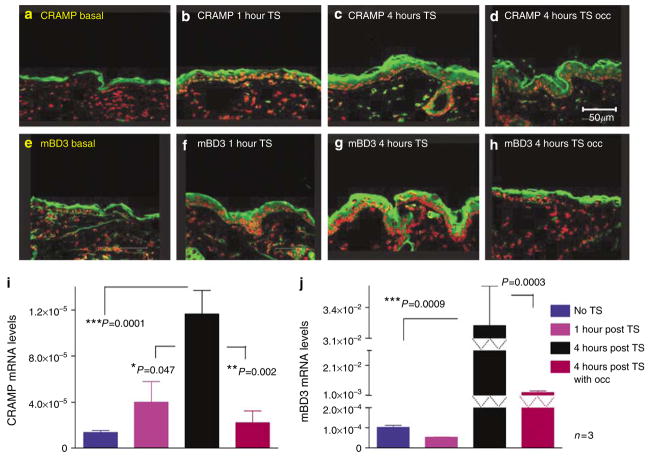
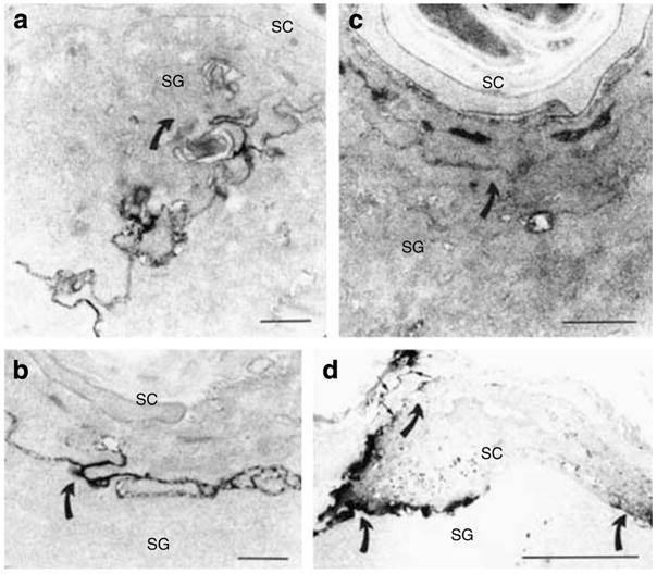
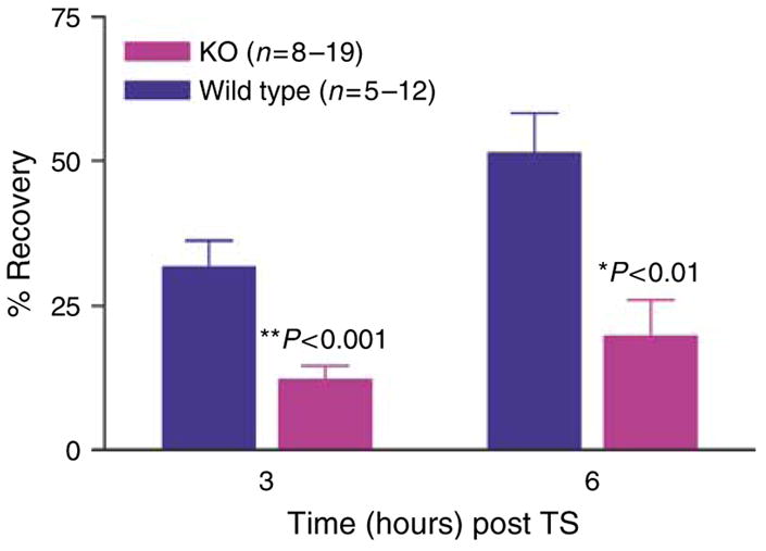
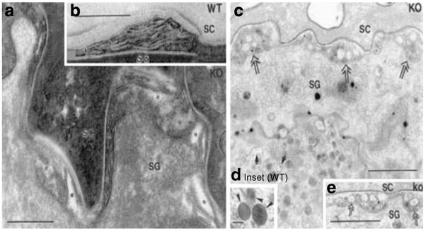
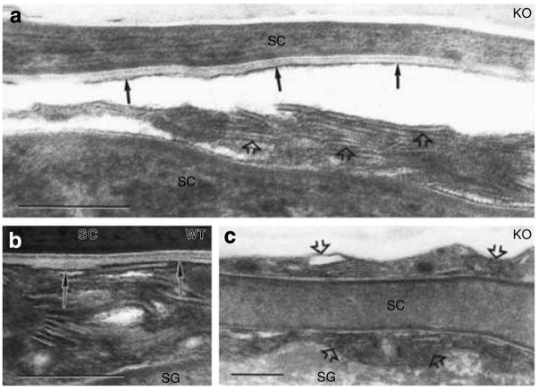
Human epidermis elaborates two small cationic, highly hydrophobic antimicrobial peptides (AMP), beta-defensin 2 (hBD2), and the carboxypeptide cleavage product of human cathelicidin (hCAP18), LL-37, which are co-packaged along with lipids within epidermal lamellar bodies (LBs) before their secretion. Because of their colocalization, we hypothesized that AMP and barrier lipid production could be coregulated by altered permeability barrier requirements. mRNA and immunostainable protein levels for mBD3 and cathelin-related antimicrobial peptide (CRAMP) (murine homologues of hBD2 and LL-37, respectively) increase 1-8 hours after acute permeability barrier disruption and normalize by 24 hours, kinetics that mirror the lipid metabolic response to permeability barrier disruption. Artificial permeability barrier restoration, which inhibits the lipid-synthetic response leading to barrier recovery, blocks the increase in AMP mRNA/protein expression, further evidence that AMP expression is linked to permeability barrier function. Conversely, LB-derived AMPs are also important for permeability barrier homeostasis. Despite an apparent increase in mBD3 protein, CRAMP-/- mice delayed permeability barrier recovery, attributable to defective LB contents and abnormalities in the structure of the lamellar membranes that regulate permeability barrier function. These studies demonstrate that (1) the permeability and antimicrobial barriers are coordinately regulated by permeability barrier requirements and (2) CRAMP is required for permeability barrier homeostasis.


Conflict of interest statement

CONFLICT OF INTEREST

The authors state no conflict of interest.

Figures

Figure 1
Figure 1. Acute permeability barrier disruption by tape stripping (TS) stimulates CRAMP and mBD3 expression; upregulation is blocked by occlusion
Flanks of normal hairless mice (n=3 in all groups) were sequentially tape stripped until TEWL≥10-fold higher than in untreated controls. Tape-stripped (TS) sites on some animals were immediately occluded with a vapor-impermeable (Latex) membrane. (ah) Frozen sections (8 μm) from biopsies of CRAMP and mBD3 were immunostained as described in Materials and Methods. (i and j) mRNA was isolated from epidermis and quantitated by reverse transcription–PCR, using 18S RNA as standard (see Materials and Methods). (b and f) The levels of CRAMP and mBD3 are at baseline, likely replenished via rapid secretion of preformed lamellar body contents. (i and j) As expected, there is no significant increase in mRNA levels at this time point. At 4 hours after TS, expression of CRAMP and mBD3 increases (c and g), which is blocked by occlusion (d and h). This correlates with increased mRNA levels for both CRAMP and mBD3 at the same time point (i and j). Bars=50 μm.
Figure 2
Figure 2. Soluble tracer moves up to and through SC interstices in CRAMP −/− epidermis
In wt epidermis, lanthanum tracer outward egress (indicated by direction of curved arrows) is blocked at level of SG (a), while tracer breaches the SG–SC interstices, and focally even above that level, in CRAMP−/− epidermis (c). Six hours after acute barrier disruption by tape stripping, tracer egress is again impeded at level of outer SG in wt epidermis (b, curved arrows), indicating restoration of normal barrier function. Yet, abundant tracer still traverses the entire SC, primarily via the interstices, in CRAMP−/− epidermis (d, arrows). (ad) Osmium tetroxide post-fixation. Bars=1 μm (a and c); 0.5 μm (b); and 5 μm (c).
Figure 3
Figure 3. CRAMP k.o. mice display a significant delay in permeability barrier recovery after tape stripping
Barrier recovery after tape stripping was compared in 6-week old CRAMP (Cnl-1-) k.o. versus wt mice (29). Mice were shaved 24 hours before tape stripping, which was repeated until TEWL rates ≥10-fold normal levels. The extent of permeability barrier recovery, as percent of the original abnormality, was assessed 3 and 6 hours after tape stripping.
Figure 4
Figure 4. Abnormalities LB secretory system in CRAMP −/− epidermis
(a, c, and e) CRAMP−/− epidermis. (b and d, inset) wt epidermis. Under basal conditions, CRAMP−/− epidermis reveals abnormalities in contents of individual LB (c, single arrows). In contrast, wt epidermis has LB with replete contents (d, inset, arrowheads). In the CRAMP−/− mouse epidermis, there are non-lamellar clefts within secreted LB contents at the SG–SC interface (a, asterisks; c and e, double arrows). In wt epidermis, transformation of secreted LB contents into lamellar bilayers already occurs at SG–SC interface, as demonstrated in (b) (arrows). (a and b) Ruthenium textroxide post-fixation; (c and d) osmium tetroxide post-fixation. Bars=0.2 μm (a and b); 1 μm (c and e); and 0.1 μm (d, inset).
Figure 5
Figure 5. Delayed and incomplete formation of lamellar bilayers in CRAMP −/− epidermis
In wt epidermis under basal conditions, secreted LB contents transform into lamellar bilayers within the SG–SC interface (b, solid arrows). In contrast, secreted lamellar contents remain partially untransformed several layers above SG-SC interface (a and c, open arrows), within SC interstices of CRAMP−/− mice. Moreover, lamellar bilayers, when present in SC of k.o. mice, fail to fill interstices (a, solid arrows). (ac) Ruthenium textroxide post-fixation. Bars=0.2 μm.

Similar articles

Cited by

References

    1. Altemus M, Rao B, Dhabhar FS, Ding W, Granstein RD. Stress-induced changes in skin barrier function in healthy women. J Invest Dermatol. 2001;117:309–17. - PubMed
    1. Bals R, Wang X, Wu Z, Freeman T, Bafna V, Zasloff M, et al. Human beta-defensin 2 is a salt-sensitive peptide antibiotic expressed in human lung. J Clin Invest. 1998a;102:874–80. - PMC - PubMed
    1. Bals R, Wang X, Zasloff M, Wilson JM. The peptide antibiotic LL-37/hCAP- 18 is expressed in epithelia of the human lung where it has broad antimicrobial activity at the airway surface. Proc Natl Acad Sci USA. 1998b;95:9541–6. - PMC - PubMed
    1. Bibel DJ, Aly R, Shinefield HR. Antimicrobial activity of sphingosines. J Invest Dermatol. 1992;98:269–73. - PubMed
    1. Braff MH, Di Nardo A, Gallo RL. Keratinocytes store the antimicrobial peptide cathelicidin in lamellar bodies. J Invest Dermatol. 2005;124:394–400. - PubMed

Publication types

LinkOut - more resources