Review
. 2007 Nov;8(11):869-83.
doi: 10.1038/nrg2136.

From microscopes to microarrays: dissecting recurrent chromosomal rearrangements

Affiliations
Review

From microscopes to microarrays: dissecting recurrent chromosomal rearrangements

Beverly S Emanuel et al. Nat Rev Genet. 2007 Nov.

Abstract

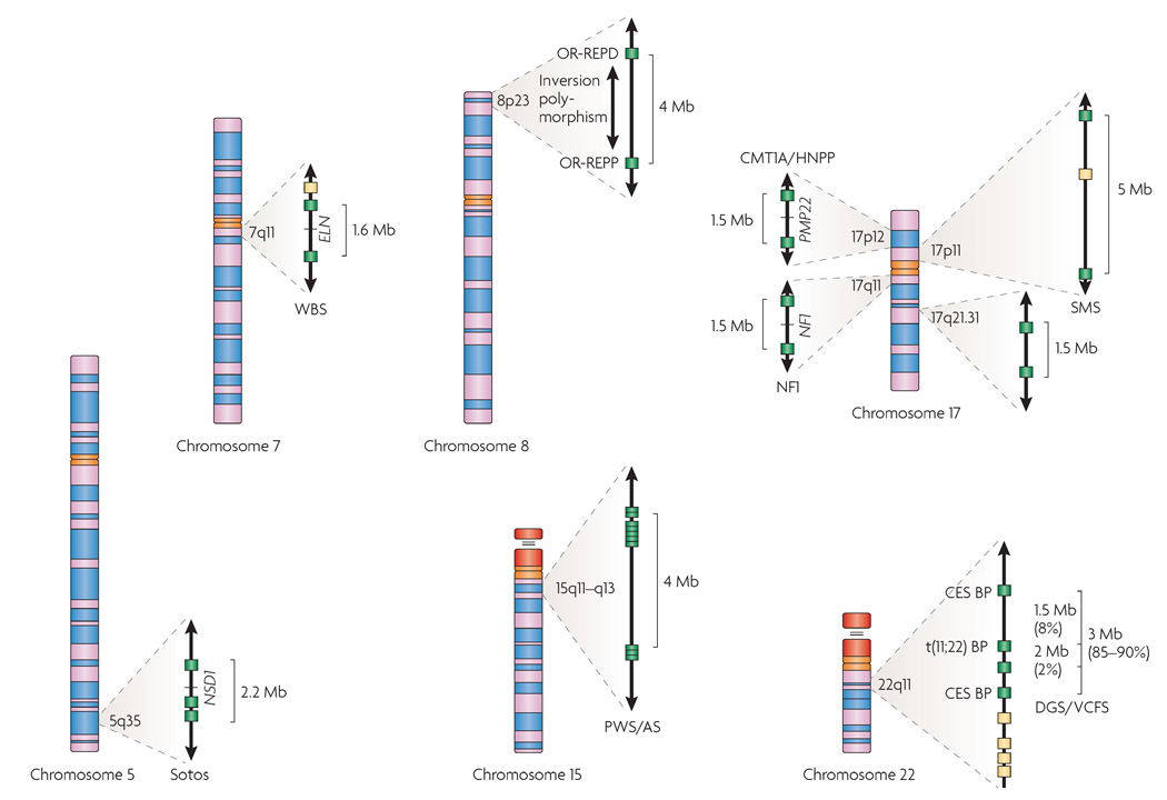
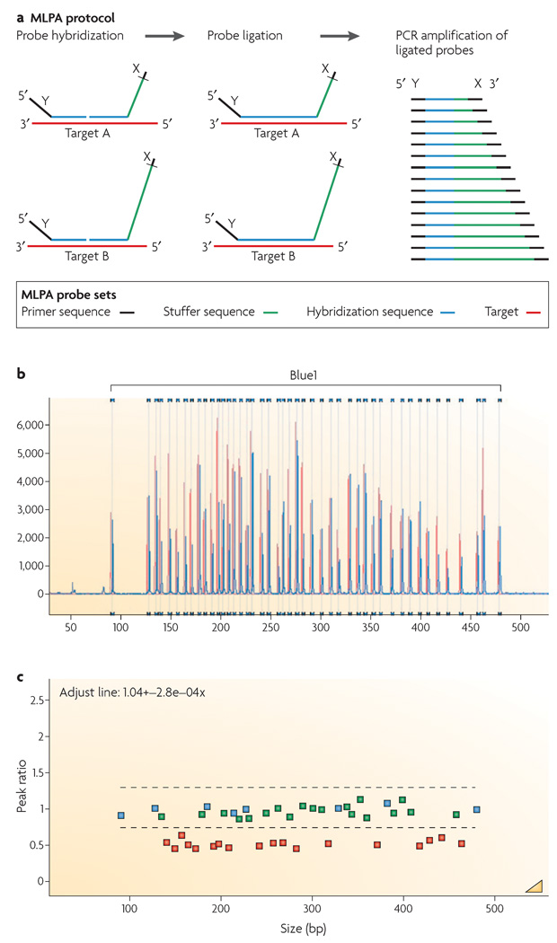
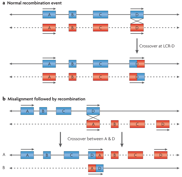
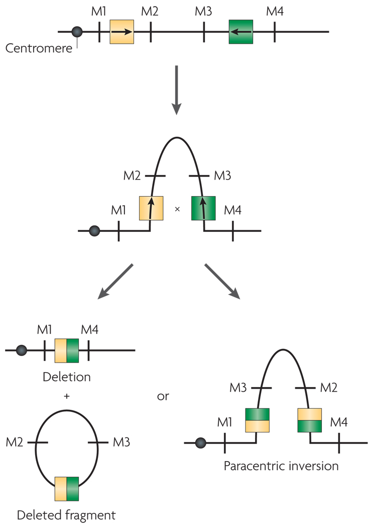
Submicroscopic chromosomal rearrangements that lead to copy-number changes have been shown to underlie distinctive and recognizable clinical phenotypes. The sensitivity to detect copy-number variation has escalated with the advent of array comparative genomic hybridization (CGH), including BAC and oligonucleotide-based platforms. Coupled with improved assemblies and annotation of genome sequence data, these technologies are facilitating the identification of new syndromes that are associated with submicroscopic genomic changes. Their characterization reveals the role of genome architecture in the aetiology of many clinical disorders. We review a group of genomic disorders that are mediated by segmental duplications, emphasizing the impact that high-throughput detection methods and the availability of the human genome sequence have had on their dissection and diagnosis.


Figures

Figure 1
Figure 1. Chromosomal rearrangements mediated by segmental duplications
The chromosomal regions involved in segmental-duplication-mediated rearrangements described in this manuscript are shown expanded. Segmental duplications are shown as filled boxes: green boxes indicate segmental duplications that are known to be involved in recurrent chromosomal rearrangements, whereas yellow boxes indicate copies that are less frequently involved in mediating known rearrangements. On chromosome 5q, the region associated with Sotos syndrome deletions is indicated along with the causative gene, nuclear-receptor-binding SET-domain protein 1 gene (NSD1). On chromosome 7q11, the deletion associated with Williams–Beuren syndrome (WBS) is shown along with the elastin gene (ELN), the main gene responsible for the supravalvular aortic stenosis of WBS. Chromosome 8 is shown with the proximal (OR-REPP) and distal (OR-REPD) olfactory-receptor gene clusters that are associated with inverted duplications of 8p and the t(4;8). Also indicated on 8p is the polymorphic inversion of the region. The region on chromosome 15q11–q13 that is deleted in Prader–Willi and Angelman syndromes (PWS/AS) is shown with its numerous duplicated hect domain and RLD 2 (HERC2) gene segments. On chromosome 17, the region on that is 17p12 duplicated or deleted in Charcot–Marie–Tooth disease Type 1A and hereditary neuropathy with pressure palsies (CMT1A/HNPP) is shown with peripheral myelin protein 22 (PMP22), the gene that is known to be involved in their aetiology. Also on 17p11 is the region involved in Smith–Magenis syndrome (SMS) deletions and the reciprocal duplication. On 17q11 is the region involved in neurofibromatosis type 1 (NF1) deletions. A recently identified segmental duplication-mediated microdeletion syndrome at 17q21.3 is indicated. On chromosome 22, recurrent deletions associated with DiGeorge and velocardiofacial syndromes (DGS/VCFS) are indicated. The proximal and distal breakpoints (BP) for the marker chromosomes in cat eye syndrome (CES) and the t(11;22) BP region are also shown.
Figure 2
Figure 2. The multiplex ligation-dependent probe amplification (MLPA) reaction
a | The figure shows two MLPA hemiprobes for each target. Each hemiprobe has a universal primer on one end. One hemiprobe is synthetic and the second, the one with the stuffer sequence (shown in green), is M13-derived. Each of the M13-derived hemiprobes has a different stuffer sequence. During the MLPA reaction, each pair of hemiprobes hybridizes to its adjacent target sequences to be enzymatically ligated. The ligation products are PCR amplified using a single primer pair (indicated as X and Y). Amplification products for each target locus have unique lengths. b | Products are separated by capillary electrophoresis. Relative probe signal strength depends on the relative amounts of target sequence that are present. c | Comparison of a test DNA sample to the copy number for control DNAs allows for calculation of the ratio. DNA from a patient with a chromosome 22 deletion indicating a 0.5 ratio for the deleted probes is used as an example. Part a modified with permission from REF. © (2002) Oxford University Press.
Figure 3
Figure 3. Model for interchromosomal recombination leading to formation of a deletion and a duplication
Chromosomes are shown as lines; solid and dotted lines are used to distinguish between the two homologues. a | Segmental duplications or low-copy repeats (LCRs) are shown as blue or red boxes with arrows to indicate the orientation of the shared modules within them. They are depicted during a normal recombination event between two properly aligned segmental duplications, A and D. b | Misalignment of segmental duplications that share sequence homology in the same orientation results in interchromosomal recombination between the two homologues of a chromosome. This results in a reciprocal duplication on homologue A and a deletion on homologue B.
Figure 4
Figure 4. Intrachromosomal recombination between segmental duplications on a single chromosome
Chromosomes are shown as lines with markers M1, M2, M3 and M4 to indicate marker order. Arrows within the segmental duplications indicate the orientation of the shared sequence elements within them. Segmental duplications that are oriented in opposite orientation to one another pair with one another based on sequence homology and loop out. A recombination event within the segmental duplications leads to either a deletion with a deleted fragment or a paracentric inversion.
Figure 5
Figure 5. Models for formation of the inv dup(15) or the cat eye syndrome marker chromosomes
Chromosomes are shown as lines; black and red are used to distinguish between the two homologues. Filled circles indicate centromeres. Segmental duplications are shown as yellow and green boxes with internal arrows to indicate the orientation of sequence blocks with high homology. a | Interchromosomal recombination between the two homologues of a particular chromosome leads to the formation of a bisatellited marker chromosome and an acentric fragment. b | Paracentric inversion within one homologue of a chromosome followed by recombination within an inversion loop leads to the formation of a bisatellited marker chromosome. Formation of an asymmetrical inv dup chromosome is shown.
Figure 6
Figure 6. Model for the translocation between 11q23 and 22q11
a | The palindromic AT-rich repeat on chromosome 22q11 or 11q23 is shown as dsDNA with the arrows indicating the head-to-head inverted repeats. DNA strand separation occurs followed by cruciform extrusion. Palindromic sequences (head-to-head arrows) are predicted to form a hairpin or cruciform structure on chromosome 11, and a similar one on chromosome 22. The tips of the cruciforms may be prone to nicking by nucleases. b | Chromosome 22 with double-strand breaks within the palindrome (coloured green) could recombine with 11q23 or with another chromosome (chromosome ‘N’) that has similar double-strand breaks. This would lead to a translocation between chromosome 22 and chromosome 11 or chromosome N, leading to the formation of der(22) and a der(11) or der(N). Most often, N is chromosome 11. cen, centromere; tel, telomere.
Figure 7
Figure 7. Ideograms and partial karyotypes of chromosome 22 abnormalities
a | The deletion of chromosome 22q11.21–11.23 (indicated by an arrow) is associated with DiGeorge and velocardiofacial syndromes. The inv dup(22) is associated with the cat eye syndrome, the +der(22)t(11;22) — a derivative chromosome 22 that is generated by the translocation between chromosomes 11 and 22 — is associated with Emanuel syndrome. The interstitial direct duplication is not visible cytogenetically. b | A copy-number diagram for the above disorders. Multiplex ligation-dependent probe amplification (MLPA) and fluorescence in situ hybridization (FISH) probes as well as copy number for the disorders shown in panel a are indicated. c | Graphical output for copy number of SNP-based probes on chromosome 22 on the Affymetrix 50K Xba Mapping GeneChip as computed by copy-number analysis software CNAG. Red dots represent raw log2 ratio values for each SNP. Blue curves represent copy-number inferences based on local mean analysis for ten consecutive SNPs. Heterozygous SNP calls are shown as green bars below the chromosome 22 ideogram at the bottom of the figure. For probes that are normal in copy number, the signal intensity ratio of the subject versus controls is expected to be 1 and the log2 ratio should be around 0.0 (log2 = 0). The deletion that has been detected in this patient with DiGeorge and velocardiofacial syndromes (DGS/VCFS) based on log2 ratio is underlined in red. Loss of copy number due to a deletion results in a negative log2 ratio (mean log2 ratio ~ −0.5).

Similar articles

Cited by

References

    1. Klein RJ, et al. Complement factor H polymorphism in age-related macular degeneration. Science. 2005;308:385–389. - PMC - PubMed
    1. Frayling TM, et al. A common variant in the FTO gene is associated with body mass index and predisposes to childhood and adult obesity. Science. 2007;316:889–894. - PMC - PubMed
    1. Dina C, et al. Variation in FTO contributes to childhood obesity and severe adult obesity. Nature Genet. 2007;39:724–726. - PubMed
    1. Helgadottir A, et al. A common variant on chromosome 9p21 affects the risk of myocardial infarction. Science. 2007;316:1491–1493. - PubMed
    1. Tishkoff SA, et al. Convergent adaptation of human lactase persistence in Africa and Europe. Nature Genet. 2007;39:31–40. - PMC - PubMed

Publication types