

Fractionated manganese-enhanced MRI
- PMID: 17944008
- PMCID: PMC4748952
- DOI: 10.1002/nbm.1211
Fractionated manganese-enhanced MRI
Abstract
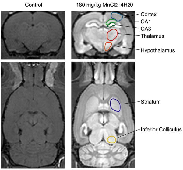
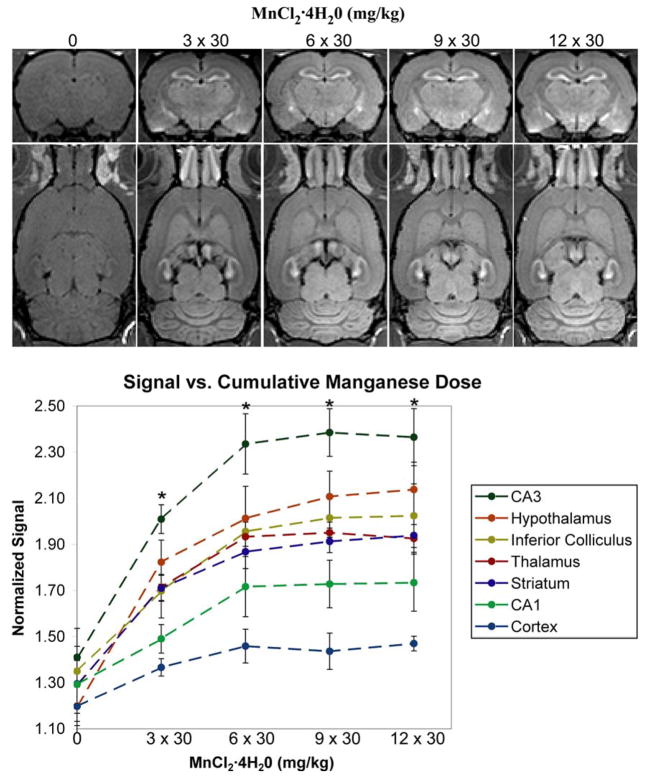
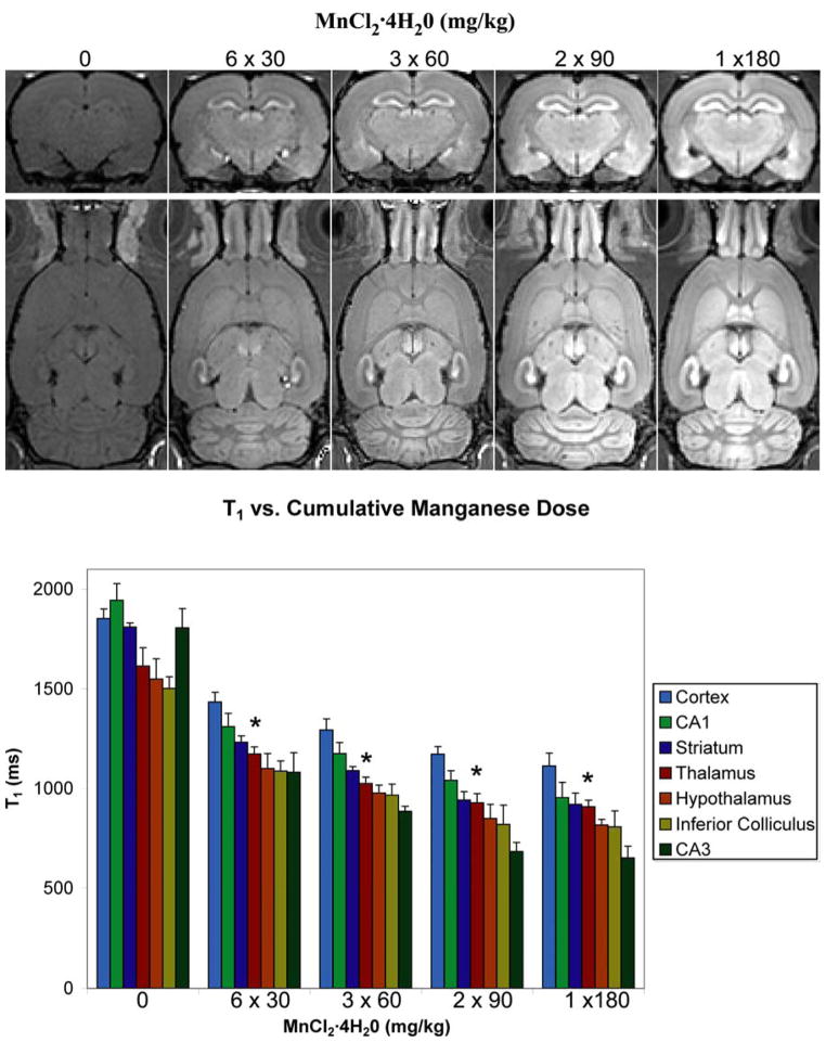
We investigated the use of manganese-enhanced MRI (MEMRI) with fractionated doses as a way to retain the unique properties of manganese as a neuronal contrast agent while lessening its toxic effects in animals. First, we followed the signal enhancement on T1-weighted images of the brains of rats receiving 30 mg/kg fractions of MnCl2 . 4H2O every 48 h and found that the signal increased in regions with consecutive fractionated doses and ultimately saturated. Second, we used T1 mapping to test whether the amount of MRI-visible manganese that accumulated depended on the concentration of manganese in the fractions. For a fixed cumulative dose of 180 mg/kg MnCl2 . 4H2O, increasing fraction doses of 6 x 30 mg/kg, 3 x 60 mg/kg, 2 x 90 mg/kg and 1 x 180 mg/kg produced progressively shorter T1 values. The adverse systemic health effects, including complications at the injection site and poor animal well-being, also rose with the fraction dose. Thus, fractionated MEMRI can be used to balance the properties of manganese as a contrast agent in animals against its toxic effects.
Figures
References
-
- Jasanoff A. Functional MRI using molecular imaging agents. Trends Neurosci. 2005;28(3):120–126. - PubMed
-
- Lauterbur PC. Image Formation by Induced Local Interactions - Examples Employing Nuclear Magnetic-Resonance. Nature. 1973;242(5394):190–191. - PubMed
-
- Koretsky AP, Silva AC. Manganese-enhanced magnetic resonance imaging (MEMRI) NMR Biomed. 2004;17(8):527–531. - PubMed
-
- Lin YJ, Koretsky AP. Manganese ion enhances T1-weighted MRI during brain activation: an approach to direct imaging of brain function. Magn Reson Med. 1997;38(3):378–388. - PubMed
-
- Takeda A, Sawashita J, Okada S. Biological half-lives of zinc and manganese in rat brain. Brain Res. 1995;695(1):53–58. - PubMed
Publication types
MeSH terms
Substances
Grants and funding
LinkOut - more resources
Full Text Sources
Medical
