. 2007 Oct 18:7:115.
doi: 10.1186/1471-213X-7-115.

Common cellular events occur during wound healing and organ regeneration in the sea cucumber Holothuria glaberrima

Affiliations

Common cellular events occur during wound healing and organ regeneration in the sea cucumber Holothuria glaberrima

José E San Miguel-Ruiz et al. BMC Dev Biol. .

Abstract

Background: All animals possess some type of tissue repair mechanism. In some species, the capacity to repair tissues is limited to the healing of wounds. Other species, such as echinoderms, posses a striking repair capability that can include the replacement of entire organs. It has been reported that some mechanisms, namely extracellular matrix remodeling, appear to occur in most repair processes. However, it remains unclear to what extent the process of organ regeneration, particularly in animals where loss and regeneration of complex structures is a programmed natural event, is similar to wound healing. We have now used the sea cucumber Holothuria glaberrima to address this question.

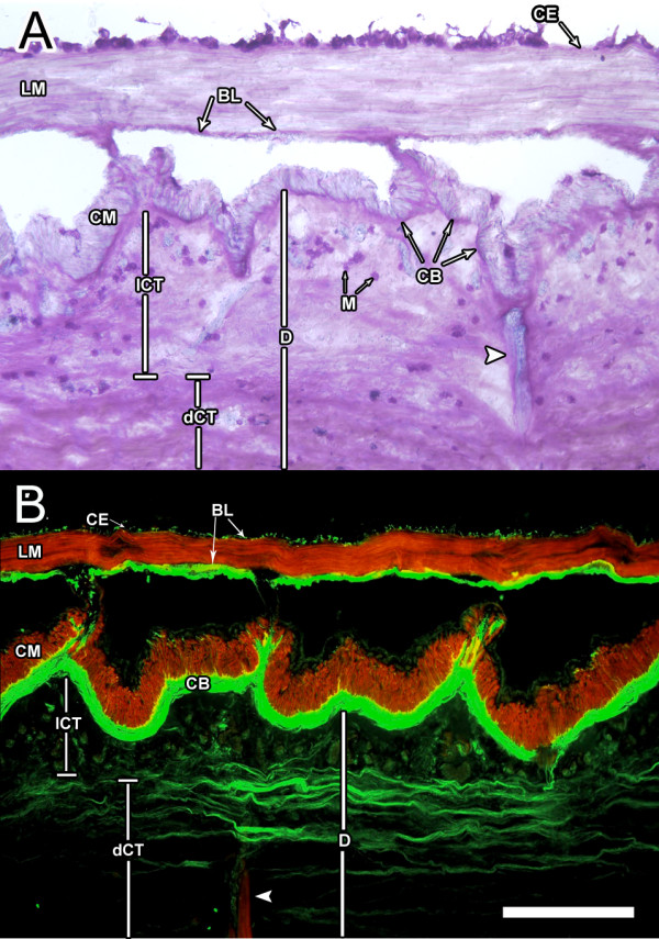
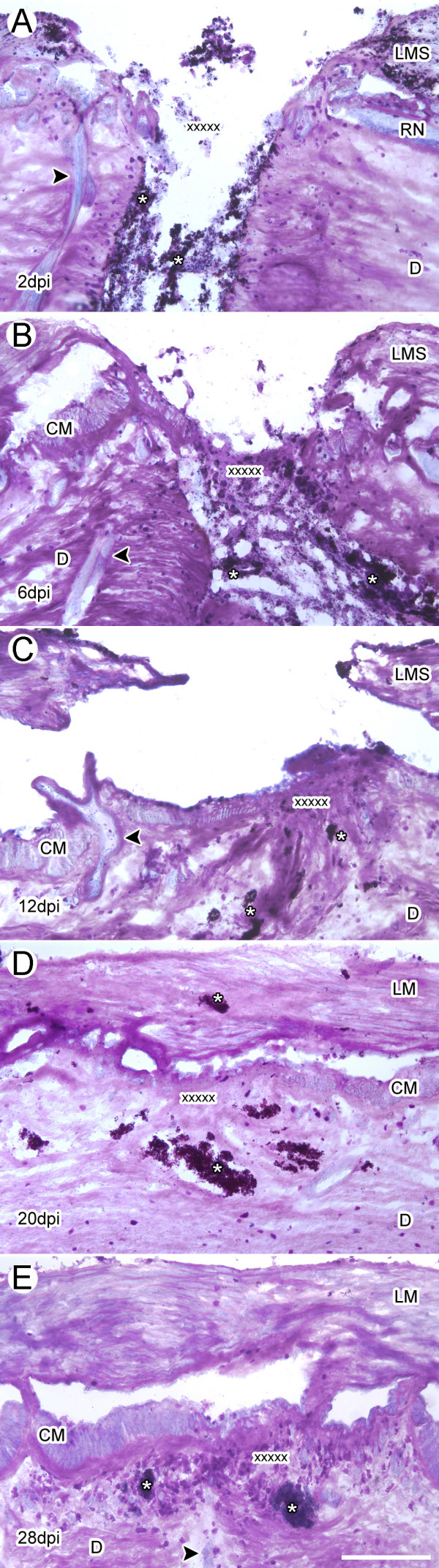
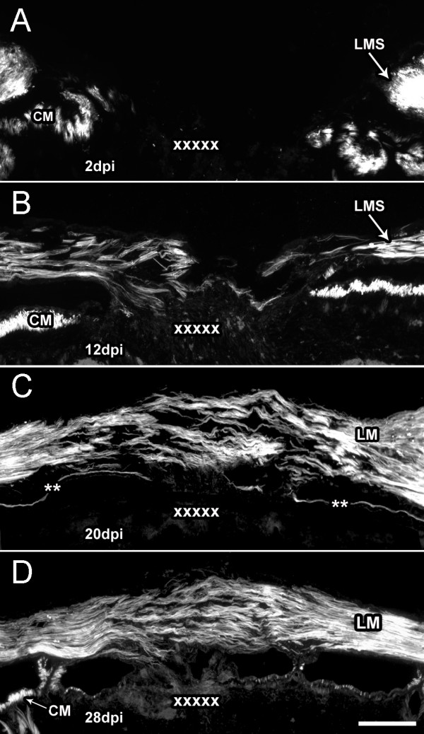
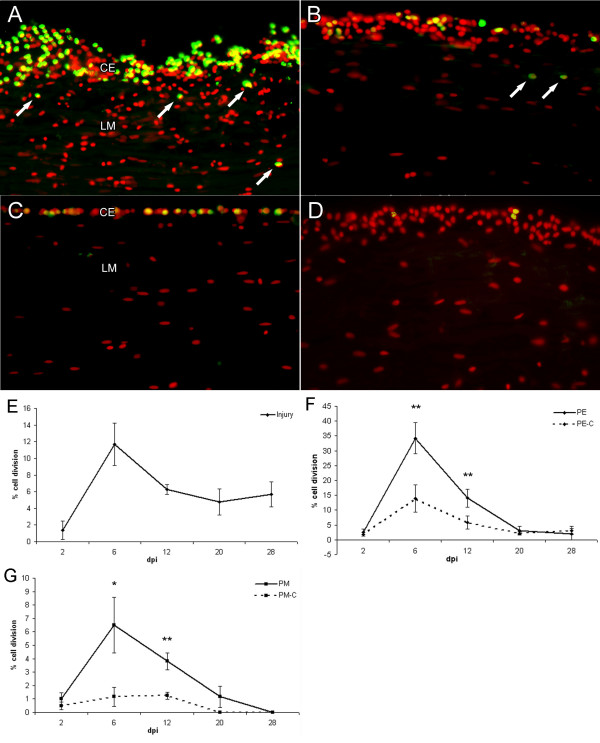
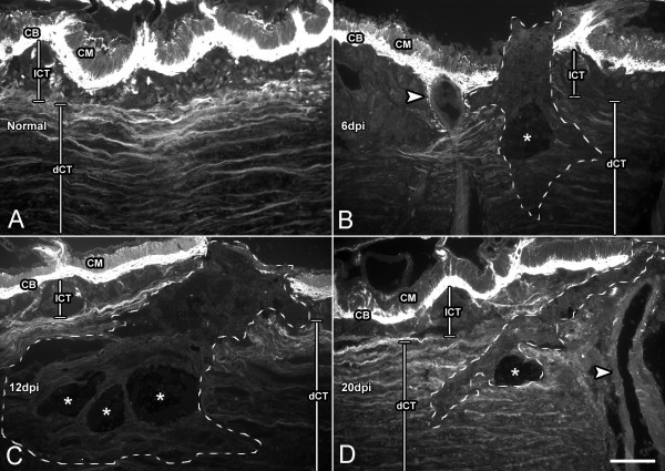
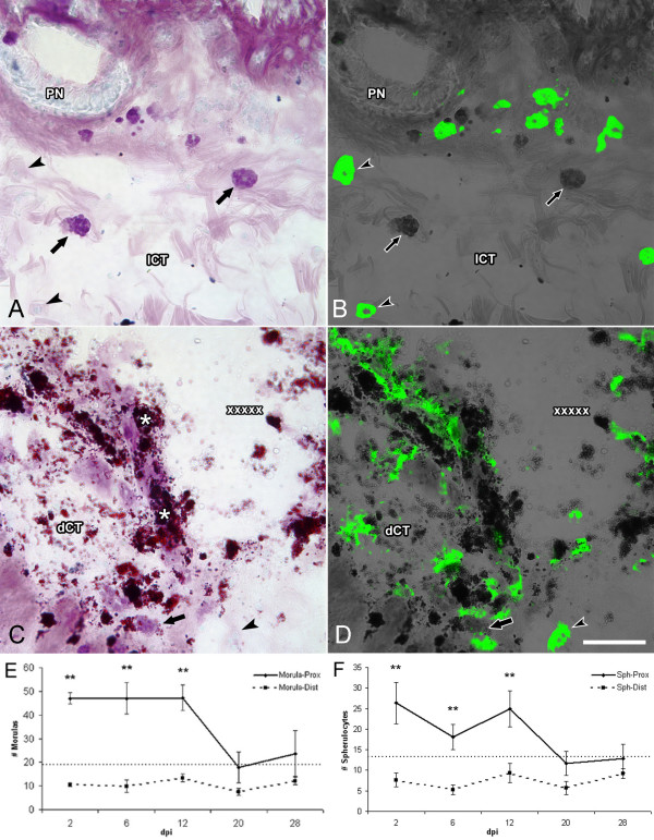
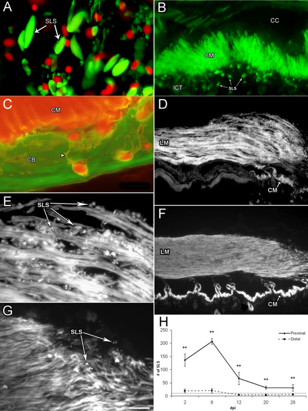
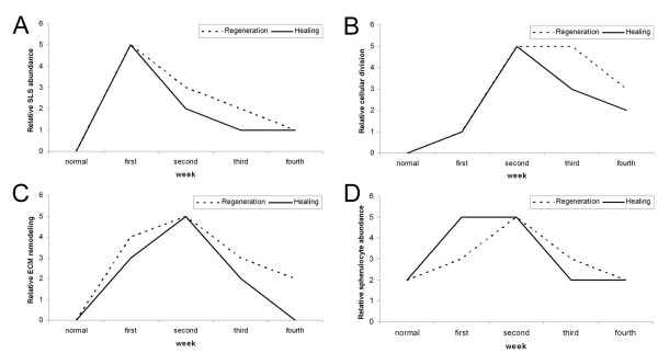
Results: Animals were lesioned by making a 3-5 mm transverse incision between one of the longitudinal muscle pairs along the bodywall. Lesioned tissues included muscle, nerve, water canal and dermis. Animals were allowed to heal for up to four weeks (2, 6, 12, 20, and 28 days post-injury) before sacrificed. Tissues were sectioned in a cryostat and changes in cellular and tissue elements during repair were evaluated using classical dyes, immmuohistochemistry and phalloidin labeling. In addition, the temporal and spatial distribution of cell proliferation in the animals was assayed using BrdU incorporation. We found that cellular events associated with wound healing in H. glaberrima correspond to those previously shown to occur during intestinal regeneration. These include: (1) an increase in the number of spherule-containing cells, (2) remodeling of the extracellular matrix, (3) formation of spindle-like structures that signal dedifferentiation of muscle cells in the area flanking the lesion site and (4) intense cellular division occurring mainly in the coelomic epithelium after the first week of regeneration.

Conclusion: Our data indicate that H. glaberrima employs analogous cellular mechanisms during wound healing and organ regeneration. Thus, it is possible that regenerative limitations in some organisms are due either to the absence of particular mechanisms associated with repair or the inability of activating the repair process in some tissues or stages.


Figures

Figure 1
Figure 1
Bodywall tissue organization. Longitudinal sections of H. glaberrima bodywall depicting its major components, (A) stained with Toluidine blue and (B) double labeled with rhodamine-labeled phalloidin (red) and monoclonal antibody against holothurian collagen (HgCol, green). The main muscle systems and their relationship to the dense and loose connective tissue of the bodywall can be seen. BL-basal lamina of the coelomic epithelia, CB-collagen band, CE-coelomic epithelia, CM-circular muscle, D-dermis, including the loose (lCT) and dense (dCT) connective tissue, LM-longitudinal muscle, M-morulas. Arrowheads identify the ampulla within the bodywall. Bar = 300 μm.
Figure 2
Figure 2
Stages in wound healing following transection of H. glaberrima bodywall. Longitudinal tissue sections of injured body wall at (A) 2, (B) 6, (C) 12, (D) 20 and (E) 28 days post injury (dpi) were stained with Toluidene Blue. (A) At 2 dpi the area is filled with debris and a clot has been formed. (B) By 6 dpi, the wound area is filled with a transitory matrix that appears to contain some of the clot material as well as new extracellular material. The muscle has healed and the stumps appear to be regenerating. (C) By 12 dpi the bodywall connective tissue has recovered some of its original organization, although its composition appears different from the non-injured tissue. The longitudinal muscle stumps have grown but have not rejoined. (D) By 20 dpi the connective tissue still appears slightly disorganized but the muscle stumps have connected forming a continuous longitudinal muscle. (E) At 28 dpi, both the muscle and the connective tissue have recovered much of the structure and organization found within non-injured tissue. CM-circular muscle, D-dermis, LM-longitudinal muscle, LMS-longitudinal muscle stump, RN-radial nerve. X's denote the injury site; Asterisks show the presence of tattoo ink used to label the injury site; arrowheads signal the ampulla structures within the bodywall. Bar = 300 μm.
Figure 3
Figure 3
Wound healing of longitudinal muscle following transection. Body wall sections from animals at (A) 2, (B) 12, (C) 20 and (D) 28 days post-injury (dpi) were labeled with rhodamine-labeled phalloidin to determine the temporal changes in the longitudinal muscle during wound healing. (A) In the 2 dpi animal, only the muscle stumps created by the wound were labeled. (B) By 12 dpi muscle fibers projecting from the wounded muscle terminals can be found across the injury site. (C) Muscle bundles from opposing stumps can be seen to make contact and cover the injury site by 20 dpi but still show a high degree of disorganization. (D) Muscle organization improves by 28 dpi and the density of the muscle bundles increases; however, organization of muscle bundles is still not fully normal, lacking the compact dense structure found in non-injured muscles (see Fig. 1). CM-circular muscle, LM-longitudinal muscle, LMS-longitudinal muscle stumps. X's mark the injury site; The presence of circular muscle depends on the plane of section since the bands of circular muscle are discontinuous. Asterisks show the area where the circular muscle can be found in nearby sections. Bar = 200 μm.
Figure 4
Figure 4
Patterns of cell proliferation in response to wound healing. Longitudinal sections were labeled with an antibody against BrdU (green) and Hoescht dye (red) to determine cell proliferation in the longitudinal muscle (LM) and coelomic epithelia (CE). (A) Actively dividing cells were mainly observed in the coelomic epithelia proximal to the injury at 6 dpi. Note that not only the number of dividing cells appears higher but the coelomic epithelium has increased in width. Some cell division is also observed in the muscle layer (arrows) (B) An area distal to the injury site shows much less cell proliferation and a thinner coelomic epithelium. (C) Control (sham-operated) animal at 6 dpi only shows modest cell division and a very thin coelomic epithelium. (D) Minimal cell division is observed at 20 dpi suggesting the stage-dependent role that cell division plays in wound healing. (E) Quantification of cell division at the injury site shows a peak in cell proliferation at 6 dpi. (F) The coelomic epithelia of experimental animals show the largest percentage of diving cells also peaking at 6 dpi. Values of control animals increase slightly but remain significantly lower than experimental animals. (G) Cell division in the longitudinal muscles follows a similar pattern with a sudden peak in cell division at 6 dpi, however the percentage of proliferating cells is much lower than for the coelomic epithelium. CE-coelomic epithelia, LM-longitudinal muscle. PE-Proximal epithelium, PE-C Proximal epithelium-Control, PM Proximal muscle, PM-C Proximal muscle-Control. Bar = 25 μm. Each point represents the mean ± S.E. of at least three animals. *p < .05, **p < .01.
Figure 5
Figure 5
Changes in collagen immunoreactivity during wound healing. Longitudinal bodywall sections of (A) non-injured animals and animals at (B) 6, (C) 12, and (D) 20 days post-injury (dpi) labeled with an antibody against holothurian collagen. The normal distribution of collagen found in non-injured animals (A) is disrupted during wound healing. (B) Initially (6 dpi), collagen is lost close to the wound site; dotted line delimits the area depleted of collagen labeling. (C) However, in subsequent stages (12 dpi) the area where collagen disappears extends over a much wider area. (D) Latter stages (20 dpi) show collagen reappearing as the wound heals. All sections were photographed using the same exposure time (30 sec) to depict the actual differences in labeling intensity. CB-collagen band, CM-circular muscle, lCT-loose connective tissue, dCT-dense connective tissue. Arrowheads signal the ampulla within the bodywall; asterisks mark areas where the tattoo ink denotes the injury site. Bar = 200 μm.
Figure 6
Figure 6
Changes in spherule-containing cells during wound healing. Tissue sections (6 dpi) stained with (A) Toluidine blue and (B) Monoclonal antibody Sph2, show two sub-populations of spherule-containing cells: (A) morulas and (B) spherulocytes within the body wall connective tissue far from the injury site. The typical round or oval morphology with well defined spherules is observed. (C-D) At the injury site both cell types undergo dramatic change in morphology, and their spherules are no longer clearly discernable. Sph2-labeled cells, in particular, appear to be releasing their cellular content into the surrounding extracellular matrix. Cell counts show that both spherulocytes (E) and morulas (F) increase in number near the injury site during the first two weeks of wound healing. Their numbers return to levels found in non-injured animals (dotted line) during the third week. A concomitant decrease in cell numbers distal to the injury site is also observed during the first 2 weeks. Dotted line denotes the number of cells found within the bodywall of non-injured animals and of controls. dCT-dense connective tissue, lCT-loose connective tissue, PN- peripheral nerve. Asterisks show the tattoo ink; used to label the injury site; X's denote the injury site; arrows indicate morulas; arrowheads indicate spherulocytes. Bar = 50 μm. Each point represents the mean ± S.E. of at least three animals. **p < .01.
Figure 7
Figure 7
Formation of spindle-like structures (SLS) by muscle cells during wound healing. (A) Double labeling of muscle tissue with rhodamine-labeled phalloidin (green) and Hoescht (red) shows the presence of SLS at the injury site. The lack of nuclei within or near the SLS shows the non-cellular nature of these structures. (B) Rhodamine-labeled phalloidin shows the circular muscle band at 6 dpi proximal to the injury site and the released SLS's (arrows) in the bodywall dermis. (C) Double labeling with rhodamine-labeled phalloidin (red) and an antibody against holothurian collagen (green) shows a magnification of the circular muscle and the SLS in the dermal connective tissue of the body wall. Cell extensions or linkers labeled with phalloidin can be observed (arrowhead). (D-G) Longitudinal muscle sections labeled with Rhodamine-labeled phalloidin. (D) At 6 dpi, proximal to the injury site, showing the loose arrangement of the muscle bundles and the presence of SLS's. (E) Magnification of D shows the large number of SLS's present around and within the muscle fibers at this stage. (F) At 28 dpi the muscle bundles are largely organized, and few SLS are observed. (G) Magnification of F showing the reduced number of SLS at the muscle terminals. (H) Quantification of SLS's shows an increase during the first week of regeneration, from 2–6 dpi. At later stages the number of SLSs decreases, although their numbers remain higher than in sham-operated controls or non-injured animals. Dotted line denotes the number of cells found within the bodywall of non-injured animals and of controls. CB-collagen band, CC-coelomic cavity, CM = circular muscle, lCT loose connective tissue, LM-longitudinal muscle, SLS's-spindle-like structure. Arrowhead indicates the SLS phalloidin-positive linker. Bar = (A&C) 10 um, (B) 25 μm, (D& F) 200 μm, (E & G) 50 μm. Each point represents the mean ± S.E. of at least three animals. **p < 0.01.
Figure 8
Figure 8
Comparison of the cellular events that occur during wound healing and organ regeneration. The four cellular events highlighted in this study were compared to those that occur during intestine regeneration [14, 16, 19, 24]. For this the magnitude of the events was assigned a relative value between 0 and 5 at the various time points where they had been studied. Each event shows a particular expression profile, nonetheless, a stunning similarity is observed between regeneration and wound healing in both the temporal pattern of the event and the peak of occurrence. Additional details provided in the text. (A) Spindle-like structure (SLS) formation, (B) cell proliferation, (C) Extracellular matrix (ECM) remodeling and (D) increase in spherulocyte population.

Similar articles

Cited by

References

    1. Brockes JP, Kumar A. Plasticity and reprogramming of differentiated cells in amphibian regeneration. Nature Rev. 2002;3:566–574. doi: 10.1038/nrm881. - DOI - PubMed
    1. Brockes JP, Kumar A, Velloso CP. Regeneration as an evolutionary variable. J Anat. 2001;199:3–11. doi: 10.1017/S0021878201008299. - DOI - PMC - PubMed
    1. Sanchez-Alvarado A. Regeneration in the metazoans: why does it happen? BioEssays. 2000;22:578–590. doi: 10.1002/(SICI)1521-1878(200006)22:6<578::AID-BIES11>3.0.CO;2-#. - DOI - PubMed
    1. Sanchez-Alvarado A. Regeneration and the need for simpler model organisms. Philos Trans R Soc Lond B Biol Sci. 2004;359:759–763. doi: 10.1098/rstb.2004.1465. - DOI - PMC - PubMed
    1. Tsonis PA. Regeneration in vertebrates. Dev Biol. 2000;221:273–284. doi: 10.1006/dbio.2000.9667. - DOI - PubMed

Publication types

LinkOut - more resources